The case of the lateral thigh, leg and knee pain

note how the bottom of the heel cup is rounded

note how the bottom of the heel cup is rounded

IMG_7238.jpg

This gal  came to see us with pain in the lateral thigh, knee, and lateral lower leg on the left. It has happened recently with skiing and alleviated only temporarily with acupuncture of the vastus lateralis and peroneal groups. She has been skiing with a foot bed (please see picture).

Evaluation reveals mild bilateral external tibial torsion, a right anatomically short  leg tibial, and bilateral medial knee fall (knee is medial of the sagittal plane) L>R during weight-bearing when the feet are pointing straight ahead. There is moderate loss of the medial longitudinal arches bilaterally, greater on the left.

We remember that when a patient has external tibial torsion, when the knees points straight the feet point to the outside. 

Translated to skiing, the feet need to be straightahead which brings the knee(s) inside of the sagittal plane. 

We remember that often times with leg length discrepancy is, the longer leg side will “pronate ” in attempt to shorten extremity and the shorter leg side “supinate”, in an attempt to lengthen the extremity.

Putting this all together:  the patient is pronating bilaterally, left greater than right with medial knee fall. The ski shop put the footbed you see in the picture in both of the patients boots. You can see that it is extremely rounded at the heel and, because orthotics are shank dependent devices, a round heel like this will just roll into pronation as there is more weight on the medial longitudinal arch. This makes the entire foot bed assembly relatively ineffective and increases the valgus moment at the knee, stressing the vastus lateralis as it is trying to pull the knee to midline as well as the peroneal group as it is trying to do the same from a closed chain position as well as supinate the foot. 

We placed a 3 mm sole lift under the right foot and added a post to the bottom the left orthotic to allow it to sit flat. This did not correct the problem completely and we needed to add a Morton’s toe extension (post under the first metatarsal) to invert the foot and bring the knee out into the sagittal plane.

Dr Ivo Waerlop, one of The Gait Guys 

#footbeds #kneepain #orthotics #skibootfit #thighpain #legpain #quadriceps

Unilateral increased tibial varum; one reason why...

Take a look at this gent in the picture. Do you notice anything peculiar? Pick a point and start either moving from above down or from the ground up.

From the ground up, the first thing you may notice is that he has a hallux abducto valgus on the right side. This could be for any number of reasons and what it actually tells you is that he is unable to anchor his first ray to the ground and have appropriate function of the adductor hallucis. Your job, during the examination process, is to sort that out.

The second thing you may notice is that he has more midfoot collapse on this same side. You would think that with that much midfoot collapse he would get his first ray to the ground but that’s obviously not the case.

Moving up from there, you may have noticed that he has significantly more tibial varum on the left-hand side. Tibial varum should be about 4-6 degrees and is largely a function of in utero positioning although diseases like osteomalacia and rickets can increase it though this is often more bilaterally symmetrical.

You need to be aware increased tibial varum means that the foot, particularly the forefoot, needs to pronate a greater degree to create a stable foot tripod on the ground. You need to ensure during the examination process that adequate range of motion in the forefoot and 1st ray are available.

You may have noticed that there is prominence of the left medial head of the gastroc which is most likely a combination of positioning as well as increased mechanical advantage secondary to the varum.

Hopefully you noticed that the knees are (relatively) in the sagittal plane and that there’s an increase progression angle on the left-hand side. If you drop a plumbline from the tibial tuberosity you’ll see the falls medial to the second metatarsal shaft indicating external tibial torsion in the lower extremity.

The unilateral increased tibial varum on the left-hand side is secondary to an anatomical leg length discrepancy where the right tibia is shorter. This has been long-standing and in compensation, the left tibia has “bowed“ to compensate for the difference, In an attempt to shorten the left leg.

Dr Ivo Waerlop, one of The Gait Guys

Like this stuff? Come and drink from the fire hydrant. Consider joining us this Wednesday evening on online CE.com for bio mechanics 326, 6 PM Mountain standard time. Likewise, you can become a Patreon supporter and get this kind of information every week.

#tibialvarum #leglengthdiscrepancy #lld #bowedlegs #pronation

Unilateral calcaneal valgus: what can it mean?

right calcaneal valgus

right calcaneal valgus

Take a good look at this picture and what do you see? Do you see the calcaneal valgus on the right side. What runs through your mind?

Possibilities for causing this condition, as well as the clinical implications are numerous.

The short list should include:

  • A shorter leg on the contralateral side: often times we will pronate more on the longer leg side to compensate for a short leg on the opposite

  • Increased rear foot and/or fore foot pronation on the valgus side. Laxity of the spring ligament or incompetency of the musculature which helps to maintain your arch (tibialis posterior, foot intrinsics, tibialis anterior to name a few) often causes more collapse on the effected side

  • A lack of available rearfoot eversion on the contralateral side. It may be that the increase calcaneovalgus is normal and the opposite side is more rigid.

  • If you were seeing this in the middle of the gait cycle it could be that that is their strategy to get around a loss of hip extension or ankle rocker

  • External tibial torsion on that side. Go ahead, stand up and spin your right foot into external rotation and keep your left foot with a normal progression angle. Can you see how your arch collapses to a greater degree on the side with the external torsion? Remember that pronation is dorsiflexion, eversion and abduction.

  • Internal tibial torsion on the contralateral side. Internal tibial torsion puts the foot into supination which makes it into more of a rigid lever rather than mobile adapter.

    And the list goes on…

    Next time you see a unilateral deformity like this, hopefully some of these things run through your mind and will help you to pinpoint where the problem actually is.

    Dr Ivo Waerlop, one of The Gait Guys

    #calcanealeversion #rearfootvalgus
    #lowerextremitydeformities

Plantar Plate Gait

This girl has a (healing) plantar plate lesion on the left hand side at the head of the second met. She also has an anatomical short leg on the same side. Her second metatarsal of both feet or longer than the first

A few things I hope you notice about the video:

  • Can you see how she “reaches“ to get to the ground with her left foot?

  • Can you see how her left foot is more inverted that strikes in the right, creating a greater amount of forefoot pronation that needs to be controlled?

  • Can you see how poor her motion control is of her pronation on the left foot with the sudden “crash” at impact?

  • Have you noticed her “crossover“ gait?


Does it make sense that because of her anatomy and running style, that the constant reach, increased forefoot inversion and lack of pronation control (which causes more abduction of the forefoot at toe off); this drives the force to the second metatarsal head which is longer and more prominent and is more than likely what led to her plantar plate lesion in the first place?


Remediation?

  • A 3 mm full length sole lift for the left foot

  • Foot intrinsic strengthening exercises

  • Hip abduction strengthening exercises/drills

  • Moving her more to a “midfoot strike” running gait with toes extended to engage the windlass


Dr Ivo Waerlop, one of The Gait Guys


#plantarplate #gaitanalysis #crossovergait #leglengthdifference #thegaitguys


When you see this, you should be thinking one of 3 possible etiologies...

Cardinal sign of either a forefoot supinatus/forefoot varus or collapsing midfoot

I was hiking behind this young chap over the weekend along with my son and friends. Note the amount of calcaneal eversion present on the right side that is not present on the left. Also note the increased progression angle of the right foot and subtle circumduction of the extremity.

In my experience, you would generally see this much calcaneal diversion and one of three scenarios:

1. Moderate leg length discrepancy with the increased calcaneovalgus occurring on the longer leg side. This would support the amount of circumduction were seeing on the right side.

2. When there is a forefoot supinatus present and and inadequate range of motion available in the midfoot and/or forefoot. This is most likely the case here.

3. In moderate To severe midfoot collapse. This is clearly not the case as the medial aspect of the shoe is usually “blown out”.

Next time you see an everting rearfoot, think about these three possible etiologies.

Dr Ivo Waerlop, on of The Gait Guys

#evertedrrarfoot #calcanealvalgus #shortleg #forefootsupinatus #forefootvarus #gaitanalysis #thegaitguys

The amazing power of compensation. Coming to a patient in your office… Maybe today

This gal has had a right sided knee replacement. She has an anatomical right short leg, a forefoot supinatus, an increased Q angle and a forefoot adductus. So, what’s the backstory?

When we have an anatomical short leg, we will often have a tendency to try to “lengthen“ that extremity and “shorten” the longer extremity. This is often accomplished through pelvic rotation although sometimes can be with knee flexion/extension or change in the Q angle. When the condition is long-standing, the body will often compensate in other ways, such as what we are seeing here.

IMG_6736.jpg

The fore foot can supinate in an attempt to lenthen the extremity. Note how the right extremity forefoot is in varus with respect to the rearfoot, effectively lengthening the extremity. As you can see from the picture, this is becoming a “hard“ deformity resulting in a forefoot varus.

IMG_6740.jpg

Over time, the forefoot has actually “adducted “ as you can see, again in an attempt to lengthen the extremity. Remember that supination is plantar flexion, abduction and inversion, all three which are visible here.


You will also see that the Q angle is less on the right side (se above), effectively lengthening that extremity, but not quite enough as we can see from the picture :-)


Dr Ivo Waerlop, one of The Gait Guys

#forefootadductus #shortleg #kneereplacement #tkr #forefootvarus #gait #thegaitguys

Neuroma! Triple Threat....

Can you guess why this patient is developing a neuroma on the left foot, between the 3rd and 4th metatarsals?

IMG_6220.jpg
IMG_6218.jpg
IMG_6219.jpg

This gal presented to the office with pain in the left foot, in the area she points to as being between the 3rd and 4th metatarsals. It has been coming on over time and has become much worse this spring with hiking long distances, especially in narrower shoes. It is relieved by rest and made worse with activity.

Note the following:

  • She has an anatomical short leg on the left (tibial)

  • internal tibial torsion on the left

  • left forefoot adductus (see the post link below if you need a refresher)

Lets think about this.

The anatomical short leg on the left is causing this foot to remain in relative supination compared the right and causes her to bear weight laterally on the foot.

The internal tibial torsion has a similar effect, decreasing the progression angle and again causing her to bear weight laterally on the foot, compressing the metatarsals together.

We have discussed forefoot adductus before here on the blog. Again, because of the metararsal varus angle, it alters the forces traveling through the foot, pushing the metatarsals together and irritating the nerve root sheath, causing hypertrophy of the epineurium and the beginnings of a neuroma.

In this patients case, these things are additive, causing what I like to a call the “triple threat”.

So, what do we do?

  • give her shoes/sandals with a wider toe box

  • work on foot mobility, especially in descending the 1st ray on the left

  • work on foot intrinsic strength, particularly the long extensors

  • treat the area of inflammation with acupuncture

Dr Ivo Waerlop, one of The Gait Guys

#forefootadductus #metatarsusadductus #neuroma #gaitanalysis #thegaitguys #internaltibialtorsion

The “Standing on Glass” Static Foot/Pedograph Assessment: Part 1

Screen Shot 2019-01-13 at 7.42.41 PM.png

The “Standing on Glass” Static Foot/Pedograph... PART 1
We hope you find this case presentation dialogue interesting.

* note: This is a static assessment dialogue. One cannot, and must not, make clinical decisions from a static assessment. As in all assessments, information is taken in, digested and them MUST be confirmed, denied and/or at the very least, folded into a functional and clinically relevant assessment of the client before the findings are accepted, dismissed and acted upon.

The “Standing on Glass” Static Foot/Pedograph Assessment: Part 1

* note: This is a static assessment dialogue. One cannot, and must not, make clinical decisions from a static assessment. As in all assessments, information is taken in, digested and them MUST be confirmed, denied and/or at the very least, folded into a functional and clinically relevant assessment of the client before the findings are accepted, dismissed and acted upon. As we always say, a gait analysis or pedograph-type assessment is never enough to make decisions on treatment to resolve problems and injuries. What is seen and represented on either are the client’s strategies around clinical problems or compensations. Today’s photo and blog post are an exercise in critical clinical thinking to get the juices flowing and to get the observer thinking about the client’s presentation and to help open up the field to questions the observer should be entertaining. The big questions should be, “why do i see this, what could be causing these observances ?”

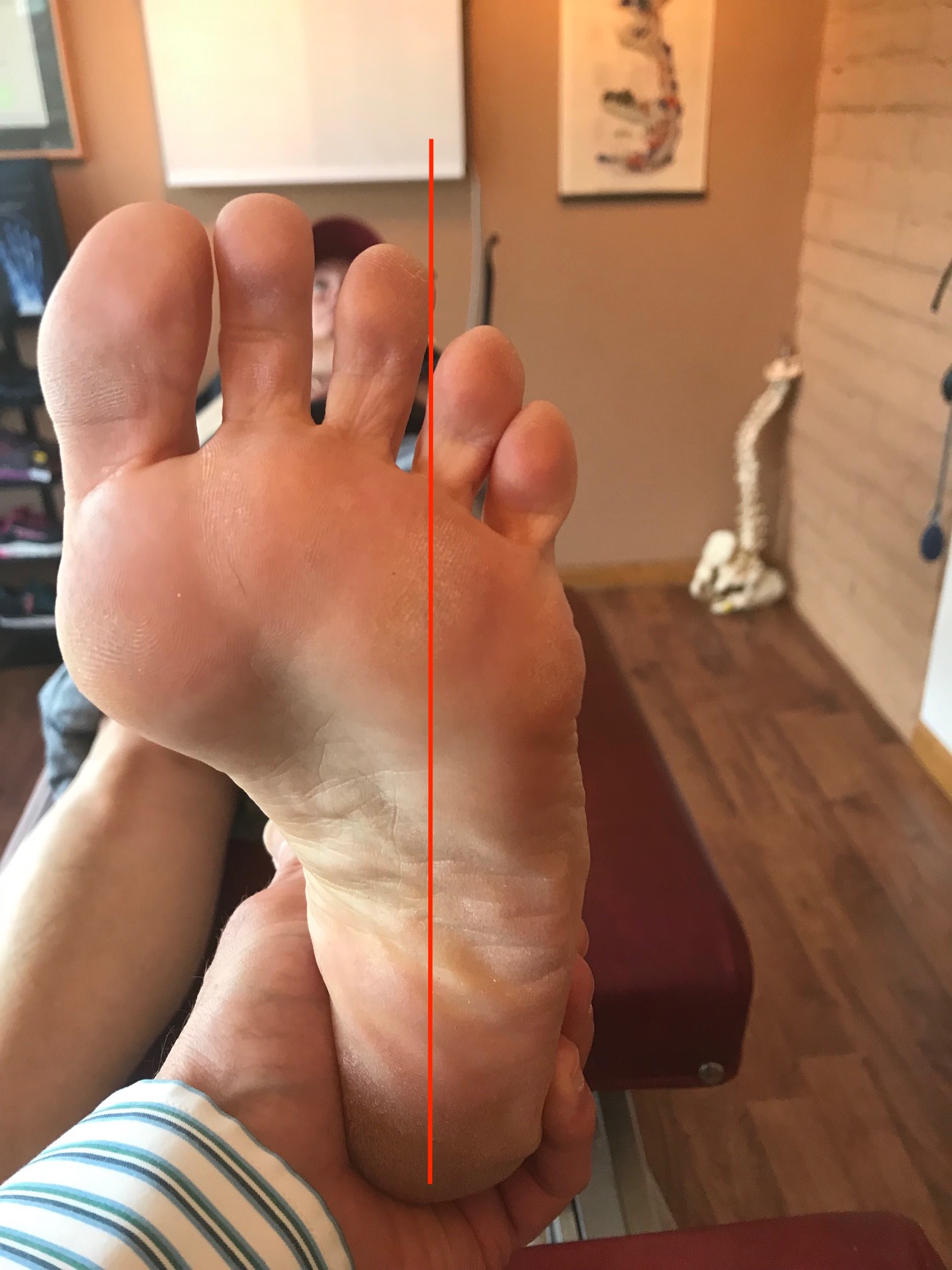
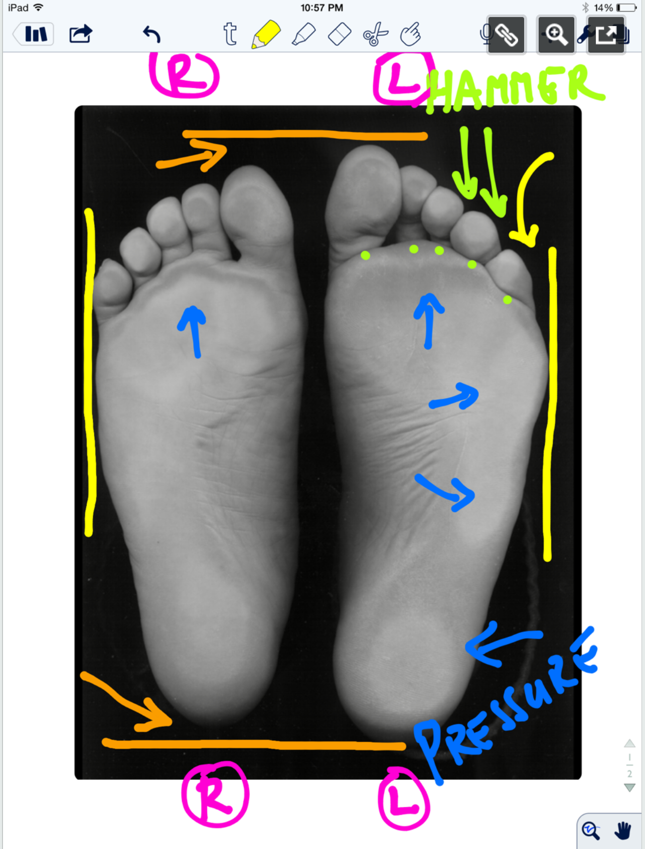
* note the right and left sides by the R and L circled in pink.

ORANGE lines: The right foot appears to be shorter, or is it that the left is longer (see the lines and arrows drawing your attention to these differences)? A shorter foot could be represented by a supinated foot (if you raise the arch via the windlass mechanism you will shorten the foot distance between the rear and forefoot). A longer foot could be represented by a more pronated foot. Is that what we have here ? There is no way to know, this is a static presentation of a client standing on glass. What we should remember is that the goal is always to get the pelvis square and level. If an anatomically or functionally short leg is present, the short leg side MAY supinate to raise the mortise and somewhat lengthen the leg. In that same client, they may try to meet the process part way by pronating the other foot to functionally “shorten” that leg. Is that what is happening here ? So, does this client have a shorter right leg ? Longer left ? Do you see a plunking down heavily onto the right foot in gait ? Remember, what you see is their compensation. Perhaps the right foot is supinating, and thus working harder at the bottom end of the limb (via more supination), to make up for a weak right glute failing to eccentrically control the internal spin of the leg during stance phase ? OR, perhaps the left foot is pronating more to drive more internal rotation on the left limb because there is a restricted left internal hip rotation from the top ? Is the compensation top-down or bottom up ? These are all viable possibilities and you must have these things flowing freely through your head during the clinical examination as you rule in/rule out your hands-on findings. Remember, just going by a FMS-type screen to drive prescription exercises from what you see on a movement screen is not going to necessarily fix the problem, it could in fact lead one to drive a deeper compensation pattern. You can be sure that Gray Cook’s turbo charged brain is juggling all of these issues (and more !) when he sees a screen impairment, although we are not speaking for him here.

Remember this critical fact. After an injury or a long standing problem, muscles and motor patterns jobs are to stabilize and manage loads (stability and mobility) for adequate and necessary movement. Injuries leave a mark on the system as a whole because adaptation was necessary during the initial healing phase. This usually spills over during the early movement re-introduction phase, particularly if movement is reintroduced too early or too aggressively. Plasticity is the culprit. Just because the injury has come and gone does not mean that new patterns of skill, endurance, strength (S.E.S -our favorite mnemonic), stability and mobility were not subsequently built onto the apparently trivial remnants of the injury. There is nothing trivial if it is abnormal. The forces must, and will, play out somewhere in the body and this is often where pain or injury occurs but it is rarely where the underlying problem lives.

Come back tomorrow, where we will open your mind into the yellow, pink, blue and lime markings on the photo. Are the hammering toes (lime) on the left a clue ? How about the width of the feet (yellow) ? The posturing differences of the 5th toe to the lateral foot border ? What about the static plantar pressure differences from side to side (blue)? Maybe, just maybe, we can bring a logical clinical assumption together and then a few clinical exam methods to confirm or dis-confirm our working diagnostic assumption. See you tomorrow friends !

Shawn and ivo, the gait guys

Here is the case link.......

https://thegaitguys.tumblr.com/post/99409232289/the-standing-on-glass-static-footpedograph?fbclid=IwAR3gd3d81Gwt3ywAB7BcTwXqST2Z_5nmieODzSb8rJQYBcJFhTs6rS_9auA

Support for visual gait analysis... with respect to leg length discrepancies


image credit: https://pixabay.com/photos/eye-blue-eyelashes-vision-make-up-691269/

image credit: https://pixabay.com/photos/eye-blue-eyelashes-vision-make-up-691269/

We talk about leg length discrepancies all the time here on the blog and sometimes, how small discrepancies cause changes in peoples biomechanics. The gold standard for measuring a leg length difference is full length lower extremity X ray, but this presents a problem due to the ionizing radiation, accessibility as well as impracticality of X rays every person with a suspected difference.

We have talked about different compensations as to how to get around a leg length discrepancies. Last week we actually did a tell a seminar on this entire subject. Your patient or client needs to “create clearance” for the longer leg side. This can be accomplished in many ways.

The 5 most common strategies (keep in mind there are many more) are:

  • lean the torso to the short leg side (essentially hip adduction of the longer side)

  • hike the torso on the long leg side

  • circumduct the longer lower extremity

  • increase plantar flexion of the calf of the short leg side

  • increase hip and knee flexion on the longer leg side

And that is exactly what this study found. They looked at kinematics in people with anatomical leg length discrepancies and found that hip adduction as well as increased hip and knee flexion were 2 variables that were consistent in folks with anatomical differences and suggest these variables are a useful screening tool.

Paying attention to how people move and looking for asymmetries. In our opinion, that’s the name of the game : )

Dr Ivo Waerlop, on of The Gait Guys

Zeitoune GNadal JBatista LAMetsavaht LMoraes APLeporace G.Prediction of mild anatomical leg length discrepancy based on gait kinematics and linear regression model. Gait Posture. 2019 Jan;67:117-121. doi: 10.1016/j.gaitpost.2018.09.027. Epub 2018 Sep 29.

#LLD #leglengthdifference #leglengthinequality #visualgaitanalysis #thegaitguys #gaitanalysis

Case Studies in Gait Analysis: Focus on the Short Leg (online video class)

Case Studies in Gait Analysis: Focus on the Short Leg
*link is below

*this is the online Continuing education class we did last week, for those of you who could not get to the Wednesday evening class.
*our entire catalogue of lectures and seminars are all here on this site for CE/CEU

Case Studies in Gait Analysis: Focus on the Short Leg
- Review anatomical vs functional short leg
-Review the kinematics and kinetics of the short leg during the gait cycle
-View and discuss case studies looking at functional and anatomical short legs
-Predict pathomechanics that will arise from a short leg
-Propose remedies for the gait abnormalities seen

Link: https://chirocredit.com/course/Chiropractic_Doctor/Biomechanics_211

Screen Shot 2019-04-22 at 1.20.16 PM.png

Wild Haggis? Leg length discrepancies on the uphill side? What?

An old Scottish myth has it that the wild haggis (given the fitting taxonomic moniker Haggis scoticus ) is a small fictitious creature (although many folks visiting Scotland believe they are real) that has legs that are longer on one side than the other. There are two varieties: in one the right fore and hind limb are shorter and the other, of course, the left. The asymmetry helps the haggis to circumnavigate the steep mountainsides of its native terrain, but only in a clockwise (if the right legs are short) or counter clockwise (if the left legs are short) direction, so as to not roll down the steep hillside and come to an untimely death; this is purported to be one of the reasons for their near extinction (the other was the introduction of sheep).

The two species coexist peacefully but are unable to interbreed in the wild because in order for the male of one variety to mate with a female of the other, he must turn to face in the same direction as his intended mate, causing him to lose his balance before he can mount her. As a result of this difficulty, differences in leg length among the haggis population are further accentuated, as is there dwindling numbers.

image source: https://en.wikipedia.org/wiki/Wild_haggis#/media/File:Haggis_scoticus.jpg

image source: https://en.wikipedia.org/wiki/Wild_haggis#/media/File:Haggis_scoticus.jpg

It’s an amusing concept, but unfortunately there’s a non-mythical human corollary: Leg-length discrepancies (LLDs), which do not discriminate and affect a wide variety of people, including children with cerebral palsy, people who’ve had hip and knee replacements, and those with scoliosis, pelvic obliquity, or certain muscle contractures/dysfunctions.

Haggis is actually a Scottish dish; lungs and liver of a sheep cooked with other ingredients inside its stomach. Yum (Not!) We are not sure why or how the two are related but it does make for an interesting post : )

Learn more about LLD’s and their compensations by joining us Wednesday, April 17th 5 PST, 6MST, 7CST and 8 EST on onlinece.com: Biomechanics 307

Dr Ivo Waerlop, one of The Gait Guys

#haggis #wildhaggis #LLD #leglengthdiscrepancy #leglengthdifference #leglengthinequality #gait #thegaitguys


https://en.wikipedia.org/wiki/Wild_haggis

https://lermagazine.com/article/limb-length-discrepancy-when-how-to-intervene

https://www.atlasobscura.com/articles/what-is-haggis

https://www.thehaggis.com/wild-haggis-all-about-haggis/

https://www.undiscoveredscotland.co.uk/usfeatures/haggis/wildhaggis.ht

There is more than one way around an LLD....

Leg length discrepancies. Love them, hate them, they happen. They can be either functional, anatomical or both.

No matter what the cause, there are numerous ways to compensate for a leg length discrepancy. Today we are going to look at one of the more common ones, "leaning" to the short leg side to create enough clearance for the opposite lower extremity. This patient has a left sided short leg. Note how he abducts his pelvis, utilizing both the stance limb gluteus medius and swing limb quadratus lumborum of the left leg to create enough space to swing the right leg through.

Want to know more about LLD’s and their compensations? Join us on onlinece.com, Wednesday, April 17th for Biomechanics 307. 6 PM Mountain time. See you there!

Dr Ivo Waerlop, one of The Gait Guys.

#LLD #leglengthdifference #leglengthdiscrepancy #leglengthinequality #compensation #gait #gait analysis #thegaitguys

Subtle clues to an LLD?

Leg length discrepancies, whether their functional anatomical, have biomechanical consequences north of the foot. This low back pain patient exhibited 2 signs. Can you tell what they are?

can you see the difference ?

can you see the difference ?

how about now?

how about now?

compare right to left

compare right to left

compare right to left

compare right to left

can you see the difference in the Q angles?

can you see the difference in the Q angles?

Look at the first picture and noticed how the left knee is hyper extended compared to the right. Sometimes we see flexion of this extremity. This is to "functionally shorten" that extremity.

Now look at the Q angles. Can you see how the left QL angle is greater than the right? This usually results from a long-term leg length discrepancy where the body is attempting to compensate by increasing the valgus angle of that knee, effectively shortening the extremity.

Dr Ivo Waerlop, one of The Gait Guys

#subtle #clues #LLD #leglengthdiscrepancy #leglengthinequality #thegaitguys #gaitabnormality

Increased unilateral foot pronation can cause cephalad asymmetries.

Screen Shot 2019-04-07 at 9.44.59 AM.png

Increased unilateral foot pronation affects lower limbs and pelvic biomechanics during walking. Nothing earth shaking here, we should all know this as fact. When a foot pronates more excessively, the arch can flatten more, and this can accentuate a leg length differential between the 2 legs. But it is important to note that when pronation is more excessive, it usually carries with it more splay of the medial tripod as the talus also excessively plantarflexes, adducts and medially rotates. This action carries with it a plantar-ward drive of the navicular, medial cuneiforms and medial metatarsals (translation, flattening of the longitudinal arch). These actions force the distal tibia to follow that medially spinning and adducting talus and thus forces the hip to accommodate to these movements. And, where the hip goes, the pelvis must follow . . . . and so much adaptive compensations.
So could a person say that sometimes a temporary therapeutic orthotic might only be warranted on just one foot ? Yes, of course, one could easily reason that out.
-Shawn Allen, one of The Gait Guys

#gait, #gaitanalysis, #gaitproblems, #thegaitguys, #LLD, #leglength, #pronation, #archcollapse, #orthotics, #gaitcompensations, #hippain, #hipbiomechanics

Gait Posture. 2015 Feb;41(2):395-401. doi: 10.1016/j.gaitpost.2014.10.025. Epub 2014 Nov 3.
Increased unilateral foot pronation affects lower limbs and pelvic biomechanics during walking.
Resende RA1, Deluzio KJ2, Kirkwood RN3, Hassan EA4, Fonseca ST5.

Increased unilateral foot pronation and its effects upward into the chain.

Increased unilateral foot pronation affects lower limbs and pelvic biomechanics during walking. Nothing earth shaking here, we should all know this as fact. When a foot pronates more excessively, the arch can flatten more, and this can accentuate a leg length differential between the 2 legs. But it is important to note that when pronation is more excessive, it usually carries with it more splay of the medial tripod as the talus also excessively plantarflexes, adducts and medially rotates. This action carries with it a plantar-ward drive of the navicular, medial cuneiforms and medial metatarsals (translation, flattening of the longitudinal arch). These actions force the distal tibia to follow that medially spinning and adducting talus and thus forces the hip to accommodate to these movements. And, where the hip goes, the pelvis must follow . . . . and so much adaptive compensations.
So could a person say that sometimes a temporary therapeutic orthotic might only be warranted on just one foot ? Yes, of course, one could easily reason that out.
-Shawn Allen, one of The Gait Guys

#gait, #gaitanalysis, #gaitproblems, #thegaitguys, #LLD, #leglength, #pronation, #archcollapse, #orthotics, #gaitcompensations, #hippain, #hipbiomechanics

Gait Posture. 2015 Feb;41(2):395-401. doi: 10.1016/j.gaitpost.2014.10.025. Epub 2014 Nov 3.
Increased unilateral foot pronation affects lower limbs and pelvic biomechanics during walking.
Resende RA1, Deluzio KJ2, Kirkwood RN3, Hassan EA4, Fonseca ST5.

What does a pedograph of a person with hallux limitus look like?

IMG_5779.jpg
IMG_5780.jpg

Take a good look at the pedographs above. Can you figure out which side has the hallux limitus from the pictures? 

You would think that with hallux limitus there would be increased printing over the distal phalanx of great toe and possibly over the distal metatarsal as seen in the print of the right foot. This would make sense as if you have limited motion here and the pressure will be more forward. However, often times Hallux limitus is painful and the patient develops a compensation to NOT load the joint, as we see on the print of the left foot. We see the lack of printing under the first metatarsal head and increased printing laterally in the foot from avoidance of that joint. Also notice a slight increased printing in the right heel teardrop (hash marks are more filled in) and slight widening of it anteriorly. He has a right sided leg length discrepancy and we would normally expect an increased amount of pronation on the longer leg side, however because of the weight shift to the left we are seeing increased pronation on the right. Now, with this valgus moment of the right foot do you understand why the printing is so heavy under the first metatarsal and distal phalanx. Note also the increased printing at the distal phalanx of toes number two, three and five on the right hand side in an attempt to stabilize as his center of gravity shifts to the right.

And now you know!

Dr Ivo, one of The Gait Guys

#halluxlimitis, #gaitanalysis, #pedograph, #leglengthdiscrepancy, #LLD

Heel lift or sole lift ?

*DO NOT USE A HEEL LIFT, please, for the love of God and all that is beautiful on this earth stop using just heel lifts to correct a length length discrepancy, and thus causing plantarflexion at the ankle by raising just the heel. What about raising the forefoot, too ?! Heel lifts are specific unicorns you only use when you are trying to get more plantarflexion at the ankle, unload a barking unresponsive achilles tendonitis, or for some strange reason you wish to rush someone to the forefoot, or want a shorter posterior compartment (amongst other stupid things you probably do not want in your client mechanics)).
Besides, many people's problems arise from insufficient ankle rocker/dorsiflexion as it is , so why are you sentencing them to the depths of hell by predisposing them to pre-plantarflexed strategies ? You should love your clients ! Using a heel lift requires smarts, deep smarts, and intimate understanding of the pitfalls of pre-positioning the heel higher than the forefoot and what it may do to your clients mechanics over time. Did decades of high heel ramp, high heel-toe drop shoes or a century of high heeled women's shoes not teach us anything? (ok, we are going overboard here to make our point :)
When do we almost exclusively use a heel lift? Very temporarily in unresponsive achilles tendonopathies, and even that can be argued. But, sometimes you have to use unicorns and black magic.
Use your noggin, daily.

shawn and ivo, the gait guys

#gait, #gaitproblems, #gaitanalysis, #thegaitguys, #heellifts, #solelifts, #anklerocker, #ankleplantarflexion, #ankledorsiflexion, #heeltoedrop, #heelrise, #shortachilles

LLD's and Achilles Tendinopathy

Sometimes, it doesn't matter whether it is long or short.

 Achilles tendinopathy .. there are many factors that can contribute. Have you considered leg length inequality? Generally speaking, People have a tendency to overpronate on the longer leg side and under prone only shorter leg side with strain on the medial and lateral aspects of the Achilles tendon respectively. It would make sense that this could be a contributing factor.

 "The mean inequality in length of legs (ILL) was 5 +/- 4 mm. Among the 48 patients with ILL > or = 5 mm, the side affected with ruptured tendon was longer in 48% of cases and shorter in 52%. "

Age and pathology can play a role with younger, healthy tender and having greater compliance.

Proprioception is impaired on the affected side of folks with Achilles tendinopathy. This is a "chicken and the egg" scenario. Did impaired proprioception cause the tendinopathy or is the tendinopathy causing the impaired proprioception? Probably, a little bit of both.

Dr Ivo, one of The Gait Guys

Leppilahti J, Korpelainen R, Karpakka J, Kvist M, Orava S. Ruptures of the Achilles tendon: relationship to inequality in length of legs and to patterns in the foot and ankle. Foot Ankle Int. 1998 Oct;19(10):683-7.

Scholes M, Stadler S, Connell D, Barton C, Clarke RA, Bryant AL, Malliaras P. Men with unilateral Achilles tendinopathy have impaired balance on the symptomatic side. J Sci Med Sport. 2018 May;21(5):479-482. doi: 10.1016/j.jsams.2017.09.594. Epub 2017 Oct 6.

Intziegianni K, Cassel M, Rauf S, White S, Rector M, Kaplick H, Wahmkow G, Kratzenstein S, Mayer F. Influence of Age and Pathology on Achilles Tendon Properties During a Single-leg Jump. Int J Sports Med. 2016 Nov;37(12):973-978. Epub 2016 Aug 8.

#achilles,#tendon, #achillestendon, #tendinopathy, #proprioception

More subtle clues..LLD's

IMG_5518.jpg
IMG_5515.jpg
IMG_5521.jpg

This gentleman presented to the office with left-sided knee pain at the medial collateral ligament following a cutting injury, moving from right to left with the left foot planted. As you can see, he has an anatomical leg length discrepancy with tibial and probable femoral length deficiencies on the left side. Can you see the subtle, increased tone of the long flexors of the toes on the left hand side as it evidenced by the increased prominence of the long extensor tendons to a greater degree on the shorter side? This is a common compensation seen in true leg length discrepancies with clawing of the toes in attempt to create stability on the shorter leg side. Often times, the progression angle on the shorter side will be increased as well.

And why does this guy have hip pain?

line up the center of the heel counters with the outsoles, and what do you see?

line up the center of the heel counters with the outsoles, and what do you see?

can you see how the heel counter is centered on the outsole, like it is supposed to be

can you see how the heel counter is centered on the outsole, like it is supposed to be

notice how the heel counter of the shoe is canted medially on the outsole of the shoe, creating a varus cant

notice how the heel counter of the shoe is canted medially on the outsole of the shoe, creating a varus cant

Take a guy with lower back and left sided sub patellar pain that also has a left anatomically short leg (tibial) and bilateral internal tibial torsion and put him in these baby’s to play pickleball and you have a prescription for disaster.

Folks with an LLD generally (soft rule here) have a tendency to supinate more on the short leg side (in an attempt to make the limb longer) and pronate more on the longer leg side (to make the limb shorter). Supination causes external rotation of the lower limb (remember, we are trying to make the foot into a rigid lever in a “normal” gait cycle). this external rotation with rotate the knee externally (laterally). Folks with internal tibial torsion usually rotate their limb externally to give them a better progression angle (of the foot) so they don’t trip and fall from having their feet pointing inward. This ALSO moves the knee into external rotation (laterally), often moving it OUTSIDE the saggital plane. In this case, the knee, because of the difference in leg length AND internal tibial torsion AND the varus cant of the shoe, has his knee WAY OUTSIDE the saggital plane, causing faulty patellar tracking and LBP.

Moral of the story? When people present with a problem ALWAYS TAKE TIME TO LOOK AT THEIR SHOES!