The “Standing on Glass” Static Foot/Pedograph Assessment: Part 1

Screen Shot 2019-01-13 at 7.42.41 PM.png

The “Standing on Glass” Static Foot/Pedograph... PART 1
We hope you find this case presentation dialogue interesting.

* note: This is a static assessment dialogue. One cannot, and must not, make clinical decisions from a static assessment. As in all assessments, information is taken in, digested and them MUST be confirmed, denied and/or at the very least, folded into a functional and clinically relevant assessment of the client before the findings are accepted, dismissed and acted upon.

The “Standing on Glass” Static Foot/Pedograph Assessment: Part 1

* note: This is a static assessment dialogue. One cannot, and must not, make clinical decisions from a static assessment. As in all assessments, information is taken in, digested and them MUST be confirmed, denied and/or at the very least, folded into a functional and clinically relevant assessment of the client before the findings are accepted, dismissed and acted upon. As we always say, a gait analysis or pedograph-type assessment is never enough to make decisions on treatment to resolve problems and injuries. What is seen and represented on either are the client’s strategies around clinical problems or compensations. Today’s photo and blog post are an exercise in critical clinical thinking to get the juices flowing and to get the observer thinking about the client’s presentation and to help open up the field to questions the observer should be entertaining. The big questions should be, “why do i see this, what could be causing these observances ?”

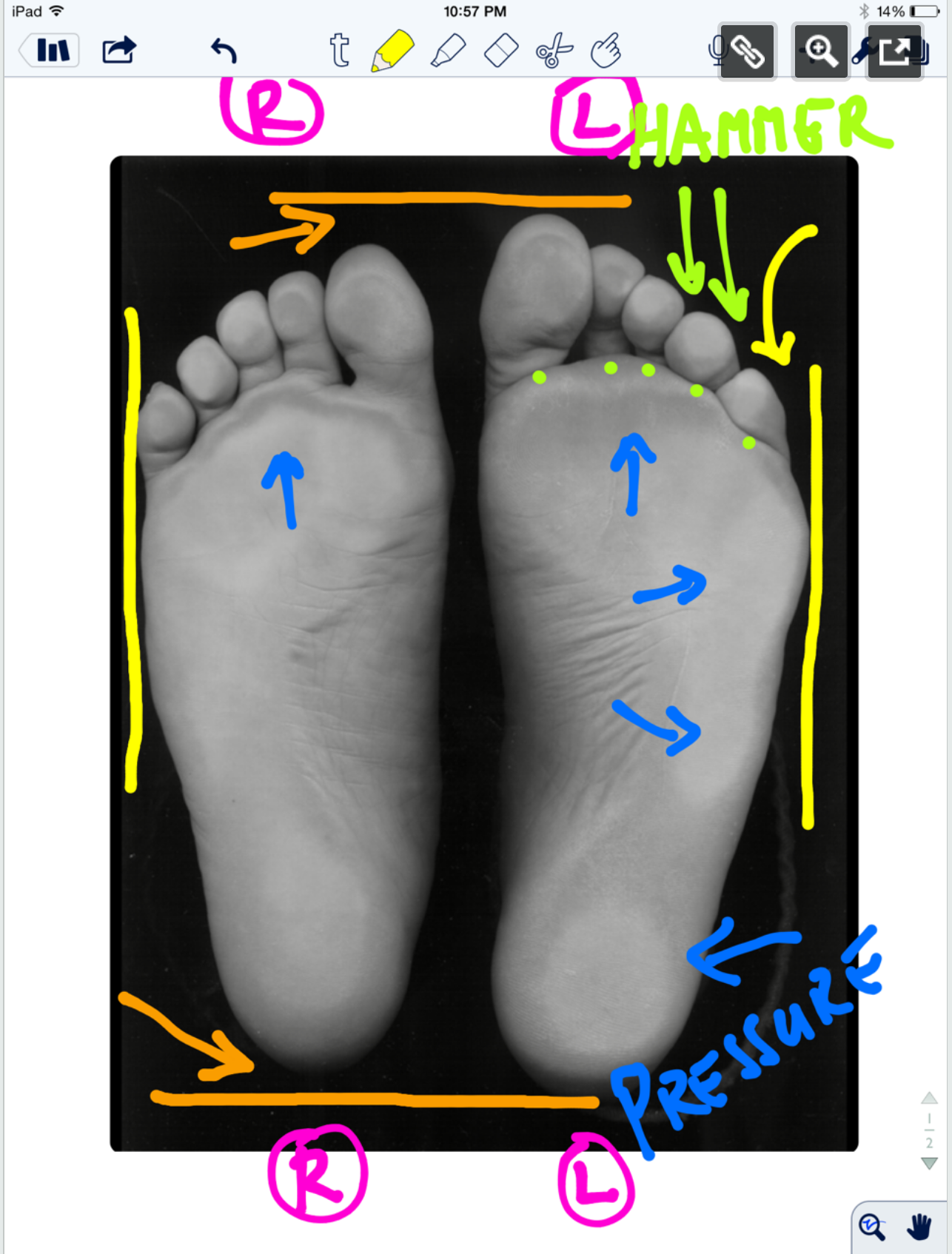
* note the right and left sides by the R and L circled in pink.

ORANGE lines: The right foot appears to be shorter, or is it that the left is longer (see the lines and arrows drawing your attention to these differences)? A shorter foot could be represented by a supinated foot (if you raise the arch via the windlass mechanism you will shorten the foot distance between the rear and forefoot). A longer foot could be represented by a more pronated foot. Is that what we have here ? There is no way to know, this is a static presentation of a client standing on glass. What we should remember is that the goal is always to get the pelvis square and level. If an anatomically or functionally short leg is present, the short leg side MAY supinate to raise the mortise and somewhat lengthen the leg. In that same client, they may try to meet the process part way by pronating the other foot to functionally “shorten” that leg. Is that what is happening here ? So, does this client have a shorter right leg ? Longer left ? Do you see a plunking down heavily onto the right foot in gait ? Remember, what you see is their compensation. Perhaps the right foot is supinating, and thus working harder at the bottom end of the limb (via more supination), to make up for a weak right glute failing to eccentrically control the internal spin of the leg during stance phase ? OR, perhaps the left foot is pronating more to drive more internal rotation on the left limb because there is a restricted left internal hip rotation from the top ? Is the compensation top-down or bottom up ? These are all viable possibilities and you must have these things flowing freely through your head during the clinical examination as you rule in/rule out your hands-on findings. Remember, just going by a FMS-type screen to drive prescription exercises from what you see on a movement screen is not going to necessarily fix the problem, it could in fact lead one to drive a deeper compensation pattern. You can be sure that Gray Cook’s turbo charged brain is juggling all of these issues (and more !) when he sees a screen impairment, although we are not speaking for him here.

Remember this critical fact. After an injury or a long standing problem, muscles and motor patterns jobs are to stabilize and manage loads (stability and mobility) for adequate and necessary movement. Injuries leave a mark on the system as a whole because adaptation was necessary during the initial healing phase. This usually spills over during the early movement re-introduction phase, particularly if movement is reintroduced too early or too aggressively. Plasticity is the culprit. Just because the injury has come and gone does not mean that new patterns of skill, endurance, strength (S.E.S -our favorite mnemonic), stability and mobility were not subsequently built onto the apparently trivial remnants of the injury. There is nothing trivial if it is abnormal. The forces must, and will, play out somewhere in the body and this is often where pain or injury occurs but it is rarely where the underlying problem lives.

Come back tomorrow, where we will open your mind into the yellow, pink, blue and lime markings on the photo. Are the hammering toes (lime) on the left a clue ? How about the width of the feet (yellow) ? The posturing differences of the 5th toe to the lateral foot border ? What about the static plantar pressure differences from side to side (blue)? Maybe, just maybe, we can bring a logical clinical assumption together and then a few clinical exam methods to confirm or dis-confirm our working diagnostic assumption. See you tomorrow friends !

Shawn and ivo, the gait guys

Here is the case link.......

https://thegaitguys.tumblr.com/post/99409232289/the-standing-on-glass-static-footpedograph?fbclid=IwAR3gd3d81Gwt3ywAB7BcTwXqST2Z_5nmieODzSb8rJQYBcJFhTs6rS_9auA

The QP....What's the deal?

tumblr_mau352bxll1qhko2so1_540.png

Possibly heard of, rarely implicated and not often treated, this is one muscle you should consider taking a look at.

The quadratus plantae is generally considered to arise from two heads of differing and variable  fiber type composition, with the lateral head having slightly more Type 1 endurance fibers (1) The two heads are separated from each other by the long plantar ligament, though it can arise from from one (somewhat more common)  to 3 heads (very rare).  The attachments can be variable, The medial head is larger and more muscular, attached to the medial calcaneus, lateral aspect of the long plantar ligament and often from the plantar calcaneocuboid ligament (2);  the lateral head is smaller and more tendinous, attaching to the lateral border of the inferior surface of the calcaneus and the long plantar ligament.  The two portions join and end in a flattened band which inserts into the lateral, upper and under surfaces of the muscles, tendons or aponeurosis of predominantly the flexor digitorum longus and usually of the second and third, and sometimes fourth toes (2,3). 

Its action can be equally as variable. In addition to augmenting the pull of the long flexor tendons along the long axis of the foot and so that the 3rd and 4th toes do not curl under the foot, the tendinous slips of the FHL may distribute the load of the great toe to the second toe to the third or fourth toe in the forefoot, especially during toe-off (3).

look at the 4th and 5th digits trying to "crawl under the foot"

look at the 4th and 5th digits trying to "crawl under the foot"

The main attachment of the QP to the tendinous slips of the FHL may provide more efficient control of the long flexor tendons in comparison with that of the QP to the tendon of the FDL (3). EMG studies suggest it resists extension of the toes during the stance phase of locomotion, which serves to increase the stability of the foot. Additional EMG studies suggest it actually acts as a primary toe flexor in voluntary movements, being preferentially recruited over flexor digitorum longus and from comparative anatomical considerations it also seems likely that quadratus plantae may be an intrinsic evertor of the foot (4).

This muscle is a major player in gait and rehabilitation of this muscle should not be overlooked. I could only find one study looking at exercise activation of the QP (5) . It was examined along with the abductor hallucis, flexor digitorum brevis, abductor digiti minimi, flexor digiti minimi, adductor hallucis oblique, flexor hallucis brevis, interossei and lumbricals during rehabilitative the short-foot exercise, toes spread out, first-toe extension, second- to fifth-toes extension.

So, what else can you do?

  • you could ignore the muscle and hope it gets better. (in all likelihood it will worsen)
  • you could give them long flexor, toe scrunching towel-curling, marble-grasping exercises, like you see all over the internet…and give the flexor digitorum longus even more of a mechanical advantage, and make the problem worse
  • you could give them exercises to increase the function of the long extensors, which would increase the mechanical advantage of the quadratus plantae. like the shuffle walk; lift, spread and reach and tripod standing exercises
  • look north of the foot to see what might be causing the problem (loss of ankle rocker, insufficient gluteal activity, loss of internal rotation of the hip, etc) 

Check out the QP on your next foot pain patient, or whenever you see the toes trying to crawl under the foot. You may be surprised at your results. 


1. Schroeder KL, Rosser BW, Kim SY. Fiber type composition of the human quadratus plantae muscle: a comparison of the lateral and medial heads. J Foot Ankle Res. 2014 Dec 13;7(1):54. doi: 10.1186/s13047-014-0054-5. eCollection 2014.

2. Pretterklieber B1. Morphological characteristics and variations of the human quadratus plantae muscle. Ann Anat. 2017 Nov 21;216:9-22. doi: 10.1016/j.aanat.2017.10.006. [Epub ahead of print]

3. Hur MS, Kim JH, Woo JS, Choi BY, Kim HJ, Lee KS. An anatomic study of the quadratus plantae in relation to tendinous slips of the flexor hallucis longus for gait analysis. Clin Anat. 2011 Sep;24(6):768-73. doi: 10.1002/ca.21170.

4. Sooriakumaran P, Sivananthan S. Why does man have a quadratus plantae? A review of its comparative anatomy. Croat Med J. 2005 Feb;46(1):30-5.

5. Gooding TM, Feger MA, Hart JM, Hertel J. ntrinsic Foot Muscle Activation During Specific Exercises: A T2 Time Magnetic Resonance Imaging Study. J Athl Train. 2016 Aug;51(8):644-650. Epub 2016 Oct 3.

Which foot exercises activate the intrinsics?

So, your goal is to strengthen the intrinsics. What exercise is best? Probably the most specific one, right? Well....maybe. These 4 exercises seem to all hit them.

This study looked at the muscle activation of the abductor hallucis, flexor digitorum brevis, abductor digiti minimi, quadratus plantae, flexor digiti minimi, adductor hallucis oblique, flexor hallucis brevis, and interossei and lumbricals with the short foot, toe spreading, big toe extension and lesser toes extension exercises with T2 weighted MRI post exercises (perhaps not the best way to look at it) and shows they all work to varying degrees.

"All muscles showed increased activation after all exercises. The mean percentage increase in activation ranged from 16.7% to 34.9% for the short-foot exercise, 17.3% to 35.2% for toes spread out, 13.1% to 18.1% for first-toe extension, and 8.9% to 22.5% for second- to fifth-toes extension."

Gooding TM, Feger MA, Hart JM, Hertel J. Intrinsic Foot Muscle Activation During Specific Exercises: A T2 Time Magnetic Resonance Imaging Study. Journal of Athletic Training. 2016;51(8):644-650. doi:10.4085/1062-6050-51.10.07.

link to full text: https://www.ncbi.nlm.nih.gov/pmc/articles/PMC5094843/

tumblr_ni72j0wHZ11qhko2so2_1280.jpg
tumblr_ni72j0wHZ11qhko2so1_1280.jpg
tumblr_ni72j0wHZ11qhko2so3_1280.jpg
tumblr_ni72j0wHZ11qhko2so4_1280.jpg
tumblr_ni72j0wHZ11qhko2so5_1280.jpg

A clear cut case of Form follows Function.  Leave a deforming force long enough and the body will accommodate. 

When the lateral quadratus plantae (QP) is weak and the flexor digitorum longus pulls unopposed (relying on the QP to properly orient the long flexor pull) for too long the 4th and 5th toes and drift medially and spin inwards toward the midline of the foot (as seen in the photo). Then, as the 4th toe presses down on the fleshy pad of the 5th toe, over time the fleshy pad is pancaked and triangulated. Then, with repeated pressure a corn like hardness becomes of the tip of that triangluted tissue, it resembles a hard callus. A corn is a coalescing of the skin cells into a tighter formation, a reaction to fend off repeated pressure and friction.  Form follows prolonged function.  Shave these things down and they will come back, unless you get to the root source of the problem, which could be all the way up the chain. 

-Dr. Allen

Toe sardines. What have we done to our feet ?

Note that form follows function. If you are observant, you will see the deformation of the 5 digit, just like in this case as the quadratus weakens and the long flexors dominate. The toe begins to spin laterally, and thus the plantar toe pad begins to deform medially, look closely, you can see that here in the video.

Does this look like your foot ? There are a few subtle issues here. 

In the foot, the toe that delineates abduction and adduction of the toes is the 2nd toe. The 2nd toe is considered the anatomic middle of the digits and forefoot. Any toe or movement that moves away from the 2nd toe is abduction and any movement towards the 2nd toe is adduction. This is obviously different than in the hand where the 3rd digit, the one you use during road rage, is the reference digit. Next time you are questioned, tell them you threw them your reference finger, not “the bird”, it is a more accurate descriptor.

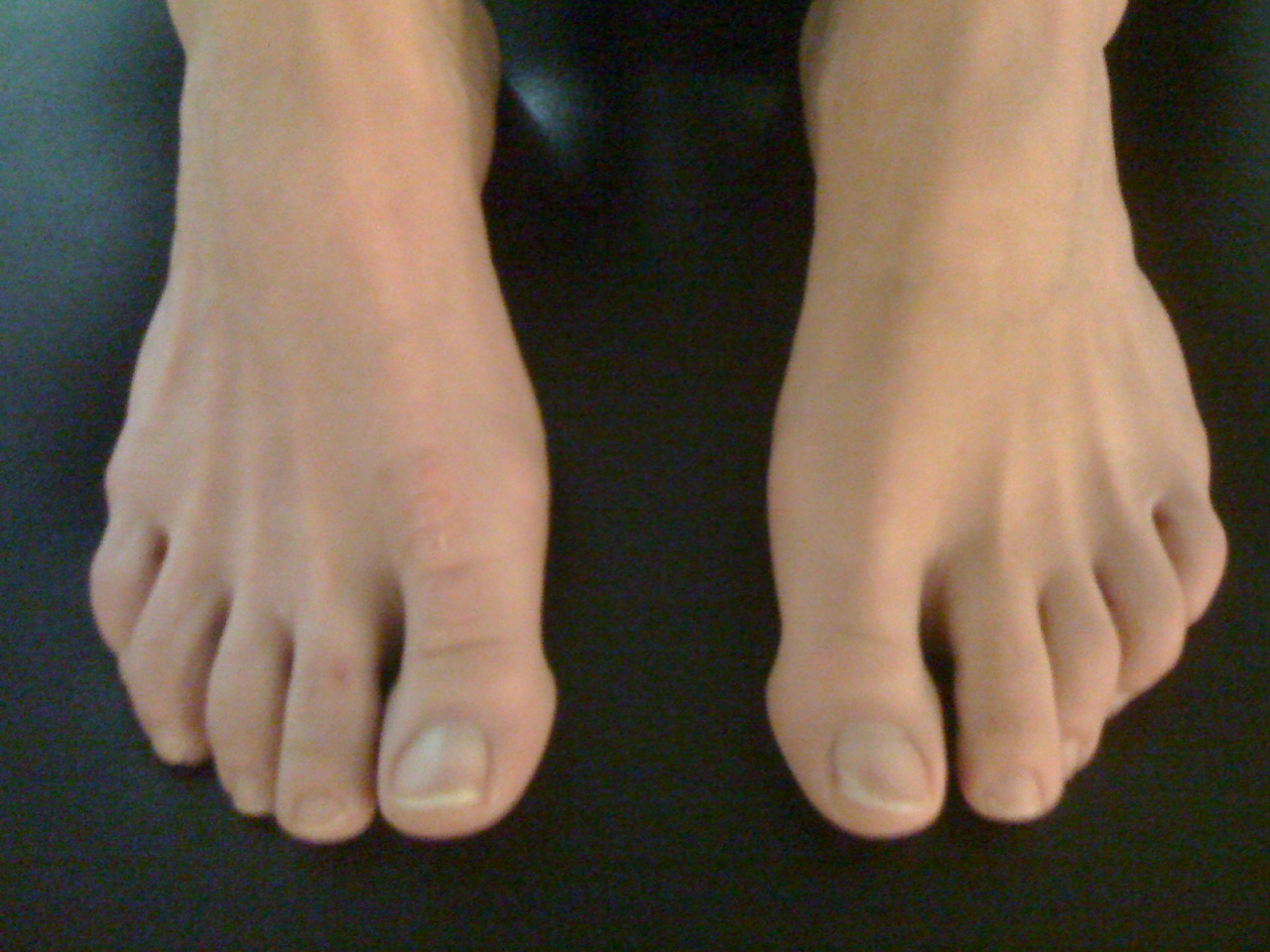
In this foot, note how neatly and tightly packed the cute little toes are, all snuggled up to their brothers and sisters. Remember, form follows function. Obviously function has been low on these fellas, at least in abduction.  This often comes from snug toe box footwear and lack of abduction (toe spread) use.  But make no mistake, this is a weak foot.

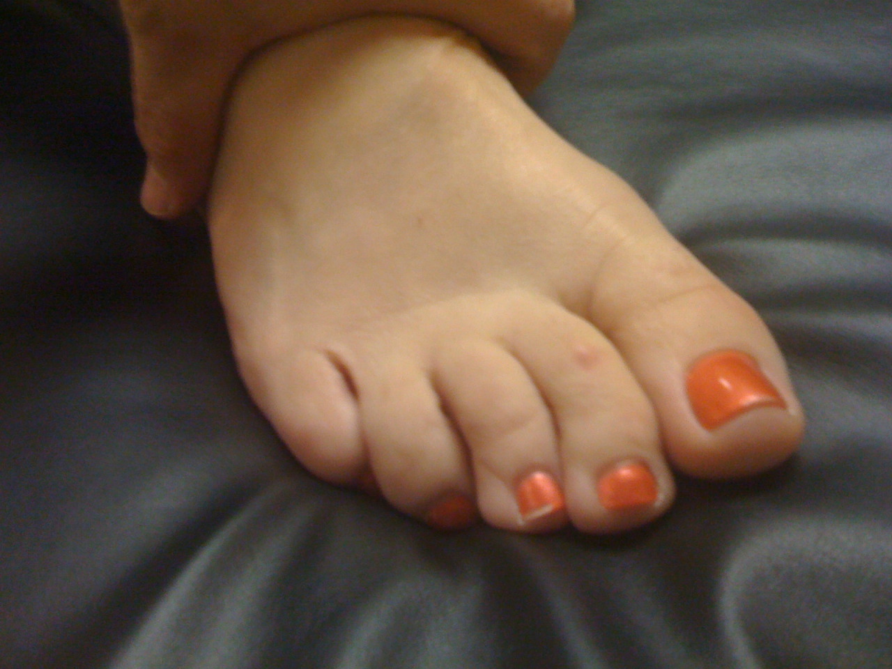
Today we wish to really focus your attention to an old topic, just a revisit. We can see the 4th and 5th toes curl under from the probably weak lateral head of the quadratus plantae thus encouraging unopposed oblique pull of the long flexors of the digits (FDL). See this post here for an explanation of this phenomenon.  There is also obvious imbalance between the long and short flexors and extensors in these toes, the long flexors are expressing more tone, and that means the long extensors are deprived. 

Note that form follows function. If you are observant, you will see the deformation of the 5 digit, just like in this case as the quadratus weakens and the long flexors dominate. The toe begins to spin laterally, and thus the plantar toe pad begins to deform medially, look closely, you can see that here in the video. This spin can carry the toe nail so far laterally sometimes that the nail can begin to touch the ground during gait and cause painful nail lifting with even some losing the nail. 

There is plenty of life left in this foot, but you have to get to it quickly and get them in lower heeled shoes if tolerable and ones with a wider toe box.  The client needs to be retaught how to access the toe extensors and abductors. Lumbrical retraining, which is a recurrent topic here on our blog, should also be instituted. 

Shawn Allen, one of the gait guys

The deeper your knowledge and experiences, the more things you will see. As in life, the more experiences you have the wiser you become and the clearer the bigger picture becomes. All these things enrich the experience or observation. These experien…

The deeper your knowledge and experiences, the more things you will see. As in life, the more experiences you have the wiser you become and the clearer the bigger picture becomes. All these things enrich the experience or observation. These experiences take simple black and white and render an infinite palate of grey tones. 

To the untrained observer, these are just two feet. With a little more experience these are two feet of different length. Deeper further, these are two different sized feet with different plantar pressure responses (helped here by increasing the greyscale contrast). Deeper yet, this represents a left foot (viewers right) that has a dysfunctional flexor digitorum longus (FDL) and lateral quadratus plantae muscle. All of these observations allow the skilled and knowledgeable viewer to extrapolate and theorize, with clear thought processes, which leg could be shorter/longer, how the pelvis might be distortioned, step length and stride length variability, foot stability and so much more.

The life long student does not need the contrast enhanced picture on the right to heighten the visibility of the plantar pressures, but it helps.  This is what wisdom and experience do, they enable you to look deeper into something and to see it for what it truly is, not what it appears to be.

Come listen to our teleseminar tonight (Wednesday March 18th, 2015) on www.onlinece.com at 7pm central. Log in early to get set up. Come listen in while we delve into one of the bigger questions, if the left foot (viewers right) is longer it has likely pronated more over a longer period of time stretching out plantar soft tissues and corrupting joint function in multiple areas. But if this is the case, why then are they presenting with plantar pressures that are more representative of supination standards ?  

This is mental gymnastics. It is good stuff to do regularly, even though this is a static presentation, many good theories and thoughts can be brought forth. Getting the answer is not the goal, getting the thought process down is.

The more you know, the more truth you will see.

See you tonight, we will break this down into a microscopic level that will challenge you all.

Shawn and Ivo, the gait guys

tumblr_nd32fvOzjx1qhko2so3_r1_1280.png
tumblr_nd32fvOzjx1qhko2so2_1280.png
tumblr_nd32fvOzjx1qhko2so1_1280.png
tumblr_nd32fvOzjx1qhko2so4_r1_500.png

Part 2: “Standing on Glass” Static Foot/Pedograph Assessment

* note (see warning at bottom): This is a static assessment dialogue. One cannot, and must not, make clinical decisions from a static assessment. The right and left sides are indicated by the R and L circled in pink. There are 4 photos here today.

Blue lines: Last time we evaluated possible ideas on the ORANGE lines here, it would be to your advantage to start there. 

We can see a few noteworthy things here in these photos. We have contrast-adjusted the photo so the pressure areas (BLUE) are more clearly noted. There appears to be more forefoot pressure on the right foot (the right foot is on the readers left), and more rearfoot pressure on the left (not only compare the whiteness factor but look at the displacement of the calcaneal fat pad (pink brackets). There is also noticeably more lateral forefoot pressure on the left. There is also more 3-5 hammering/flexion dominance pressure on the left.  The metatarsal fat pad positioning (LIME DOTS represent the distal boundary) is intimately tied in with the proper lumbrical muscle function  (link) and migrates forward toward the toes when the flexors/extensors and lumbricals are imbalanced. We can see this fat pad shift here (LIME DOTS). The 3-5 toes are clearly hammering via flexor dominance (LIME ARROWS), this is easily noted by visual absence of the toe shafts, we only see the toe pads. Now if you remember your anatomy, the long flexors of the toes (FDL) come across the foot at an angle (see photo). It is a major function of the lateral head of the Quadratus plantae (LQP) to reorient the pull of those lesser toe flexors to pull more towards the heel rather than on an angle. One can see that in the pressure photos that this muscle may be suspicious of weakness because the toes are crammed together and moving towards the big toe because of the change in FDL pull vector (YELLOW LINES). They are especially crowding out the 2nd toe as one can see, but this can also be from weakness in the big toe, a topic for another time. One can easily see that these component weaknesses have allowed the metatarsal fat pad to migrate forward. All of this, plus the lateral shift weight bearing has widened the forefoot on the left, go ahead, measure it. So, is this person merely weight bearing laterally because they are supinating ? Well, if you read yesterday’s blog post we postulated thoughts on this foot possibly being the pronated one because of its increased heel-toe and heel-ball length. So which is it ? A pronated yet lateral weight bearing foot  or a normal foot with more lateral weight bearing because of the local foot weaknesses we just discussed ? Or is it something else ? Is the problem higher up, meaning, are they left lateral weight bearing shift because of a left drifted pelvis from weak glute medius/abdominal obliques ?  Only a competent clinical examination will enlighten us.

Is the compensation top-down or bottom up, or both in a feedback cycle trying to find sufficient stability and mobility ? These are all viable possibilities and you must have these things flowing freely through your head during the clinical examination as you rule in/rule out your hands-on findings.  Remember, just going by a screen to drive prescription exercises from what you see on the movement screen is not going to necessarily fix the problem, it could in fact lead one to drive a deeper compensation pattern. 

Remember this critical fact.  After an injury or a long standing problem, muscles and motor patterns jobs are to stabilize and manage loads (stability and mobility) for adequate and necessary movement. Injuries leave a mark on the system as a whole because adaptation was necessary during the initial healing phase. This usually spills over during the early movement re-introduction phase, particularly if movement is reintroduced too early or too aggressively.  Plasticity is the culprit. Just because the injury has come and gone does not mean that new patterns of skill, endurance, strength (S.E.S -our favorite mnemonic), stability and mobility were not subsequently built onto the apparently trivial remnants of the injury.  There is nothing trivial if it is abnormal. The forces must, and will, play out somewhere in the body and this is often where pain or injury occurs but it is rarely where the underlying problem lives.

Come back tomorrow.  We will try to bring this whole thing together, but remember, it will just be a theory for without an exam one cannot prove which issues are true culprits and which are compensations. Remember, what you see is often the compensatory illusion, it is the person moving with the parts that are working and compensating not the parts that are on vacation.  See you tomorrow friends !

Shawn and ivo, the gait guys

* note: This is a static assessment dialogue. One cannot, and must not, make clinical decisions from a static assessment. As in all assessments, information is taken in, digested and then MUST be confirmed, denied and/or at the very least, folded into a functional and clinically relevant assessment of the client before the findings are accepted, dismissed and acted upon. As we always say, a gait analysis or static pedograph-type assessment (standing force plate) is never enough to make decisions on treatment to resolve problems and injuries. What is seen and represented on either are the client’s strategies around clinical problems or compensations.  Today’s photo and blog post are an exercise in critical clinical thinking to get the juices flowing and to get the observer thinking about the client’s presentation and to help open up the field to questions the observer should be entertaining.  The big questions should be, “why do i see this, what could be causing these observances ?”right foot supinated ? or more rear and lateral foot……avoiding pronation ?

The “Standing on Glass” Static Foot/Pedograph Assessment: Part 1
* note: This is a static assessment dialogue. One cannot, and must not, make clinical decisions from a static assessment. As in all assessments, information is taken in, di…

The “Standing on Glass” Static Foot/Pedograph Assessment: Part 1

* note: This is a static assessment dialogue. One cannot, and must not, make clinical decisions from a static assessment. As in all assessments, information is taken in, digested and them MUST be confirmed, denied and/or at the very least, folded into a functional and clinically relevant assessment of the client before the findings are accepted, dismissed and acted upon. As we always say, a gait analysis or pedograph-type assessment is never enough to make decisions on treatment to resolve problems and injuries. What is seen and represented on either are the client’s strategies around clinical problems or compensations.  Today’s photo and blog post are an exercise in critical clinical thinking to get the juices flowing and to get the observer thinking about the client’s presentation and to help open up the field to questions the observer should be entertaining.  The big questions should be, “why do i see this, what could be causing these observances ?”

* note the right and left sides by the R and L circled in pink.

ORANGE lines: The right foot appears to be shorter, or is it that the left is longer (see the lines and arrows drawing your attention to these differences)? A shorter foot could be represented by a supinated foot (if you raise the arch via the windlass mechanism you will shorten the foot distance between the rear and forefoot). A longer foot could be represented by a more pronated foot.  Is that what we have here ? There is no way to know, this is a static presentation of a client standing on glass. What we should remember is that the goal is always to get the pelvis square and level.  If an anatomically or functionally short leg is present, the short leg side MAY supinate to raise the mortise and somewhat lengthen the leg.  In that same client, they may try to meet the process part way by pronating the other foot to functionally “shorten” that leg.  Is that what is happening here ? So, does this client have a shorter right leg ? Longer left ?  Do you see a plunking down heavily onto the right foot in gait ? Remember, what you see is their compensation.  Perhaps the right foot is supinating, and thus working harder at the bottom end of the limb (via more supination), to make up for a weak right glute failing to eccentrically control the internal spin of the leg during stance phase ? OR, perhaps the left foot is pronating more to drive more internal rotation on the left limb because there is a restricted left internal hip rotation from the top ? Is the compensation top-down or bottom up ? These are all viable possibilities and you must have these things flowing freely through your head during the clinical examination as you rule in/rule out your hands-on findings.  Remember, just going by a FMS-type screen to drive prescription exercises from what you see on a movement screen is not going to necessarily fix the problem, it could in fact lead one to drive a deeper compensation pattern. You can be sure that Gray Cook’s turbo charged brain is juggling all of these issues (and more !) when he sees a screen impairment, although we are not speaking for him here.

Remember this critical fact.  After an injury or a long standing problem, muscles and motor patterns jobs are to stabilize and manage loads (stability and mobility) for adequate and necessary movement. Injuries leave a mark on the system as a whole because adaptation was necessary during the initial healing phase. This usually spills over during the early movement re-introduction phase, particularly if movement is reintroduced too early or too aggressively.  Plasticity is the culprit. Just because the injury has come and gone does not mean that new patterns of skill, endurance, strength (S.E.S -our favorite mnemonic), stability and mobility were not subsequently built onto the apparently trivial remnants of the injury.  There is nothing trivial if it is abnormal. The forces must, and will, play out somewhere in the body and this is often where pain or injury occurs but it is rarely where the underlying problem lives.

Come back tomorrow, where we will open your mind into the yellow, pink, blue and lime markings on the photo. Are the hammering toes (lime) on the left a clue ? How about the width of the feet (yellow) ? The posturing differences of the 5th toe to the lateral foot border ?  What about the static plantar pressure differences from side to side (blue)? Maybe, just maybe, we can bring a logical clinical assumption together and then a few clinical exam methods to confirm or dis-confirm our working diagnostic assumption.  See you tomorrow friends !

Shawn and ivo, the gait guys

tumblr_mwzu9gTBzS1qhko2so1_1280.jpg
tumblr_mwzu9gTBzS1qhko2so2_400.gif
tumblr_mwzu9gTBzS1qhko2so3_r1_1280.jpg
tumblr_mwzu9gTBzS1qhko2so4_r1_1280.jpg

Is your 5th toe curled under ? What do you do when “this little piggy” can’t go wee wee wee all the way home.

Have a look at the 4 photos above.  You will see this curling of the lesser toes quite often in your practice, and when you know what it means it can help to guide your thinking, both from a diagnostic and treatment perspective.  

You should have noticed in the photos that the 4th and 5th toes curl under and are hyper-flexed, and this is at rest.  So, what does this mean ?

It means that the long flexors are overactive, the extensors are underactive, and the adduction pull of the long flexors is unopposed by the under appreciated quadratus plantae muscle.

Look at the clinical drawing. The quadratus plantae has 2 heads, a medial head and a lateral head.  Being able to clinically test these two heads will give you much insight into the function of the foot and when you see these outer two toes curling under, as you see in the photo, you will always see weakness of the lateral head of the quadratus plantae.  

The quadratus plantae arises from two heads separated from each other by the long plantar ligament. The medial head is larger and more muscular, attached to the medial calcaneus;  the lateral head is smaller and more tendinous, attaching to the lateral border of the inferior surface of the calcaneus and the long plantar ligament.  The two portions join and end in a flattened band which inserts into the lateral, upper and under surfaces of the tendons of the flexor digitorum longus, usually the second, third, and fourth toes.

But this time, if you have studied the drawing, you should notice the oblique line of pull of the long flexors.  This should in fact create this undesirable curling effect of the lateral two toes since they are so far out on the oblique line of pull. However, if you look at the insertion of the lateral head of the quadratus plantae you should be able to conclude that this head is designed to offset this oblique pull of the outer two long flexor tendons.  The quadratus creates a posterior pull on the outer long flexor tendons ensuring that the curling effect (as seen in the photo) is nullified. Thus, we have a clinical presentation of a weak lateral head of the quadratus plantae (and probably a few others which we will not discuss here so as to not dilute the purpose of today’s post). Now you just have to figure out why it is weak or if there is a biomechanical reason for its insufficiency

  • is there a foot type presenting itself that makes it difficult for this muscle to create sufficient posterior pull to offset the tremendous leverage of the long flexors? Maybe a forefoot varus, which gives the flexor tendons a mechanical advantage or a forefoot valgus which puts the quadratus plantae at a mechanical disadvantage? (Taking our National Shoe Fit Certification Program will help you get closer to understanding many of these issues.)
  • Are their other anatomical variants like an increased forefoot width or bunions (medial or tailor’s)
  • is there excessive rear or midfoot pronation?
  • Shoe choice problem ?

Some folks do have adequate function of the quadratus plantae. Note the lovely feet in the last picture … .  they must have strong lateral quadratus plantae and abductors of the lateral foot and toes ! And, they have great toe separation, thus great intrinsic interossei muscles, and nice flat toes (great balance between flexors and extensors).

So, what do you do?

  • you could do a surgery, amputate or fuse some of the joints to make them look better. Extreme for a problem like this
  • you could ignore the issue and hope it goes away. (in all likelihood it will worsen)
  • you could give them long flexor, toe scrunching Towel-curling, marble-grasping exercises , like you see all over the internet…and give the flexor digitorum longus even more of a mechanical advantage, and make the problem worse
  • you could give them exercises to increase the function of the long extensors, which would increase the mechanical advantage of the quadratus plantae. like the shuffle walk; lift, spread and reach and tripod standing exercises (hmm…sounding better)
  • be a real clinician and in addition to looking at the foot, look north of the foot to see what might be causing the problem (loss of ankle rocker, insufficient gluteal activity, loss of internal rotation of the hip, etc) Hmmm; sounding like a good idea too…

The Gait Guys. Hammering it home, day after day, about the importance of gait and giving you clues to be a better _________ (insert athlete, coach, trainer, clinician, shoe fitter, rehab specialist…).

tumblr_mau352bxll1qhko2so1_540.png
tumblr_mau352bxll1qhko2so2_400.jpg

Foot Talk

It’s all about communication. In this case, compartmental communication. There has not been alot on consensus about how many compartments the foot has, but it is known that all the compartments talk to one another. This study identified six compartments: dorsal, medial, lateral, superficial central, deep forefoot, and deep hindfoot. It goes on to say: Communication was evident between the deep hindfoot compartment and the superficial central and deep central forefoot compartments.

This should not be that surprising. In this case, the deep hindfoot intrinsic muscles would include the quadratus plantae (seen above attaching to the calcaneus), which augments the pull of the long the long flexor muscles and helps to keep the toes flat on the ground.

The superficial central compartment would include the short flexors (flexor digitorum brevis), another stance phase muscle that is also important in keeping the toes flat on the ground.

The deep central forefoot compartment would include the transverse head of the adductor hallucis. important in maintaining 1st ray stability and keeping the head of the 1st metatarsal on the ground and maintaining an adequate foot tripod.

Another point worth mentioning was this: In the hindfoot, the neurovascular bundles were located in separate tissue sheaths between the central hindfoot compartment and the medial compartment. In the forefoot, the medial and lateral bundles entered the deep central forefoot compartment.

This tells us that in the rearfoot, the important neurology is in the muscles which help to invert the rearfoot, and help create supination. In the central forefoot, information is fed from the lateral and medial aspects of the foot tripod to the transverse head of the adductor longus. this muscle, when biomechanics are appropriate and the head of the 1st metatarsal is anchored, assists in supination. It seems all roads leaad to assisting in supination and propelling us forward in the gravitational plane…

Communication. Not just for interpersonal relationships : )

The Gait Guys: communicating with you daily and keeping you current on all things feet.

Surg Radiol Anat. 2012 May 26. [Epub ahead of print] Compartments of the foot: topographic anatomy. Faymonville C, Andermahr J, Seidel U, Müller LP, Skouras E, Eysel P, Stein G. Source

Department of Orthopaedic and Trauma Surgery, University Hospital of Cologne, Kerpener Str. 62, 50924, Cologne, Germany, christoph@faymonville.de.

Abstract

Recent publications have renewed the debate regarding the number of foot compartments. There is also no consensus regarding allocation of individual muscles and communication between compartments. The current study examines the anatomic topography of the foot compartments anew using 32 injections of epoxy-resin and subsequent sheet plastination in 12 cadaveric foot specimens. Six compartments were identified: dorsal, medial, lateral, superficial central, deep forefoot, and deep hindfoot compartments. Communication was evident between the deep hindfoot compartment and the superficial central and deep central forefoot compartments. In the hindfoot, the neurovascular bundles were located in separate tissue sheaths between the central hindfoot compartment and the medial compartment. In the forefoot, the medial and lateral bundles entered the deep central forefoot compartment. The deep central hindfoot compartment housed the quadratus plantae muscle, and after calcaneus fracture could develop an isolated compartment syndrome.

tumblr_lkowdng74B1qhko2so2_1280.jpg
tumblr_lkowdng74B1qhko2so1_540.png

The Quadratus plantae (Flexor accessorius) muscle. Do you have foot pain ?

(*There are two pictures here on the blog. Move your cursor over to the side of the photo and you will see that you can toggle between the photo and anatomy pic).

This is a great, but highly overlooked, muscle.  The QP acts to assist in flexing the 2nd to 5th toes.  Equally important is its effect of offsetting the oblique pull of the long toe flexor group (flexor digitorum longus). It has two heads, medial and lateral.  The medial head is attached to the calcaneus, while the lateral head originates from the lateral border of the calcaneus, in front of the lateral process of the calcaneal tuberosity and the long plantar ligament.

The fact that we just love, and one that we believe is often overlooked is the acute angle at which the muscle heads attach into the tendons of the flexor digitorum longus (see picture) and has a rather dramatic alignment effect on the lateral 3 digits (since the line of pull on the long flexor tendons to these 3 digits is most dramatically changed by the purely posterior pull of the Quadratus Plantae.  As you can see in this stripped down anatomy picture, without the QP pulling on the tendons of the FDL to these 3 lateral toes, those toes will have to curl medially and gently flex (*see the photo, a classic presentation!)  By having a competent and active QP that oblique line of pull of the FDL /long flexors is rearranged to be more of a pure posterior pull and you will not see this classic lateral 3 digit curl and medial drift. This action is accentuated in a cavus foot type, where the pull of the FDL will be accentuated, due to the mechanical advantage afforded it and relative adduction of the forefoot with respect to the rear foot.

In the photo you can see a classic representation of a deficient Quadratus Plantae, in this case the patients lateral head was dramatically weaker than the medial, but both were weak.  So, summary time….if you know your anatomy, know your biomechanics, and if you can test the muscle bundles specifically……..then you can see why form follows function (and in this case, why form has followed dysfunction).  As we always say, “ya gotta know your stuff”, and you have to test what you suspect……there are other things that could also do this……so, let your eyes gain info, let your brain process and prove or disprove the information.

we are…….the gait guys !