Unilateral increased tibial varum; one reason why...

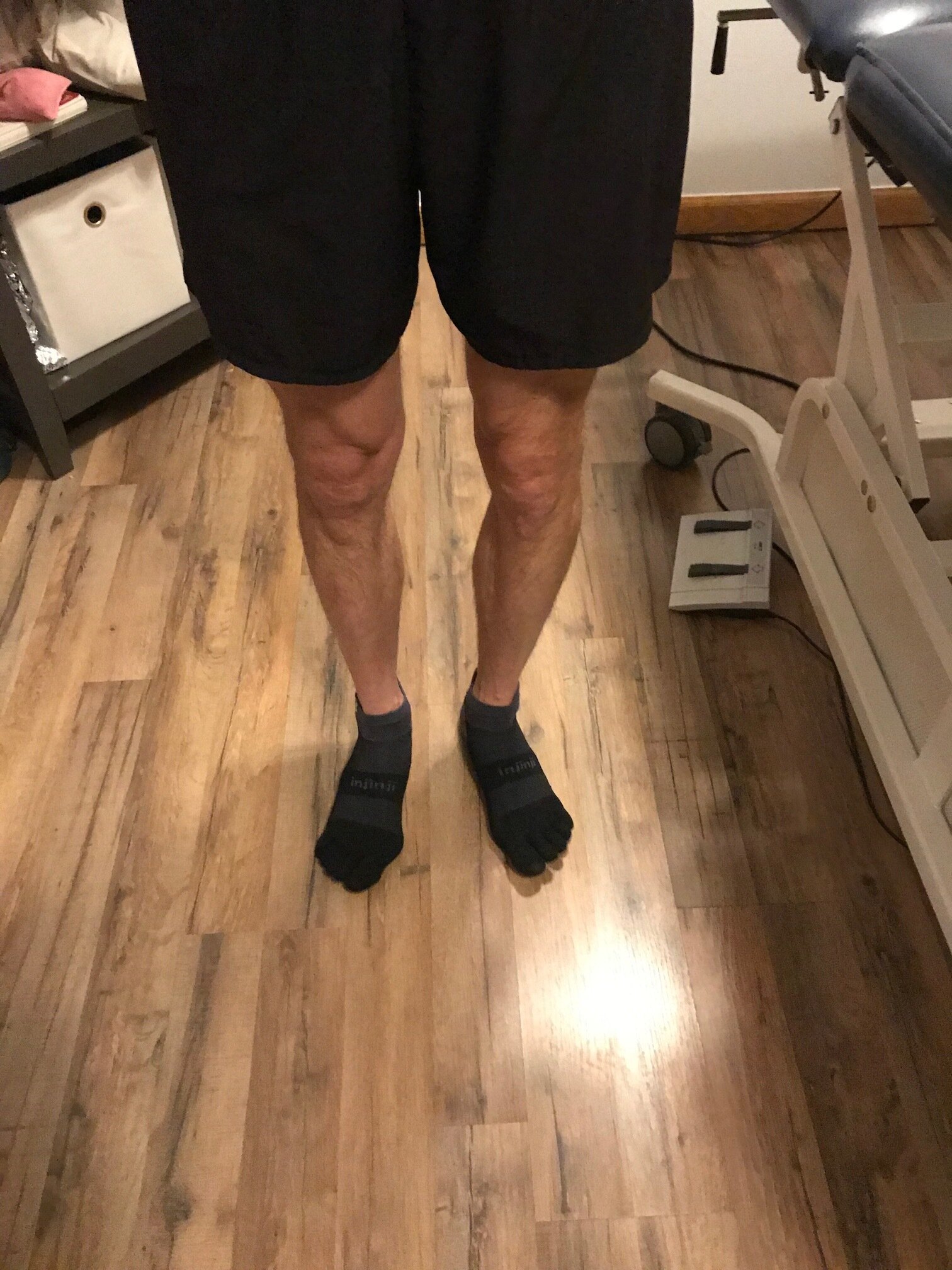
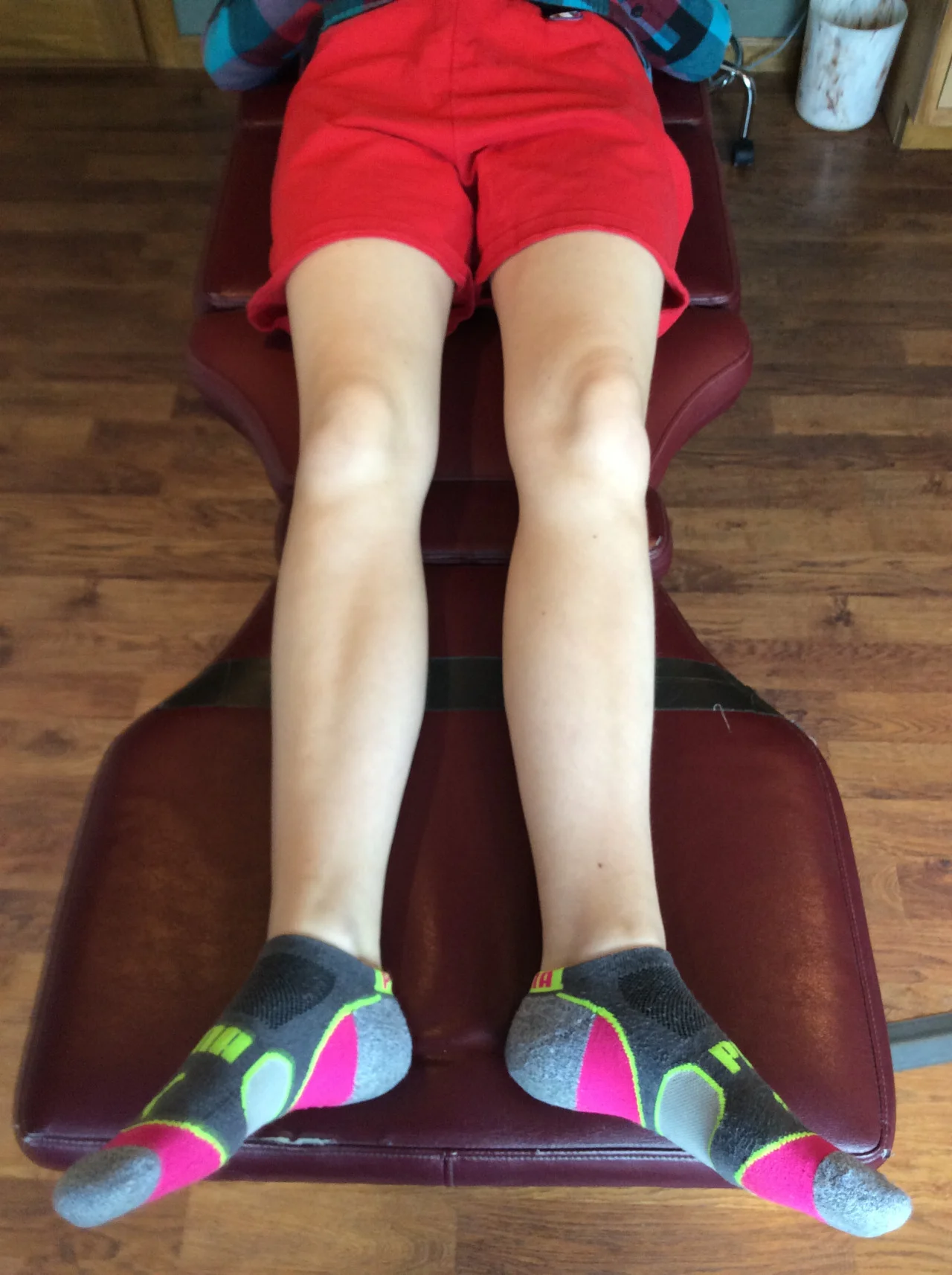
Take a look at this gent in the picture. Do you notice anything peculiar? Pick a point and start either moving from above down or from the ground up.

From the ground up, the first thing you may notice is that he has a hallux abducto valgus on the right side. This could be for any number of reasons and what it actually tells you is that he is unable to anchor his first ray to the ground and have appropriate function of the adductor hallucis. Your job, during the examination process, is to sort that out.

The second thing you may notice is that he has more midfoot collapse on this same side. You would think that with that much midfoot collapse he would get his first ray to the ground but that’s obviously not the case.

Moving up from there, you may have noticed that he has significantly more tibial varum on the left-hand side. Tibial varum should be about 4-6 degrees and is largely a function of in utero positioning although diseases like osteomalacia and rickets can increase it though this is often more bilaterally symmetrical.

You need to be aware increased tibial varum means that the foot, particularly the forefoot, needs to pronate a greater degree to create a stable foot tripod on the ground. You need to ensure during the examination process that adequate range of motion in the forefoot and 1st ray are available.

You may have noticed that there is prominence of the left medial head of the gastroc which is most likely a combination of positioning as well as increased mechanical advantage secondary to the varum.

Hopefully you noticed that the knees are (relatively) in the sagittal plane and that there’s an increase progression angle on the left-hand side. If you drop a plumbline from the tibial tuberosity you’ll see the falls medial to the second metatarsal shaft indicating external tibial torsion in the lower extremity.

The unilateral increased tibial varum on the left-hand side is secondary to an anatomical leg length discrepancy where the right tibia is shorter. This has been long-standing and in compensation, the left tibia has “bowed“ to compensate for the difference, In an attempt to shorten the left leg.

Dr Ivo Waerlop, one of The Gait Guys

Like this stuff? Come and drink from the fire hydrant. Consider joining us this Wednesday evening on online CE.com for bio mechanics 326, 6 PM Mountain standard time. Likewise, you can become a Patreon supporter and get this kind of information every week.

#tibialvarum #leglengthdiscrepancy #lld #bowedlegs #pronation

Motion control Shoes + Internal Tibial Torsion = Knee Pain

Thinking about putting a motion control shoe under that foot to control pronation? You had better make sure you make friends with the knee, as it will often (depending on the compensation) be placed OUTSIDE the SAGGITAL PLANE. Like Dr Allen has said many times before , the knee is basically a hinge joint placed between 2 ball and socket joints, and it is usually the one to start grumbling...

Learn more as Dr Ivo Waerlop of The Gait Guys explains in this brief video

#gait #Gaitanalysis #gaitguys #thegaitguys #kneepain #motioncontrolshoes #internaltibialtorsion

https://vimeo.com/154496722

tumblr_o471xcV05d1qhko2so1_1280.jpg
tumblr_o471xcV05d1qhko2so3_1280.jpg
tumblr_o471xcV05d1qhko2so2_1280.jpg
tumblr_o471xcV05d1qhko2so4_1280.jpg

Holy twisted tibias Batman! What is going here in this R sided knee pain patient?

In the 1st picture note this patient is in a neutral posture. Note how far externally rotated her right foot is compared to the left. Note that when you drop a plumbline down from the tibial tuberosity it does not pass-through or between the second and third metatarsals. Also note the incident left short leg
In the next picture both of the patients legs are fully externally rotated. Note the large disparity from right to left. Because of the limited extra rotation of the right hip this patient most likely has femoral retro torsion. This means that the angle of her femoral head is at a greater than 12° angle. We would normally expect approximately 40° of external Rotation. 4 to 6° is requisite for normal gait and supination.

In the next picture the patients knees are fully internally rotated you can see that she has an excessive amount of internal rotation on the right compare to left, confirming her femoral antetorsion.

When this patient puts her feet straight (last picture), her knees point to the inside causing the patello femoral dysfunction right greater than left. No wonder she has right-sided knee pain!

Because of the degree of external tibial torsion (14 to 21° considered normal), activity modification is imperative. A foot leveling orthotic with a modified UCB, also inverting the orthotic is helpful to bring her foot somewhat more to the midline (the orthotic pushes the knee further outside the sagittal plane and the patient internally rotate the need to compensate, thus giving a better alignment).

a note on tibial torsion. As the fetus matures, The tibia then rotates externally, and most newborns have an average of 0- 4° of internal tibial torsion. At birth, there should be little to no torsion of the tibia; the proximal and distal portions of the bone have little angular difference (see above: top). Postnatally, the tibia should twist outward (externally) a total of 15 degrees until adult values are reached between ages 8 and 10 years of 23° of external tibial torsion (range, 0° to 40°). more cool stuff on torsions here

Wow, cool stuff, eh?

tumblr_n2aypmQpun1qhko2so1_1280.jpg
tumblr_n2aypmQpun1qhko2so2_1280.jpg
tumblr_n2aypmQpun1qhko2so3_1280.jpg
tumblr_n2aypmQpun1qhko2so4_1280.jpg

Holy Hand Grenades! What kind of shoe do I put these feet in?

Take a look at these feet. (* click on each of the photos to see the full photo, they get cropped in the viewer) Pretty bad, eh? How about a motion control shoe to help things along? NOT! OK. but WHY NOT? Let’s take a look and talk about it.

To orient you:

  • top photo: full internal rotation of the Left leg
  • 2nd photo: full internal rotation of the Right leg
  • 3rd photo: full external rotation of the Left leg
  • last photo: full external rotation of the Right leg

Yes, this gal has internal tibial torsion (yikes! what’s that? click here for a review).

Yes, it is worse on the Left side

Yes, she has a moderate genu valgus, bilaterally.

If someone has internal tibial torsion, the foot points inward when the knee is in the saggital plane (it is like a hinge). The brain will not allow us to walk this way, as we would trip, so we rotate the feet out. This moves the knee out of the saggital plane (ie. now it points outward).

What happens when we place a motion control shoe (with a generous arch and midfoot and rearfoot control) under the foot? It lifts the arch (ie it creates supination and it PREVENTS pronation). This creates EXTERNAL rotation of the leg and thigh, moving the knee EVEN FURTHER outside the saggital plane. No bueno for walking forward and bad news for the menisci.

Another point worth mentioning is the genu valgus. What happens when you pick up the arch? It forces the knee laterally, correct? It does this by externally rotating the leg. This places more pressure/compression on the medial aspect of the knee joint (particularly the medial condyle of the femur). Not a good idea if there is any degeneration present, as it will increase pain. And this is no way to let younger clients start out their life either.

So, what type of shoe would be best?

  • a shoe with little to no torsional rigidity (the shoe needs to have some “give”)
  • a shoe with no motion control features
  • a shoe with less of a ramp delta (ie; less drop, because more drop = more supination of the foot (supination is plantarflexion, inversion and adduction)
  • a shoe that matches her sox, so as not to interfere with the harmonic radiation of the colors (OK, maybe not so much…)

Sometimes giving the foot what it appears to need can wreak  havoc elsewhere. One needs to understand the whole system and understand what interventions will do to each part. Sometimes one has to compromise to a partial remedy in one area so as not to create a problem elsewhere. (Kind of like your eye-glass doctor. Rarely do they give you the full prescription you need, because the full prescription might be too much for the brain all at once.  Better to see decent and not fall over, than to see perfectly while face down in the dirt.) 

Want to know more? Consider taking the National Shoe Fit Certification Program. Email us for details: thegaitguys@gmail.com.

We are the Gait Guys, and yes, we like her sox : )

tumblr_mnhmmspqeA1qhko2so1_1280.jpg
tumblr_mnhmmspqeA1qhko2so2_1280.jpg

Hmmm. We are fully internally rotating this gentleman’s lower leg (and thus hip) on each side. What can you tell us?

Look at the upper picture. Does the knee go past midline? NO! So we have limnited internal rotation of the hip. What are the possible causes?

  • femoral retro torsion
  • tight posterior capsule of hip
  • OA of hip
  • tight gluteal group (max or posterior fibers of medius)
  • labral derangement

Now line up the tibial tuberosity and the foot. What do you see? The foot is externally rotated with respect to the leg. What are the possible causes?

  • external tibial torsion
  • subtalar valgus
  • fracture/derangement causing this position

Now look at the bottom picture. Awesome forearm and nice choice of watch. Good thing we didn’t wear Mickey Mouse!

Look at upper leg. Hmm. Same story as the right side.

Look at the lower leg and line up the tibial tuberosity and the foot. What do you see? The foot is internally rotated with respect to the leg. What are the possible causes?

  • internal tibial torsion
  • subtalar varum
  • fracture/derangement causing this position

So this individual will have very different lower leg mechanics on the right side compared to the left (external torsion right, internal left). We refere to this as “windswept” biomechanics, as it looks like the wind came in from the right and “swept” the feet together to the left.

What will this look like? Most likely increased pronation on the right and supination on the left. What may we see?

  • calcaneal (rearfoot) valgus on right
  • calcaneal (rearfoot) varum on the left
  • bilateral knee fall to midline
  • knee fall to midline on right occurring smoother than on left
     (the patient has an uncompensated forefoot varus bilaterally; he is already partially pronated on the right, so it may appear to be less abrupt)
  • toeing off in supination more pronounced on the left (due to the internal torsion and forefoot varus)

The Gait Guys. Increasing your foot and gait IQ with each and every post.