Malleolar Fracture

What does a post lateral malleolar fracture patient look like?

Can you say loss of proprioception? Can you say loss of triceps surae strength? Can you say loss of hip abductor strength?


Whenever we have a fracture and that segment is subsequently immobilized, we usually have damage to joint and muscle Mechanoreceptors as well as some atrophy of the receptors because of the immobilization. Lucky for us, and this patient in particular, those changes are rapidly reversed due largely to initially neural adaptation which is responsible for most of the early strength changes and then later, after a few weeks, muscular hypertrophy.

The Hip "Airplane"

Here’s another great hip strengthening exercise for the gluteus medius, minimus, deep six external rotators as well as proprioceptive components about the hip. It is often used in conjunction with the hip helicopter exercises.

Dr Ivo Waerlop, one of The Gait Guys

#hipairplaneexercise #hippain #deep6extrenalrotators #gaitguys #proprioception #proprioceptiveexercises #thegaitguys


https://vimeo.com/371217385

The effects of aging on the proprioceptive system

When the nervous system breaks down, there are predictable patterns that we can see. Aging isn't that much different in the grand scale of things than some neurological disorders. Here is a brief video of a gentleman that presented to us with neck discomfort and limited range of motion. Step through it several times before proceeding.

Hopefully, you noted the following:

  • Increased arm swing on the right (or, decreased on Left)

  • Pelvic shift to the left on Left stance phase

  • Decreased step length on the left

  • Hip hike on Left during Right stance phase


The patient DOES NOT have a leg length deficiency.

We remember that there are 3 systems that keep us upright in the gravitational plane:

1. vision
2. vestibular system

3. proprioceptive system

We also remember that as one of these systems become impaired, the others will usually increase their function to help maintain homeostasis. All these systems are known to decline in function with aging. So we have 3 systems breaking down simultaneously.

Did you also note the head forward posture, to move the center of gravity forward? How about the subtle head tilt to the right and “bobble” right and left? Motions which have to do with the head are functions of the vestibular system. He is attempting to increase the input to these areas (by exaggerating movements) to increase input.

How about the glasses? Presbyopia (hardening of the lens) makes it more difficult to focus. Movement (detected largely by rods in the eyes have a much higher density than cones, which are for visual acuity). By moving the head, he provides more input to the visual (and thus nervous system)

Amplified extremity movements provide greater input to the proprioceptive system (muscle spindles and golgi tendon organs (GTO’s), as well as joint mechanoreceptors).

Think of the cortical implications (and effects on the cerebellum, the queen of motor activity and important component for learning). You are witnessing the cognitive effects of aging playing out on the ability to ambulate and its effect on gait.


So what do we do?

  • Improve quality of joint motion, whether that is mobilization or manual methods to improve motion where motion is lost. Perhaps acupuncture to help establish homeostasis and improve muscular function. There are many options.

  • Postural advice and exercises

  • Core work

  • Proprioceptive exercises (like head repositioning accuracy, heel to toe and heel to shin)

  • Gait retraining


You get the idea. Providing some of that increased input for him and helping the system to better process the information will be the key to improving his function and helping to counteract and maybe slow the effects of aging on the locomotor system.

We are the Gait Guys; giving you the info so we can all make a difference, every day


We will be talking about some principals of proprioceptive rehabilitation along with 2 cases of neurological disorders Wednesday evening for our "3rd Wednesdays" talk on online.com: Biomechanics 321. 5 PST, 6MST, 7CST, 8EST

Special thanks to RM, who allowed us to use this video for this discussion.

When the nervous system breaks down, gait becomes more primitive.

Whether we are looking at an injury or a neurological disorder, when something goes awry, we can almost always predict that the gait pattern will start to decompose. We can learn a lot about gait from watching this kiddo walk. An immature nervous system is very similar to one which is compensating meaning there will often "cheat" around a more proper and desirable movement pattern; we often resort to a more primitive state when challenges beyond our ability are presented. This is very common when we lose some aspect of proprioception, particularly from some peripheral joint or muscle, which in turn, leads to a loss of cerebellar input (and thus cerebellar function). Remember, the cerebellum along with the upper brainstem is a temporal pattern generating center so a loss of cerebellar sensory input leads to poor pattern generation output.

Watch this clip several times and then try and note each of the following:

  • Wide based gait; this is because proprioception is still developing (joint and muscle mechanoreceptors and of course, the spino cerebellar pathways and motor cortex)

  • increased progression angle of the feet: this again is to try and retain stability. External rotation allows them to access a greater portion of the glute max and the frontal plane (engaging an additional plane is always more stable).

  • Shortened step length: this keeps the center of gravity close to the body and makes corrections for errors that much easier This immature DEVELOPING system is very much like a mature system that is REGRESSING. This is a paramount learning point !)

  • Decreased speed of movement; this allows more time to process proprioceptive clues, creating accuracy of motion

  • Sometimes we see increased arm and accessory movements, again to try and increase proprioceptive input and provide additional stability.


Proprioceptive clues are an important aspect of gait analysis, in both the young and old, especially since we tend to revert back to an earlier phase of development when we have an injury or dysfunction.

We will be talking about these principals along with 2 cases of neurological disorders and more this Wednesday evening for our "3rd Wednesdays" talk on online.com: Biomechanics 321. 5 PST, 6MST, 7CST, 8EST


Dr Ivo Waerlop, one of The Gait Guys

#gaitanalysis #decompositionofgait #proprioception #neurologicaldisorder #thegaitguys


The Mighty Multifidus

The multifidi are important proprioceptive sentinels for the low back, as well as the rest of the body, for virtually every activity you do weight bearing, including gait. They are implicated in many instances of low back pain, especially folks with flexion or extension intolerance, since their fiber orientation and thus mechanical advantage (or disadvantage) is dependent upon whether or not you are maintaining a normal lumbar lordosis.

Modalities which boost their function are an excellent adjunct to the rehabilitation process. Since they are not under volitional control (go ahead, try and contract your L2/L3 multifidus), they are innervated by the vestibulospinal tract and we must use proprioceptive work to engage them. Dry Needling is one modality that can help them to become functional again.

RESULTS and CONCLUSION:

"Significant difference was found in the percentage of change of muscle activation post needling between groups on the right side at level L4-5. A slight increase in the percentage of muscle activity, post procedure was observed in the dry needling group compared with the control group, although not significant in other segments examined. An improvement of back muscle function following dry needling procedure in healthy individuals was found. This implies that dry needling might stimulate motor nerve fibers and as such increase muscle activity."

see also our post here: https://tmblr.co/ZrRYjx14tXWrD

Dr Ivo Waerlop, one of The Gait Guys

J Back Musculoskelet Rehabil. 2015 Sep 6. [Epub ahead of print] The immediate effect of dry needling on multifidus muscles' function in healthy individuals. Dar G1,2, Hicks GE3.

#gait, #gaitanalysis, #multifidus, #lowbackpain, #proprioception,#thegaitguys

Hearing IS related to balance

Footnotes 7 - Black and Red.jpg

"Hearing had a clearly beneficial effect of auditory inputs on vestibulospinal coordination, especially for distance of displacement and angle of rotation." 

Given that these 2 systems, hearing as well as balance, are contained within the same location; “the organ of corti”, this is not surprising. The cochlea is the organ for hearing and the semicircular canals, utricle of saccule componenents of the vestibular apparatus. They share a common internal "fluid" called endolymph in the membranous labrynth of the inner ear that can flow freely from the vestibular apparatus to the cochlea. You will remember that the vestibular apparatus controls the vestibula spinal pathway which keeps us upright in the gravitational plain, along with vision and the joint mechanoreceptors. Involvement of 1 system will usually affect the other.

Dr Ivo, one of The Gait Guys

Seiwerth I, Jonen J, Rahne T, Schwesig R, Lauenroth A, Hullar TE, Plontke SK. Influence of hearing on vestibulospinal control in healthy subjects. HNO. 2018 Jul;66(Suppl 2):49-55. doi: 10.1007/s00106-018-0520-7.

#hearing, #balance, #gait, #thegaitguys, #gaitanalysis, #gaitcompensations, #vestibular

Testing the Spinocerebellar Pathways

Though we know about the importance of the neck and gait function, like where we discussed here and here, we cannot leave out the integrity of the spincerebellar pathways. Can you test them and discern their functions? This quick video demonstrates how. Some great clinical pearls in this short segment.

Dr Ivo, one of the Gait Guys

#spinocerebellar, #pathways, #clinicaltesting, #gait, #gaitanalysis

LLD's and Achilles Tendinopathy

Sometimes, it doesn't matter whether it is long or short.

 Achilles tendinopathy .. there are many factors that can contribute. Have you considered leg length inequality? Generally speaking, People have a tendency to overpronate on the longer leg side and under prone only shorter leg side with strain on the medial and lateral aspects of the Achilles tendon respectively. It would make sense that this could be a contributing factor.

 "The mean inequality in length of legs (ILL) was 5 +/- 4 mm. Among the 48 patients with ILL > or = 5 mm, the side affected with ruptured tendon was longer in 48% of cases and shorter in 52%. "

Age and pathology can play a role with younger, healthy tender and having greater compliance.

Proprioception is impaired on the affected side of folks with Achilles tendinopathy. This is a "chicken and the egg" scenario. Did impaired proprioception cause the tendinopathy or is the tendinopathy causing the impaired proprioception? Probably, a little bit of both.

Dr Ivo, one of The Gait Guys

Leppilahti J, Korpelainen R, Karpakka J, Kvist M, Orava S. Ruptures of the Achilles tendon: relationship to inequality in length of legs and to patterns in the foot and ankle. Foot Ankle Int. 1998 Oct;19(10):683-7.

Scholes M, Stadler S, Connell D, Barton C, Clarke RA, Bryant AL, Malliaras P. Men with unilateral Achilles tendinopathy have impaired balance on the symptomatic side. J Sci Med Sport. 2018 May;21(5):479-482. doi: 10.1016/j.jsams.2017.09.594. Epub 2017 Oct 6.

Intziegianni K, Cassel M, Rauf S, White S, Rector M, Kaplick H, Wahmkow G, Kratzenstein S, Mayer F. Influence of Age and Pathology on Achilles Tendon Properties During a Single-leg Jump. Int J Sports Med. 2016 Nov;37(12):973-978. Epub 2016 Aug 8.

#achilles,#tendon, #achillestendon, #tendinopathy, #proprioception

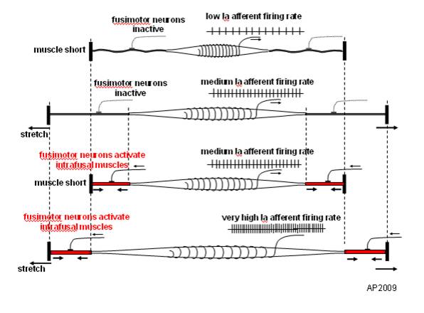
Muscle Spindles and Proprioception

image source: https://en.wikipedia.org/wiki/File:Fusimotor_action.jpg

image source: https://en.wikipedia.org/wiki/File:Fusimotor_action.jpg

And what have we been saying for the last 6 years? 

Connected to the nervous system by large diameter afferent (sensory) fibers, they are classically thought of as appraising the nervous system of vital information like length and rate of change of length of muscle fibers, so we can be coordinated. They act like volume controls for muscle sensitivity. Turn them up and the muscle becomes more sensitive to ANY input, especially stretch (so they become touchy…maybe like you get if you are hungry and tired and someone asks you to do something); turn them down and they become less or unresponsive.

Their excitability is governed by the sum total (excitatory and inhibitory) of all neurons (like interneuron’s) acting on them (their cell bodies reside in the anterior horn of the spinal cord).

Along with with Golgi tendon organs and joint mechanoreceptors, they also act as proprioceptive sentinels, telling us where our body parts are in space. We have been teaching this for years. Here is a paper that exemplifies that, identifying several proteins responsible for neurotransduction including the Piezo2 channel as a candidate for the principal mechanotransduction channel. Many neuromuscular diseases are accompanied by impaired  muscle spindle function, causing a decline of motor performance and coordination. This is yet another key finding in the kinesthetic system and its workings. 

Remember to include proprioceptive exercises and drills (on flat planar surfaces, like we talked about here) in your muscle rehab programs


Kröger S Proprioception 2.0: novel functions for muscle spindles. Curr Opin Neurol. 2018 Oct;31(5):592-598. 

Woo SH, Lukacs V, de Nooij JC, Zaytseva D, Criddle CR, Francisco A, Jessell TM, Wilkinson KA, Patapoutian A. Piezo2 is the principal mechanotransduction channel for proprioception.Nat Neurosci. 2015 Dec; 18(12):1756-62. Epub 2015 Nov 9.

Fusimotor control of proprioceptive feedback during locomotion and balancing: can simple lessons be learned for artificial control of gait?

Hulliger M. Fusimotor control of proprioceptive feedback during locomotion and balancing: can simple lessons be learned for artificial control of gait? Prog Brain Res. 1993; 97:173-80.

Sometimes, it doesn't matter whether it is long or short.

Achilles tendinopathy (AT) .. there are many factors that can contribute.

Have you considered leg length inequality? Generally speaking, People have a tendency to overpronate on the longer leg side and under prone only shorter leg side with strain on the medial and lateral aspects of the Achilles tendon respectively. It would make sense that this could be a contributing factor.

 "The mean inequality in length of legs (ILL) was 5 +/- 4 mm. Among the 48 patients with ILL > or = 5 mm, the side affected with ruptured tendon was longer in 48% of cases and shorter in 52%. " (1)

Age and pathology can play a role with younger, healthy tendons having greater compliance, with compliance being considered a risk factor for AT.(2)

Proprioception is impaired on the affected side of folks with Achilles tendinopathy (3). This is a "chicken and the egg" scenario. Did impaired proprioception cause the tendinopathy or is the tendinopathy causing the impaired proprioception? Probably, a little bit of both.

All this and more to be discussed in one of the 3 cases we will discuss tomorrow evening, Wednesday, August 15th on onlinece.com: Biomechanics 320   5PST, 6 MST, 7 CST, 8 EST

Hope to see you there!

1. Leppilahti J, Korpelainen R, Karpakka J, Kvist M, Orava S. Ruptures of the Achilles tendon: relationship to inequality in length of legs and to patterns in the foot and ankle. Foot Ankle Int. 1998 Oct;19(10):683-7.

2. Intziegianni K, Cassel M, Rauf S, White S, Rector M, Kaplick H, Wahmkow G, Kratzenstein S, Mayer F. Influence of Age and Pathology on Achilles Tendon Properties During a Single-leg Jump. Int J Sports Med. 2016 Nov;37(12):973-978. Epub 2016 Aug 8.

3. Scholes M, Stadler S, Connell D, Barton C, Clarke RA, Bryant AL, Malliaras P. Men with unilateral Achilles tendinopathy have impaired balance on the symptomatic side. J Sci Med Sport. 2018 May;21(5):479-482. doi: 10.1016/j.jsams.2017.09.594. Epub 2017 Oct 6.

Gait: How we stay upright in the gravitational plane.

We remember that we have 3 systems that keep us upright in the gravitational plane: The visual system, The vestibular system and the proprioceptive system. As we age, we seem to become more dependent upon the visual system to maintain stability of the head (which is largely under the purview of the vestibular system). Older folks seem to be less stable than younger ones....At least when it comes to head stability.

This study looked at the neuromechanical mechanisms of head stability in younger and older women during gait initiation, a time when we employ the visual system for things like balance and foot placement. The visual system in this case is king and trumps the other systems in that it will do whatever it need to to keep the eyes level with the horizon.

They used a stereophotogrammetric system to measure angular displacement and acceleration and surface emg (no, not the best) to look at activation latencies of neck (particularly the SCM), trunk and pelvic muscles. Older women had higher variability in angular displacement of the head (possibly age related breakdown of the vestibular system?), decreased ability to attenuate accelerations in the sagittal (forward) plane, and increased SCM activation latencies.

The bottom line?

Make sure the older women you are working with have:

- better functioning joints
- better functioning muscles (appropriate skill, endurance and strength)
- better proprioception

You could help these by:

- manipulating and mobilizing joints that have pathomechanics
- improving muscular function through endurance and strength based exercises of the neck as well as core
-improving muscular function through modalities you use
-give them more proprioceptive based exercises, especially ones which incorporate the head, like head repositioning exercises,
-encourage them to engage in proprioceptive heavy activities, like rock climbing (which also works the axial extensors), cross country skiing, bike riding, etc

Maslivec A, Bampouras T, Dewhurst S, Vannozzi G, Macaluso A, Laudani L. Mechanisms of head stability during gait initiation in young and older women: A neuro-mechanical analysis.
J Electromyogr Kinesiol. 2017 Nov 23;38:103-110. doi: 10.1016/j.jelekin.2017.11.010. [Epub ahead of print]

link to free full text: https://www.sciencedirect.com/science/article/pii/S1050641116302036?via%3Dihub

Proprioceptive Clues in Children’s Gait.

This goes along with Mondays post. We can learn a lot about gait from watching our children walk. An immature nervous system is very similar to one which is compensating meaning cheating around a more proper and desirable movement pattern; we often resort to a more primitive state when challenges beyond our ability are presented. This is very common when we lose some aspect of proprioception, particularly from some peripheral joint or muscle, which in turn, leads to a loss of cerebellar input (and thus cerebellar function). Remember, the cerebellum is a temporal pattern generating center so a loss of cerebellar sensory input leads to poor pattern generation output. Watch this clip several times and then try and note each of the following:

  • wide based gait; this is because proprioception is still developing (joint and muscle mechanoreceptors and of course, the spino cerebellar pathways and motor cortex)
  • increased progression angle of the feet: this again is to try and retain stability. External rotation allows them to access a greater portion of the glute max and the frontal plane (engaging an additional plane is always more stable).
  • shortened step length; this keeps the center of gravity close to the body and makes corrections for errors that much easier (remember our myelopathy case from last week ? LINK. This immature DEVELOPING system is very much like a mature system that is REGRESSING. This is a paramount learning point !)
  • decreased speed of movement; this allows more time to process proprioceptive clues, creating accuracy of motion


Remember that Crosby, Still, Nash and young song “Teach Your Children”? It is more like, “teach your parents”…


Proprioceptive clues are an important aspect of gait analysis, in both the young and old, especially since we tend to revert back to an earlier phase of development when we have an injury or dysfunction.


Functional Ankle Instability and the Peroneals

tumblr_mgcawrDlDn1qhko2so1_400.jpg

Lots of links available here with today’s blog post. please make sure to take your time and check out each one (underlined below) 

As you remember, the peroneii (3 heads) are on the outside of the lower leg (in a nice, easy to remember order of longus, brevis and tertius, from top to bottom) and help to stabilize the lateral ankle. The peroneus brevis and tertius dorsiflex and evert the foot while the peroneus longus plantarflexes and everts the foot. We discuss the peroneii more in depth here in this post. It then is probably no surprise to you that people with ankle issues, probably have some degree of peroneal dysfunction. Over the years the literature has supported notable peroneal dysfunction following even a single inversion sprain event. 

Functional ankle instability (FAI) is defined as “ the subjective feeling of ankle instability or recurrent, symptomatic ankle sprains (or both) due to proprioceptive and neuromuscular deficits." 

Arthrogenic muscle inhibition (AMI) is a neurological phenomenon where the muscles crossing a joint become "inhibited”, sometimes due to effusion (swelling) of the joint (as seen here) and that may or may not be the case with the ankle (see here), or it could be due to nociceptive input altering spindle output or possibly higher centers causing the decreased muscle activity. 

This paper (see abstract below) merely exemplifies both the peroneals and FAI as well as AMI.

Take home message?

Keep the peroneals strong with lots of balance work!                                                             


2009 May;37(5):982-8. doi: 10.1177/0363546508330147. Epub 2009 Mar 6.

Peroneal activation deficits in persons with functional ankle instability.

Palmieri-Smith RM, Hopkins JT, Brown TN.

Source

School of Kinesiology, University of Michigan, 401 Washtenaw Avenue, Ann Arbor, MI 48109, USA. riannp@umich.edu

Abstract

BACKGROUND:

Functional ankle instability (FAI) may be prevalent in as many as 40% of patients after acute lateral ankle sprain. Altered afference resulting from damaged mechanoreceptors after an ankle sprain may lead to reflex inhibition of surrounding joint musculature. This activation deficit, referred to as arthrogenic muscle inhibition (AMI), may be the underlying cause of FAI. Incomplete activation could prevent adequate control of the ankle joint, leading to repeated episodes of instability.

HYPOTHESIS:

Arthrogenic muscle inhibition is present in the peroneal musculature of functionally unstable ankles and is related to dynamic peroneal muscle activity.

RESULTS:

The FAI patients had larger peroneal H:M ratios in their nonpathological ankle (0.399 +/- 0.185) than in their pathological ankle (0.323 +/- 0.161) (P = .036), while no differences were noted between the ankles of the controls (0.442 +/- 0.176 and 0.425 +/- 0.180). The FAI patients also exhibited lower EMG after inversion perturbation in their pathological ankle (1.7 +/- 1.3) than in their uninjured ankle (EMG, 3.3 +/- 3.1) (P < .001), while no differences between legs were noted for controls (P > .05). No significant relationship was found between the peroneal H:M ratio and peroneal EMG (P > .05).

CONCLUSION:

Arthrogenic muscle inhibition is present in the peroneal musculature of persons with FAI but is not related to dynamic muscle activation as measured by peroneal EMG amplitude. Reversing AMI may not assist in protecting the ankle from further episodes of instability; however dynamic muscle activation (as measured by peroneal EMG amplitude) should be restored to maximize ankle stabilization. Dynamic peroneal activity is impaired in functionally unstable ankles, which may contribute to recurrent joint instability and may leave the ankle vulnerable to injurious loads.


Try THIS at home...

Screen Shot 2018-02-07 at 11.23.24 AM.png

Cool test, results you can see and some literature to back it up. If you are interested at all in proprioception, this is an interesting read.

So, the question for us is: "Does ankle dorsiflexion actually create more stability, like is purported?"

“The point is that if I make their ankle rigid this way, then they can more effectively use the balance mechanisms at their knees, hips, and proximally, because they’re on a stable base. My proposition is that their balance is actually normal, apart from that distal segment. When their ankle is stabilized, they use their knees more effectively, and they become less dependent on their eyesight to maintain their balance.”

http://lermagazine.com/article/afos-and-balance-issues-in-peripheral-neuropathy

FAI: ankle instability. Proprioceptive issues

We have been saying this over and over, sorry for the repeated nature of this concept. But ankle sprains should not be taken lightly. This study showed:
"Conclusions: Individuals with unilateral FAI had increased error ipsilaterally (injured limb) for inversion movement detection (kinesthesia) and evertor force sense and increased error contralaterally (uninjured limb) for evertor force sense."

No only do they have loss of kinesthesia on the injured side, but this presents along with a reduced evertor force sense as well as contralateral processing deficits. The Brain is paying close attention to the first things that hits the ground, and noting how stable/unstable it is.
Ankle sprains cannot be taken lightly, even the mild ones. Plus, do not forget about the corruption of the frontal plane at the hip that often occurs after these events.


Bilateral Proprioceptive Evaluation in Individuals With Unilateral Chronic Ankle Instability

Andreia S. P. Sousa, PhD; João Leite, BSc; Bianca Costa, BSc; Rubim Santos, PhD

Escola Superior de Saúde do Porto, Centro de Estudos de Movimento e Actividade Humana, Instituto Politécnico do Porto, Portugal

Andreia S. P. Sousa, João Leite, Bianca Costa, and Rubim Santos (2017) Bilateral Proprioceptive Evaluation in Individuals With Unilateral Chronic Ankle Instability. Journal of Athletic Training: April 2017, Vol. 52, No. 4, pp. 360-367.

Want to be faster?

superhero.jpg

Take this simple test. 

If you want to be faster, you had better incorporate some proprioceptive training into your plan. It is the 1st part of our mantra: Skill, Endurance, and Strength (in that order). Proprioceptive training appears to be more important that strength or endurance training from an injury rehabilitation perspective injury rehabilitation perspective as well part of an injury prevention program

 What is proprioception? It is body position awareness; ie: knowing what your limbs are doing without having to look at them.

Take this simple test:

  • Stand in a doorway with your shoes off. Keep your arms up at your sides so that you can brace yourself in case you start to fall. Lift your toes slightly so that only your foot tripod remains on the ground (ie the base of the big toe, the base of the little toe and the center of the heel.). Are you able to balance without difficulty? Good, all 3 systems (vision, vestibular and proprioceptive) are go.
  • Now close your eyes, taking away vision from the 3 systems that keep us upright in the gravitational plane. Are you able to balance for 30 seconds? If so, your vestibular and proprioceptive systems are intact.
  • Now open your eyes and look up at the ceiling. Provided you can balance without falling, now close your eyes. Extending your neck 60 degrees just took out the lateral semicircular canals of the vestibular system (see here for more info). Are you still able to balance for 30 seconds? If so, congrats; your proprioceptive system (the receptors in the joints, ligaments and muscles) is working great. If not, looks like you have some work to do. You can begin with exercises we use every day by clicking here.

Proprioception should be the 1st part of any training and/or rehabilitation program. If you don’t have a good framework to hang the rest of your training on, then you are asking for trouble. 


Timothy E. Hewett, PhD, , Kevin R. Ford, MS, Gregory D. Myer, MS, CSCS Anterior Cruciate Ligament Injuries in Female Athletes: Part 2, A Meta-analysis of Neuromuscular Interventions Aimed at Injury Prevention The American Journal of Sports Medicine Vol 34, Issue 3, pp. 490 - 498   link to free full text: http://journals.sagepub.com/doi/abs/10.1177/0363546505282619

Lephart SM1, Pincivero DM, Giraldo JL, Fu FH. The role of proprioception in the management and rehabilitation of athletic injuries. Am J Sports Med. 1997 Jan-Feb;25(1):130-7.


Want more stability, NOW?

balancing-stones.jpg

Try this...

While walking or running running (or watching a client walk, amble or run) you may be thinking  “I need to do something to improve my (their) proprioception, or they are going to fall (again)” If you were to increase your (their) surface area, and make yourself (theirself)vless top heavy, I (they) would be more stable. How can we accomplish that?

Here is what you can do:

First, spread your toes.; why not maximize the real estate available to your feet?

Next,  widen your stance (or base of gait). Spreading your weight over a larger surface area would be more stable and provide stability.

Third, raise your arms out from your sides (no don't try to fly) to provide more input from your upper extremities to your proprioceptive system (more input from peripheral joint and muscle mechanoreceptors = more input to cerebellum = better balance)

Lastly, Slow down from your blistering pace. this will give your (aging) nervous system more time to react.

All these actions were all “primitive” reactions of the nervous system when learning to walk. We did a post on that when my youngest son was learning to walk.

Want to have better balance?

  • Spread your toes
  • Widen your stance
  • Raise your arms
  • Slow down

Notice I didn’t say this would make you faster. Who is more likely to fall on a corner when being chased by a predator; the tortoise or the hare?

A little practical neurology for you this morning brought to you by the geeks of gait. Ivo and Shawn.

Something you can do NOW to help Parkinsons folks

199px-Sir_William_Richard_Gowers_Parkinson_Disease_sketch_1886.jpg

Anything that can help get more balance and coordination info to the higher centers is a plus for Parkinsons folks, and the texture of the surface they are walking on or the texture of the insole is no different. The more afferent info we can get in through the tactile receptors, joint mechanoreceptors and muscle receptors like the spindles and golgi tendon organs, the better. These all feed (eventually or sometimes directly) to the cerebellum, the king of balance.

In this study they placed little half spheres at the distal phalanx of the hallux, heads of metatarsophalangeal joints and heel. Theses are all areas of increased cortical representation when you look at the sensory homunculus.  They wore them for a week and plantar sensation and stride length both improved, but only the increased plantar sensation remained. Neuroplasticity takes time and we are willing to bet that if they wore them longer, the results would have been more profound.

Sometimes the simplest interventions can go a long way.


Lirani-Silva E, Vitorio R, Barbieri FA, et al. Continuous use of textured insole improve plantar sensation and stride length of people with Parkinson disease: A pilot study. Gait Posture 2017;58:495-497. [

Headbonking and gait

A great article (see reference below) just came out looking at the gait changes that come along with a concussion. Basically it says that folks that have concussions have more coronal plane (i.e. side to side) sway and they walk slower. This reminded us of some of the "decomposition of gait" pieces that we have done and one post on proprioceptive clues in children gait that we did about 5 years ago. Having a concussion causes decomposition of gait, and we move toward a more primitive pattern, just like we see in kids. Here was the post:

We can learn a lot about gait from watching our children walk. An immature nervous system is very similar to one which is compensating meaning cheating around a more proper and desirable movement pattern; we often resort to a more primitive state when challenges beyond our ability are presented. This is very common when we lose some aspect of proprioception, particularly from some peripheral joint or muscle, which in turn, leads to a loss of cerebellar input (and thus cerebellar function). Remember, the cerebellum is a temporal pattern generating center so a loss of cerebellar sensory input leads to poor pattern generation output. Watch this clip several times and then try and note each of the following:

  • wide based gait; this is because proprioception is still developing (joint and muscle mechanoreceptors and of course, the spino cerebellar pathways and motor cortex)
  • increased progression angle of the feet: this again is to try and retain stability. External rotation allows them to access a greater portion of the glute max and the frontal plane (engaging an additional plane is always more stable).
  • shortened step length; this keeps the center of gravity close to the body and makes corrections for errors that much easier (remember our myelopathy case from last week ? LINK.  This immature DEVELOPING system is very much like a mature system that is REGRESSING.  This is a paramount learning point !)
  • decreased speed of movement; this allows more time to process proprioceptive clues, creating accuracy of motion

Remember that Crosby, Still, Nash and young song “Teach Your Children”? It is more like, “teach your parents”…

Proprioceptive clues are an important aspect of gait analysis, in both the young and old, especially since we tend to revert back to an earlier phase of development when we have an injury or dysfunction.


Manaseer TSGross DPDennett LSchneider KWhittaker JL1. Gait Deviations Associated With Concussion: A Systematic Review.  Clin J Sport Med. 2017 Nov 21. doi: 10.1097/JSM.0000000000000537. [Epub ahead of print]

Party over the Weekend?


So, the more you drink, the more you impair the CNS. The more you impair the CNS, the more dependent you become on peripheral mechanisms. A good reason to keep your vestibular system (alcohol changes the specific gravity of the endolymph), your visual system (long term use affects the option nerve directly and can cause involuntary saccades) and proprioceptive systems including the cutaneous mechanoreceptors (because you are relying on them more) intact. Watch what and how much you drink...

"Standing postural stability relies on input from visual, vestibular, proprioceptive and mechanoreceptive sensors. When the information from any of these sensors is unavailable or disrupted, the central nervous system maintains postural stability by relying more on the contribution from the reliable sensors, termed sensory re-weighting. Alcohol intoxication is known to affect the integrity of the vestibular and visual systems. The aim was to assess how mechanoreceptive sensory information contributed to postural stability at 0.00% (i.e. sober), 0.06% and 0.10% blood alcohol concentration (BAC) in 25 healthy subjects (mean age 25.1 years). The subjects were assessed with eyes closed and eyes open under quiet standing and while standing was perturbed by repeated, random-length, vibratory stimulation of the calf muscles. Plantar cutaneous mechanoreceptive sensation was assessed for both receptor types: slowly adapting (tactile sensitivity) and rapidly adapting (vibration perception). The correlation between recorded torque variance and the sensation from both mechanoreceptor types was calculated. The recorded stability during alcohol intoxication was significantly influenced by both the tactile sensation and vibration perception of the subjects. Moreover, the study revealed a fluctuating association between the subjects' vibration perception and torque variance during balance perturbations, which was significantly influenced by the level of alcohol intoxication, vision and adaptation. Hence, one's ability to handle balance perturbations under the influence of alcohol is strongly dependent on accurate mechanoreceptive sensation and efficient sensory re-weighting. 

Modig F, Patel M, Magnusson M, Fransson PA.Study II: mechanoreceptive sensation is of increased importance for human postural control under alcohol intoxication. Gait Posture. 2012 Mar;35(3):419-27. doi: 10.1016/j.gaitpost.2011.11.001. Epub 2011 Dec 27.
weighting.