The consequences of an inverted forefoot

A forefoot that is inverted with respect to the rearfoot. Whether it is a forefoot varus, forefoot supinatus or an everted rearfoot ( because the forefoot is still inverted with respect to the rearfoot), what are the biomechanical sequelae?

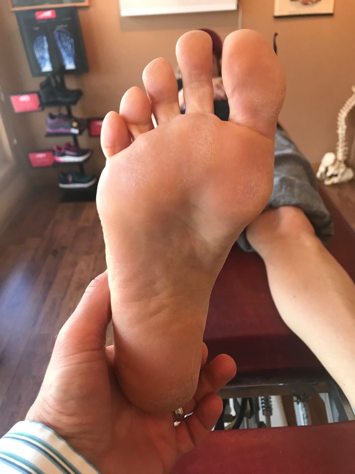
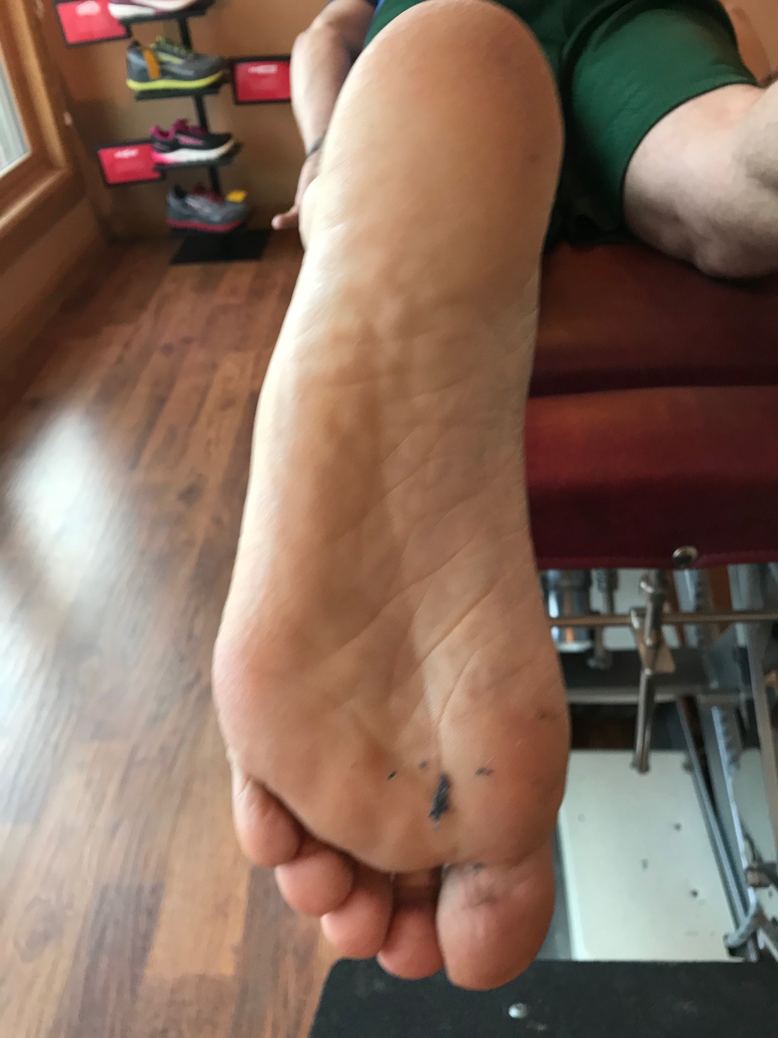
If we accept the premise that the foot is basically a tripod between the calcaneus, base of the first and base of the fifth metatarsal‘s, we know that all of these parts needs to be on the ground at certain points in the gait cycle. Forces should travel from the calcaneus, up the lateral aspect of the foot, across the metatarsal heads to the first metatarsal head and hopefully out through the hallux.

The foot should hit the ground in slight inversion of the entire foot at initial contact and pronate through the middle of mid stance and then supinate through the remainder of the gait cycle. There’s an intricate balance of biomechanical events that must occur, especially in the latter half of the gait cycle when the rear foot is inverting where the forefoot is everting, so that we can have high gear push off through the distal first ray.

If the forefoot remains inverted then somehow the head of the first metatarsal needs to be brought down to the ground. If there’s not adequate range of motion in the foot, particularly the first ray, then you may pronate through the midfoot, rearfoot or in cases where this is insufficient, bring them immediately over the foot to get it down. This of course shifts center of gravity to midline and the body above must compensate in someway.

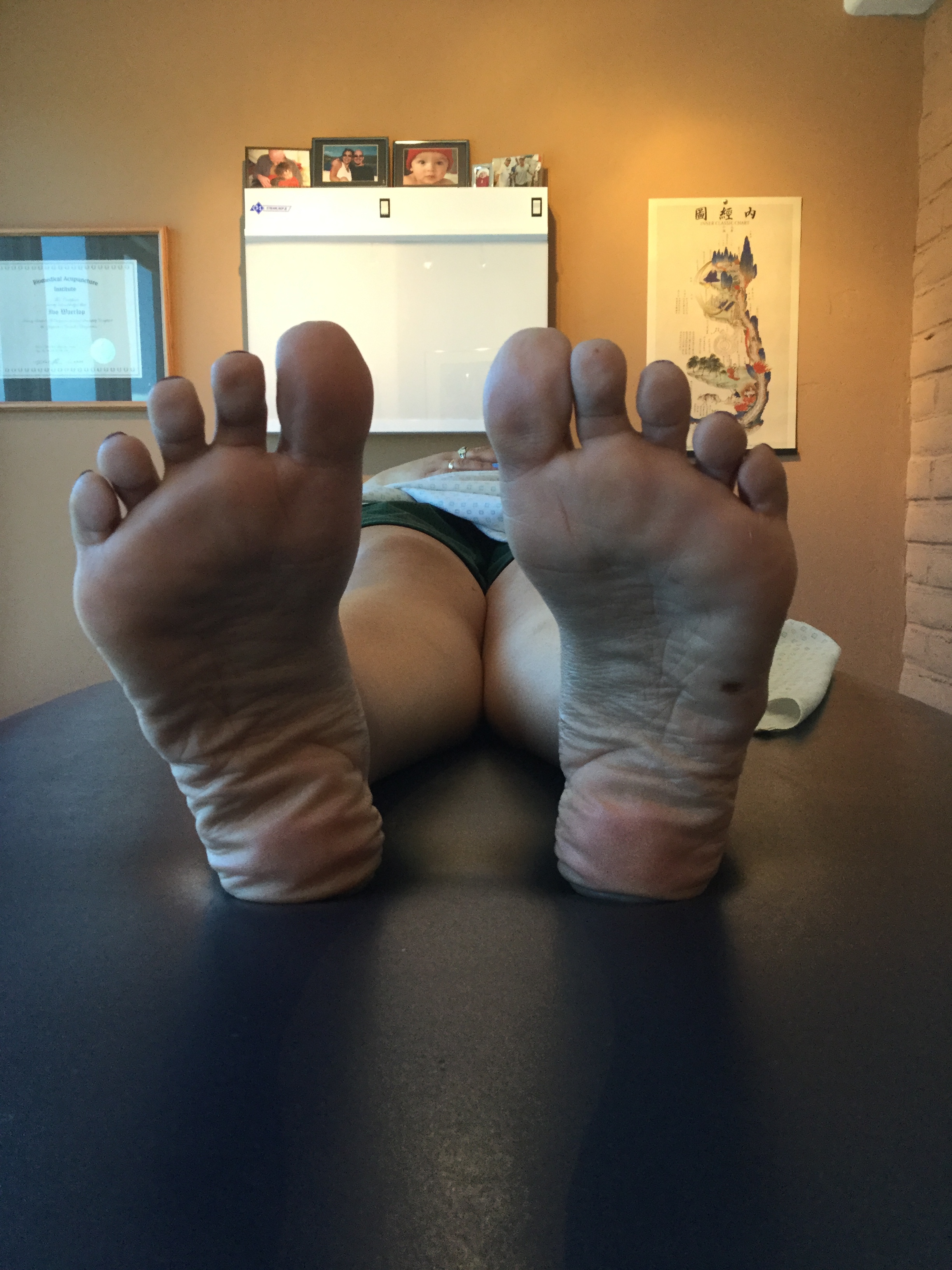
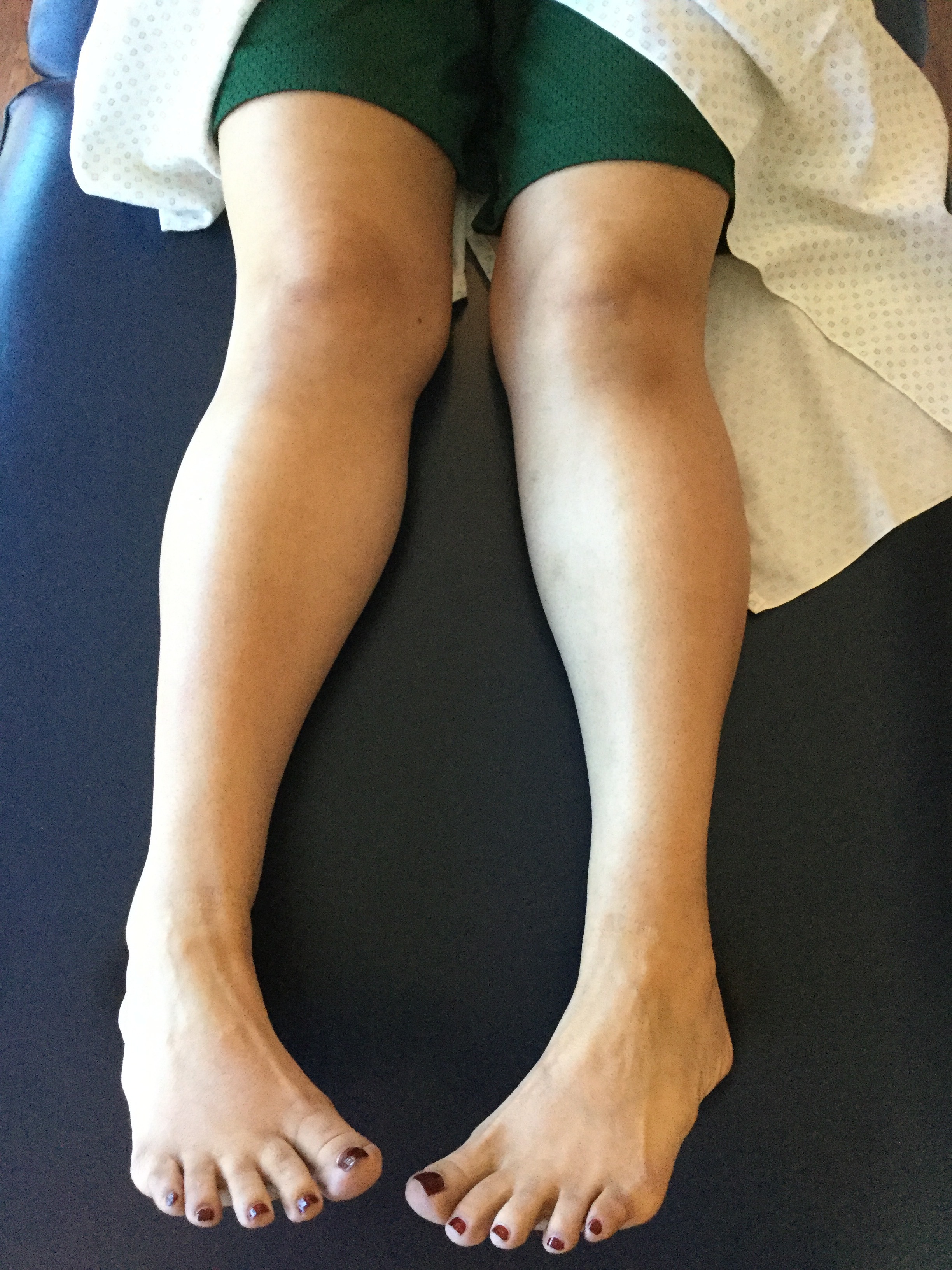
Take a look at this video footage and what do you see? She strikes on the outside of her foot but does not have adequate motion in her forefoot and therefore “crashes“ down on the forefoot, forcing a valgus moment into the ankle and the need to shift immediately by the pelvis attempts to dampen it. Notice how this is worse on the right side with more medial knee shift, pelvic shift as well as a lateral bending of the body to the right. Notice also how the upper body twists more to the left than to the right.

So what’s the fix? Well the answer is, “what’s bothering the patient?” We don’t necessarily fix what we see; we correlate what we see with what the patient’s symptoms are because that’s usually why they show up in your office. Yes, we do get people from time to time that come in strictly for “performance enhancement“ but this is pretty rare.

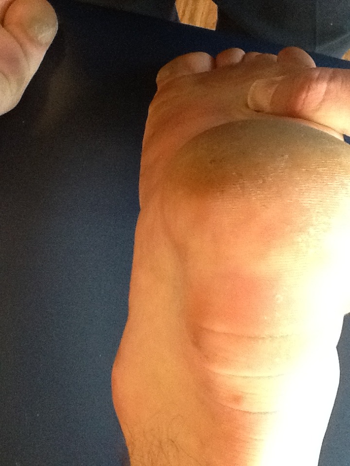
This woman has very little motion and plantar flexion of the first Ray complex so our primary goal was to get her to descend the first ray. We accomplished this by the following:

1. Manipulation in plantar and dorsiflexion of the first ray complex
2. Soft tissue work in the first intermetatarsal interval
3. Exercises of muscles to assist in descending the first ray including the following: extensor hallucis brevis, peroneus longus, flexor digitorum brevis
4. Pelvic stability work to improve the skill, endurance and strength of the gluteus medius complex as well as abdominal endurance work.

Your rehab program should change as the patient has more functional gains, tailoring it to the patient’s deficiencies.


Dr Ivo Waerlop, one of The Gait Guys

#invertedforefoot #forefootsupinatus #forefootvarus #pronation #forefoot #gaitanalysis

The “ banana foot”

IMG_7018.jpg

So, you see at foot that looks like this and what do you think? What are some of the biomechanical characteristics of people with the foot that when, you bisect the calcaneus, the line passing forward passes lateral to the second metatarsal or a line between the second and third?

This condition can be congenital, in conditions like forefoot adductus or compensatory.

The first thing that springs to mind when we see deformities like this is “things usually occur in threes“. So we would expect to see other anatomical and/or genetic abnormalities. An adducted forefoot, like you see here, often occurs as the result of lack of internal rotation of the hip on that side so therefore will often be present with conditions like internal tibial torsion and femoral retrotorsion, which we often, but not always, see together. Because of the increased gait and foot progression angle in these individuals, the forefoot compensates and adducts to bring the center of gravity more to midline.

Feet like this are often, but not always, cavus and rigid. If it remains in relative supination (plantarflexion, abduction and inversion) it is an excellent level but poor shock absorber.

Forefoot adduction can also be a compensation pattern if an individual is unable to get the head of their first ray completely down to the ground. It could be a true forefoot varus or more commonly, a forefoot supinatus; either results in an inability to get the first ray down. This often causes the foot to adduct in compensation, and, due to the tarsal articulations, often raises the base of the first metatarsal increasing the inclination angle of the first ray. This frequently leads to limited dorsiflexion of the first metatarsophalangeal articulation.

So what is a clinician to do?

Ensure that the mechanics of the foot are clean through manipulation and mobilization

Make sure there are appropriate flexors/extensor ratios of skill, endurance, and strength of the foot musculature both intrinsically and extrinsically. This means making sure that the long flexors and extensors are in some degree of balance.

Work on balance and coordination of the lower extremity. This can be impeded if they’re unable to get ahead of the first right down to the ground. Exercises for the peroneus longus, extensor hallucis brevis and short flexors of the foot will often help with this.

“Banana foot”. Coming to your clinic, or a clinic near you. Maybe today…

Dr. Ivo Waerlop, one of The Gait Guys.

#forefootadductus #bananafoot #supination #thegaitguys

Foot Types? Do they really matter?

IMG_6218 copy.jpg

The answer is " yes, often times".

Did you miss our 3rd Wednesdays presentation last week on foot types and obligate biomechanics (and pathomechanic) that ensue? Here is the video feed that you can watch and get ce credits for:

https://www.chirocredit.com/course/Chiropractic_Doctor/Biomechanics_214

#foottypes #biomechanics #thegaitguys

Determining foot types...In a nutshell

Screen Shot 2019-10-15 at 8.31.27 PM.png

We talked yesterday about how foot types (i.e., the forefoot to rear foot relationship) can often produce predictable pathomechanics. Here's How to do it. Pretty basic, eh? Its the characteristics, along with the other anatomical goodies they may have that helps to clinch the diagnosis and dictate treatment.

To find out about how to apply your newfound knowledge, join us tomorrow night on our 3rd Wednesdays tele seminar: Biomechanics 314 on online.com

5 PST, 6MST, 7CST, 8EST

When you see this, you should be thinking one of 3 possible etiologies...

Cardinal sign of either a forefoot supinatus/forefoot varus or collapsing midfoot

I was hiking behind this young chap over the weekend along with my son and friends. Note the amount of calcaneal eversion present on the right side that is not present on the left. Also note the increased progression angle of the right foot and subtle circumduction of the extremity.

In my experience, you would generally see this much calcaneal diversion and one of three scenarios:

1. Moderate leg length discrepancy with the increased calcaneovalgus occurring on the longer leg side. This would support the amount of circumduction were seeing on the right side.

2. When there is a forefoot supinatus present and and inadequate range of motion available in the midfoot and/or forefoot. This is most likely the case here.

3. In moderate To severe midfoot collapse. This is clearly not the case as the medial aspect of the shoe is usually “blown out”.

Next time you see an everting rearfoot, think about these three possible etiologies.

Dr Ivo Waerlop, on of The Gait Guys

#evertedrrarfoot #calcanealvalgus #shortleg #forefootsupinatus #forefootvarus #gaitanalysis #thegaitguys

The amazing power of compensation. Coming to a patient in your office… Maybe today

This gal has had a right sided knee replacement. She has an anatomical right short leg, a forefoot supinatus, an increased Q angle and a forefoot adductus. So, what’s the backstory?

When we have an anatomical short leg, we will often have a tendency to try to “lengthen“ that extremity and “shorten” the longer extremity. This is often accomplished through pelvic rotation although sometimes can be with knee flexion/extension or change in the Q angle. When the condition is long-standing, the body will often compensate in other ways, such as what we are seeing here.

IMG_6736.jpg

The fore foot can supinate in an attempt to lenthen the extremity. Note how the right extremity forefoot is in varus with respect to the rearfoot, effectively lengthening the extremity. As you can see from the picture, this is becoming a “hard“ deformity resulting in a forefoot varus.

IMG_6740.jpg

Over time, the forefoot has actually “adducted “ as you can see, again in an attempt to lengthen the extremity. Remember that supination is plantar flexion, abduction and inversion, all three which are visible here.


You will also see that the Q angle is less on the right side (se above), effectively lengthening that extremity, but not quite enough as we can see from the picture :-)


Dr Ivo Waerlop, one of The Gait Guys

#forefootadductus #shortleg #kneereplacement #tkr #forefootvarus #gait #thegaitguys

Sixth toe disease...That growth on the outside of your foot… Or on somebody’s foot is coming to see you…

IMG_6704.JPG

You know what we’re talking about. That extra growth on the lateral aspect of the foot that happens way too often and many of your clients. A Taylor’s bunion or sometimes referred to as a “bunionette”. 

What is the usual fix?

Usually in a ski boot or hiking boot, they blow out the lateral side of the shoe. This is usually not a good fix because most of these folks have internal tibial torsion and somewhat of a forefoot supinatus/varus.

IMG_6706.JPG
IMG_6707.JPG

The internal tibial torsion places the knee outside the saggital plane and an arch support without a forefoot valgus post will just push it further out, creating a conflict at the knee. The forefoot supinatus and/or varus places them on the outside of the foot as well. Remember, most of these folks are ALREADY on the outside of the foot and the foot wants to migrate laterally...so creating more space just means it migrates farther. Good thought, doesn’t work that way.

IMG_6710.JPG

So what did we do?

  • We created a valgus post for the forefoot (see picture above) tapering from lateral to medial and to help “push“ the distal aspect of the first ray down (because there was motion available that was not being used)

  • We gave him exercises to help descend the first ray like the extensor hallucis brevis exercise, toe waving as well as peroneus longus exercises

  • We gave him plenty of balance and coordination work

    Dr Ivo Waerlop, one of The Gait Guys


#6thtoe #internaltibialtorsion #forefootvarus # forefootsupinatus #gaitanalysis #thegaitguys


Things seem to come in 3's...

Things tend to occur in threes. This includes congenital abnormalities. Take a look this gentleman who came in to see us with lower back pain.

Highlights with pictures below:

  • bilateral femoral retrotorsion

  • bilateral internal tibial torsion

  • forefoot (metatarsus) adductus

So why LBP? Our theory is the lack of internal rotation of the lower extremities forces that motion to occur somewhere; the next mobile area just north is the lumbar spine, where there is limited rotation available, usually about 5 degrees.

Dr Ivo Waerlop, one of The Gait Guys.

#tibialtorsion #femoraltorsion #femoralretrotorsion #lowbackpain #thegaitguys #gaitproblem

this is his left hip in full internal rotation. note that he does go past zero.

this is his left hip in full internal rotation. note that he does go past zero.

full internal rotation of the right hip; note he does not go past zero

full internal rotation of the right hip; note he does not go past zero

note the internal tibial torsion. a line dropped from the tibial tuberosity should go through the 2nd metatarsal or between the 2nd and 3rd.

note the internal tibial torsion. a line dropped from the tibial tuberosity should go through the 2nd metatarsal or between the 2nd and 3rd.

ditto for the keft

ditto for the keft

a line bisecting the calcaneus should pass between the 2nd and 3rd metatarsal shafts. If talar tosion was present, the rearfoot would appear more adducted

a line bisecting the calcaneus should pass between the 2nd and 3rd metatarsal shafts. If talar tosion was present, the rearfoot would appear more adducted

less adductus but still present

less adductus but still present

look at that long flexor response in compensation. What can you say about the quadratus plantae? NO bueno…

look at that long flexor response in compensation. What can you say about the quadratus plantae? NO bueno…

Ditto!

Ditto!

Forefoot Varus vs Forefoot Supinatus

tumblr_nry1ywvvjb1qhko2so1_250.jpg

We talked about forefoot varus, forefoot supinatus and subsequent biomechanics in a recent onlinece.com course. Here is a great commentary on a review article we discussed as well as a great explanation about thew tru differences between at forefoot varus (rare) and the more common forefoot supinatus.

Take home message? FROM THE ARTICLE:

" In summary: both look the same, but they are totally different beasts:

    a forefoot varus is bony and a forefoot supinatus is soft tissue
    a forefoot varus is a cause of ‘overpronation’ and a forefoot supinatus is the result of ‘overpronation’
    a forefoot varus is rare and a forefoot supintus is common
    a forefoot varus cannot be corrected and a forefoot supinatus can be corrected"

http://www.runresearchjunkie.com/the-effect-of-forefoot-varus-on-the-hip-and-knee-and-the-effect-of-the-hip-and-knee-on-forefoot-supinatus/

More Foot Rocker Pathology Clues.

Is ankle rocker normal and adequate or is it limited ?  Is it limited in early midstance or late midstance ? How about at Toe off?  Is it even possible to distinguish this ? Well, we are splitting hairs now but we do think that it is possible. It is important to understand the pathologies on either end of the foot that can impact premature ankle rocker. 

Look at the photo above. You can see the clinical hint in the toe wear that this runner may have a premature heel rise. However, this is not solid evidence that every time you see this you must assume pathologic ankle rocker. The question is obviously, what is the cause.

Considerations:

1- weak anterior compartment, which is quite often paired with the evil neuroprotective tight calf-achilles posterior complex to offer the necessary sagittal protection at the ankle mortise.  This will cause premature heel rise from a posterior foot aspect.

2- rigid acquired blocked ankle rocker from something like “Footballer’s ankle”. This will also cause premature heel rise from a relatively posterior foot aspect.

3- there are multiple reasons for late midstance ankle rocker pathology. The client could completely avoid the normal pronation/supination phase of gait because of pain anywhere in the foot. For example, they could have plantar fascial pain, sesamoiditis, a weak first ray complex from hallux vaglus, they could have a painful bunion, they could be avoiding the collapse of a forefoot varus. There are many reasons but any of them can impair the timely pronation-supination phase in attempting to gain a rigid lever foot to toe off the big toe-medial column in “high gear” fashion. And when this happens the preparatory late midstance phase of gait can be delayed or rushed causing them to move into premature heel rise for any one of several reasons.  Rolling off to the outside and off of the lesser toes creates premature heel rise.  

4- And now for one anterior aspect cause of premature heel rise. This is obviously past the midstance phase but it can also cause premature heel rise. Turf toe, Hallux rigidus/limitus or even the dreaded fake out, the often mysterious Functional Hallux limitus (FnHL) can cause the heel to come up just a little early if the client cannot get to the full big toe dorsiflexion range.  

We could go on and on and include other issues such as altered Hip Extension Patterning, loss of hip extension range of motion, weak glutes, or even loss of terminal knee extension (from things like an incompleted ACL rehab, Osteoarthritis etc) but these are things for another time. Lets stay in the foot today.

All of these causes, with their premature heel rise component, will rush the foot to the forefoot and likely create Metatarsal head plantar loading and could cause forces appropriate enough to create stress responses to the bone. This abrupt forefoot loading thrust will often cause a reactive hammer toe effect.  Quite often just looking at the resting nature of a clients toes while they are lying down will show the underlying increase in neuro-protective hammering pattern (increased long toe flexor and short toe extensor activity paired with shortness of the opposing pairs which we review here in this short video link).  The astute observer will also note the EVA foam compressing of the shoe’s foot bed, and will also note the distal displacement of the MET head fat pad rendering the MET head pressures even greater osseously. 

Premature ankle rocker and heel rise can occur for many reasons. It can occur from problems with the shoe, posterior foot, anterior foot, toe off, ankle mortise, knee, hip or even arm swing pathomechanics.  

When premature heel rise and impaired ankle rocker rushes us to the front of the foot we drive the front half of the shoe into the ground as the foot plantarflexion is imparted into the shoe.  The timing of the normal biomechanical events is off and the pressures are altered.  instead of rolling over the forefoot and front half of the shoe after our body has moved past the foot these forces are occurring more so as our body mass is still over the foot. And the shoe can show us clues as to the torture it has sustained, just like in this photo case.

You must know the normal biomechanical gait events if you are going to put together the clues of each runner’s clinical mystery.  If you do not know normal how will you know abnormal when you see it ? If all you know is what you know, how will you know when you see something you don’t know ?

Shawn and Ivo, The Gait Guys … .  stomping out the world’s pathologic gait mechanics one person at a time. 

What do we have here and what type of shoe would be appropriate?

You are looking at a person with a fore foot varus. This means that the fore foot (ie, plane of the metatarsal heads) is inverted with respect to the rear foot (ie, the calcaneus withe the subtalar joint in neutral). Functionally translated, this means that they will have difficulties stabilizing the medial tripod (1st MET head) to the ground making the forefoot and arch unstable and likely rendering the rate and degree of pronation increased.

The incidence of this condition is 8% of 116 female subjects (McPoil et al, 1988) and 86% of 120 male and female subjects (Garbalosa et al, 1994), so it seems to happen happen more in males. We think this second number is inflated and those folks actually had a forefoot supinatus, which is much more common.

Fore foot varus occurs in 3 flavors:

  • compensated
  • uncompensated
  • partially compensated

What is meant by compensated, is that the individual is able to get the head of the 1st ray to the ground completely (compensated), partially, or, when not at all, uncompensated.What this means from a gait perspective ( for partially and uncompensated conditions) is that the person will pronate through the fore foot to get the head of the 1st ray down and make the medial tripod of the foot (ie, they pronate through the subtalar joint to allow the 1st metatarsal to contact the ground). This causes the time from mid-stance to terminal stance to lengthen and will inhibit resupination of the foot. 

Today we are looking at a rigid, uncompensated forefoot varus, most likely from insufficient talar head derotation during fetal development and subsequent post natal development. They will not get to an effective foot tripod. They will collapse the whole foot medially. These people look like severely flat-footed hyperpronators.


So, what do you do and what type of shoe is appropriate? Here’s what we did:

  • try and get the 1st ray to descend as much as possible with exercises for the extensor hallucis brevis and short flexors of the toes (see our videos on youtube)
  • create more motion in the foot with manipulation, massage mobilization to optimize what is available
  • strengthen the intrinsic muscles of the feet (particularly the interossei)
  • increase strength of the gluteus maximus and posterior fibers of the gluteus medius to slow internal rotation of the leg during initial contact to midstance
  • put them in a flexible shoe for the 1st part of the day, to exercise the feet and a more supportive; medially posted (ideally fore foot posted) shoe for the latter part of the day as the foot fatigues
  • monitor his progress at 3-6 month intervals
  • a rigid orthotic will likely not help this client and they will find it terribly uncomfortable because this is a RIGID deformity for the most part (the foot will not accommodate well to a corrective orthotic. Besides, the correction really has to be made at the forefoot anyways. 

Lost? Having trouble with all these terms and nomenclature? Take our national shoe fit program, available by clicking here.

The Gait Guys. Uber foot geeks. Separating the wheat from the chaff, with each and every post.

Metatarsus Adductus: The Basics

Metatarsus Adductus: The Basics

A few points to remember:

  • Metatarsus adductus deformity is a forefoot which is adducted in the transverse plane with the apex of the deformity at LisFranc’s (tarso-metatarsal) joint. The fifth metatarsal base will be prominent and the lateral border of the foot which is convex in shape . The medial foot border is concave with a deep vertical skin crease located at the first metatarso cuneiform joint level. The hallux (great toe) may be widely separated from the second digit and the lesser digits will usually be adducted at their bases (se below). ln some cases the abductor hallucis tendon may be palpably taut just proximal to its insertion into the inferomedial aspect of the proximal phalanx (1)
  • To measure the deviation of the metatarsals, the midline of the foot correspondingto bisecting the heel is used as a reference. This is the line that divides the heel pad into equal parts and, when extended, runs through the second toe or the second web space. In mild deformities, the midline of the foot runs through the third toe. In moderate adductus deformities, it falls between the third and fourth toes. In severe deformities the line is lateral to the third web space.(2)
  • If detected early, stretching is a common and effective treatment for mild and some moderate cases. The heel is steadied with one hand while the forefoot is abducted in relation to the hind foot. This is done for 5 reps, 5-7 times per day. (2)
  • 85% will resolve spontaneously, is caused by intrauterine position, is flexible & resolves spontaneously in more than 90 % of children. (3)
  • Though often used interchangeably, the term "metatarsus adductus" is usually reserved for milder cases, where the forefoot is adducted on the hindfoot at the tarso-metatarsal articulation. Metatarsus varus is often reserved for conditions where the matatrsals are actually curved AND the forefoot is adducted on the hindfoot. (4) The term "Metatarsus primus varus" is reserved for feet which have the same neutral or valgus hindfoot and varus forefoot but, in addition, increased divergence of the first and second metatarsals. (5)
  • It is interesting to note that along with forefoot adductus, hip dysplasia and internal tibial torsion are common (6) and this patient has the latter
  • Gait abnormalities seen with this deformity include a decreased progression angle, in toed gait, excessive supination of the feet with low gear push off from the lesser metatarsals. 


1.  Bleck E: Metatarsus adductus: classification and relationship to outcomes of treatment. J Pediatric Orthop 3:2-9,1983.

2. Bohne W. Metatarsus adductus. Bulletin of the New York Academy of Medicine. 1987;63(9):835-838.  link to FREE full text: https://www.ncbi.nlm.nih.gov/pmc/articles/PMC1629274/

3. http://www.wheelessonline.com/ortho/metatarsus_adductus

4. Peabody, C.W. and Muro, F.: Congenital metatarsus varus. J. Bone Joint Surg. 15:171-89, 1933.

5. Truslow, W.: Metatarsus primus varus or hallux valgus? J. Bone Joint Surg.23:98-108, 1925.

6. Jacobs J: Metatarsus varus and hip dysplasia. C/inO rth o p 16:203-212, 1960


additional references:

Kane R. Metatarsus varus. Bulletin of the New York Academy of Medicine. 1987;63(9):828-834. link to FREE full text: https://www.ncbi.nlm.nih.gov/pmc/articles/PMC1629282/

Wynne-Davies R, Littlejohn A, Gormley J. Aetiology and interrelationship of some common skeletal deformities. (Talipes equinovarus and calcaneovalgus, metatarsus varus, congenital dislocation of the hip, and infantile idiopathic scoliosis). Journal of Medical Genetics. 1982;19(5):321-328. link to FREE full text: https://www.ncbi.nlm.nih.gov/pmc/articles/PMC1048914/


Forefoot Varus or Forefoot Supinatus?Forefoot varus is a fixed, frontal plane deformity where the forefoot is inverted with respect to the rearfoot. Forefoot varus is normal in early childhood, but should not persist past 6 years of age (i.e. when d…

Forefoot Varus or Forefoot Supinatus?

Forefoot varus is a fixed, frontal plane deformity where the forefoot is inverted with respect to the rearfoot. Forefoot varus is normal in early childhood, but should not persist past 6 years of age (i.e. when developmental valgus rotation of forefoot on rearfoot is complete, and plantar aspects of fore- and rearfoot become parallel to, and on same plane as, one another (1)

Forefoot supinatus is the supination of the forefoot that develops with adult acquired flatfoot deformity. This is an acquired soft tissue adaptation in which the forefoot is inverted on the rearfoot. Forefoot supinatus is a reducible deformity. Forefoot supinatus can mimic, and often be mistaken for, a forefoot varus. (2)

A forefoot varus differs from forefoot supinatus in that a forefoot varus is a congenital osseous where a forefoot supinatus is acquired and develops because of subtalar joint pronation.

“Interestingly, only internal rotation of the hip was increased in subjects with FV – no differences were present in hip adduction and knee abduction between subjects with and without FV. The authors nevertheless conclude that FV causes significant changes in mechanics of proximal segments in the lower extremity and speculate that during high-speed weight-bearing tasks such as running, the effects of FV on proximal segments in the kinetic chain might be more pronounced.”

We wonder if the folks in this study had a true forefoot varus, or actually a forefoot supinatus (3).


The Gait Guys


1. Illustrated Dictionary of Podiatry and Foot Science by Jean Mooney © 2009 Elsevier Limited.

2. Evans EL1, Catanzariti AR2. Forefoot supinatus.
Clin Podiatr Med Surg. 2014 Jul;31(3):405-13. doi: 10.1016/j.cpm.2014.03.009.

3. Scattone Silva R1, Maciel CD2, Serrão FV3. The effects of forefoot varus on hip and knee kinematics during single-leg squat. Man Ther. 2015 Feb;20(1):79-83. doi: 10.1016/j.math.2014.07.001. Epub 2014 Jul 12.

The debate continues. More support for mid and forefoot strikers.…
“Forefoot and midfoot strikers had significantly shorter ground contact times than heel strikers. Forefoot and midfoot strikers had significantly faster average race spe…

The debate continues. More support for mid and forefoot strikers.

“Forefoot and midfoot strikers had significantly shorter ground contact times than heel strikers. Forefoot and midfoot strikers had significantly faster average race speed than heel strikers.”

We are not saying “better”, but according to this study “faster”!

What is the ideal?  We wish we knew…Biomechanics seem to point to less impact is better, but what is actually best for the individual is probably due to genetics, training, practice, running surface and that individuals neuromuscular competence and ability to compensate.

The Gait Guys. bringing you the facts, even if you or we don’t like them…


J Sports Sci. 2012;30(12):1275-83. doi: 10.1080/02640414.2012.707326. Epub 2012 Aug 2.

Foot strike patterns and ground contact times during high-calibre middle-distance races.

Source

Department of Sport and Exercise Sciences, School of Life Sciences, Northumbria University, Newcastle-upon-Tyne NE1 8ST, UK. phil.hayes@northumbria.ac.uk

Abstract

The aims of this study were to examine ground contact characteristics, their relationship with race performance, and the time course of any changes in ground contact time during competitive 800 m and 1500 m races. Twenty-two seeded, single-sex middle-distance races totaling 181 runners were filmed at a competitive athletics meeting. Races were filmed at 100 Hz. Ground contact time was recorded one step for each athlete, on each lap of their race. Forefoot and midfoot strikers had significantly shorter ground contact times than heel strikers. Forefoot and midfoot strikers had significantly faster average race speed than heel strikers. There were strong large correlations between ground contact time and average race speed for the women’s events and men’s 1500 m (r = -0.521 to -0.623; P < 0.05), whereas the men’s 800 m displayed only a moderate relationship (r = -0.361; P = 0.002). For each event, ground contact time for the first lap was significantly shorter than for the last lap, which might reflect runners becoming fatigued.

PMID:22857152[PubMed - indexed for MEDLINE]

http://www.ncbi.nlm.nih.gov/pubmed/22857152

Support for a midfoot strike?
&ldquo;Running with a midfoot strike pattern resulted in a significant increase in gastrocnemius lateralis pre-activation (208 ± 97.4 %, P &lt; 0.05) and in a significant decrease in tibialis anterior EMG activity (56.2…

Support for a midfoot strike?

Running with a midfoot strike pattern resulted in a significant increase in gastrocnemius lateralis pre-activation (208 ± 97.4 %, P < 0.05) and in a significant decrease in tibialis anterior EMG activity (56.2 ± 15.5 %, P < 0.05) averaged over the entire stride cycle. The acute attenuation of foot-ground impact seems to be mostly related to the use of a midfoot strike pattern and to a higher pre-activation of the gastrocnemius lateralis. ”

Do these results surprise you? They didn’t really surprise us.

The lateral head of the gastroc is a midstance to preswing stabilizer and works synergistic with the medial head, with the medial head firing earlier. Sutherland talks about these muscles not being propulsive in nature, but rather maintainers of forward progression, step length and gait symmetry. Thinking this through in a closed chain (foot up) fashion, this would counter the inversion moment created by the medial gastroc for supination in the second half of contact phase. If the foot is already partially supinated (as we believe it would be in a midfoot strike), it would have to pre activate.

A decrease in tibialis anterior activity? Sure. If the foot is striking more parallel to the ground, the anterior compartment (including the tibialis anterior, extensor hallucis longus, and extensor digitorum longus) would not have to eccentrically contract to decelerate the lowering of the foot to the ground.

Better? Maybe, maybe not. We are seeing more and more literature about foot strike (if you missed our last few posts, click here, here, here and here), We still maintain that you need a competent lower kinetic chain, including the foot and an intact nervous system to drive the boat.

We remain, handsome, bald and nerdy…Ivo and Shawn


Eur J Appl Physiol. 2012 Aug 9. [Epub ahead of print]

Impact reduction during running: efficiency of simple acute interventions in recreational runners.

Source

University of Lyon, 42023, Saint-Etienne, France.

Abstract

Running-related stress fractures have been associated with the overall impact intensity, which has recently been described through the loading rate (LR). Our purpose was to evaluate the effects of four acute interventions with specific focus on LR: wearing racing shoes (RACE), increasing step frequency by 10 % (FREQ), adopting a midfoot strike pattern (MIDFOOT) and combining these three interventions (COMBI). Nine rearfoot-strike subjects performed five 5-min trials during which running kinetics, kinematics and spring-mass behavior were measured for ten consecutive steps on an instrumented treadmill. Electromyographic activity of gastrocnemius lateralis, tibialis anterior, biceps femoris and vastus lateralis muscles was quantified over different phases of the stride cycle. LR was significantly and similarly reduced in MIDFOOT (37.4 ± 7.20 BW s(-1), -56.9 ± 50.0 %) and COMBI (36.8 ± 7.15 BW s(-1), -55.6 ± 29.2 %) conditions compared to NORM (56.3 ± 11.5 BW s(-1), both P < 0.001). RACE (51.1 ± 9.81 BW s(-1)) and FREQ (52.7 ± 11.0 BW s(-1)) conditions had no significant effects on LR. Running with a midfoot strike pattern resulted in a significant increase in gastrocnemius lateralis pre-activation (208 ± 97.4 %, P < 0.05) and in a significant decrease in tibialis anterior EMG activity (56.2 ± 15.5 %, P < 0.05) averaged over the entire stride cycle. The acute attenuation of foot-ground impact seems to be mostly related to the use of a midfoot strike pattern and to a higher pre-activation of the gastrocnemius lateralis. Further studies are needed to test these results in prolonged running exercises and in the long term.

PMID:22875194 [PubMed - as supplied by publisher]


All material copyright 2013 The Gait Guys/The Homunculus Group, yada, yada, yada…

Looks like Newbies are heel strikers 
&ldquo;Nearly all novice runners utilize a rearfoot strike when taking up running in a conventional running shoe. Hereby, the footstrike patterns among novice runners deviate from footstrike patterns among elite…

Looks like Newbies are heel strikers

“Nearly all novice runners utilize a rearfoot strike when taking up running in a conventional running shoe. Hereby, the footstrike patterns among novice runners deviate from footstrike patterns among elite and sub-elite runners.”


please take some time to explore the links we put in, as they are germane to the post


The question begs, “Why?”

  • do they believe running is merely an extension of walking, and just “speed up” the process?
  • are they afraid of going too fast and are using the heel strike to “brake”?
  • do they learn to strike differently with more experience? at least one paper eludes to “yes”
  • is it “more comfortable” as this paper says it may be?
  • If there is a rear foot strike, the foot is poised to be able to pronate to a greater degree. This theoretically means it (ie, the foot) can absorb more shock through this mechanism, although this seemingly contradicts the Lieberman study

This paper certainly had a nice cohort size (> 900 runners) so we can state, at least for this group, that this is not by chance.  When there is a fore foot strike, the foot is more supinated and makes a seemingly “rigid lever”, does this mean there is less shock (perceived or actual) with this foot posture?

Lots of questions. This is only 1 part of the puzzle.

The Gait Guys. Sifting through the literature and giving you the beef


Gait Posture. 2012 Dec 29. pii: S0966-6362(12)00448-1. doi: 10.1016/j.gaitpost.2012.11.022. [Epub ahead of print]

Footstrike patterns among novice runners wearing a conventional, neutral running shoe.

Bertelsen ML, Jensen JF, Nielsen MH, Nielsen RO, Rasmussen S.

Aarhus University Hospital, Aalborg Hospital, Orthopaedic Surgery Research Unit, Science and Innovation Center, Aalborg DK-9000, Denmark. Electronic address: miclejber@gmail.com.

Abstract

INTRODUCTION:

It has been suggested that striking on the midfoot or forefoot, rather than the rearfoot, may lessen injury risk in the feet and lower limb. In previous studies, a disparity in distribution in footstrike patterns was found among elite-, sub-elite, and recreational runners.

PURPOSE:

The purpose of this study was to investigate the footstrike patterns among novice runners.

METHODS:

All runners were equipped with the same conventional running shoe. Participants were video filmed at 300 frames per second and the footstrike patterns were evaluated by two observers. The footstrike was classified as rearfoot, midfoot, forefoot, or asymmetrical.

RESULTS:

A total of 903 persons were evaluated. The percentages of rearfoot-, midfoot-, forefoot-, and asymmetrical footstrike among men were 96.9%, 0.4%, 0.9%, and 1.8%, respectively. Among women the percentages were 99.3%, 0%, 0%, and 0.7%, respectively.

CONCLUSION:

Nearly all novice runners utilize a rearfoot strike when taking up running in a conventional running shoe. Hereby, the footstrike patterns among novice runners deviate from footstrike patterns among elite and sub-elite runners.

Copyright © 2012 Elsevier B.V. All rights reserved.


PMID: 23280125 [PubMed - as supplied by publisher]


all material copyright 2013 The Gait Guys/The Homunculus group. Please don’t lift our stuff without asking and giving credit.

New Study Finds Group of Heel Striking Barefoot Kenyan Runners.
Not all that is barefoot is necessarily forefoot&hellip;
You may have seen our tweet yesterday and have read this article. Or maybe, because you are a foot geek, you have seen it alread…

New Study Finds Group of Heel Striking Barefoot Kenyan Runners.

Not all that is barefoot is necessarily forefoot…

You may have seen our tweet yesterday and have read this article. Or maybe, because you are a foot geek, you have seen it already.

Here’s the summary: “Jan. 9, 2013 — A recently published paper by two George Washington University researchers shows that the running foot strike patterns vary among habitually barefoot people in Kenya due to speed and other factors such as running habits and the hardness of the ground. These results are counter to the belief that barefoot people prefer one specific style of running.”

The study reported a 72 percent rearfoot landing when running barefoot at endurance pace speeds supporting the notion that speed affects landing choice (faster speeds transitioned  the runners into more midfoot / forefoot landing).  Lieberman’s Harvard study which brought much of the forefoot strike principle to the western world was often based off of sub 5 mile paced runs.

It raises the question “ If barefoot IS better, and forefoot impact IS BETTER, then, what gives?”

We think the better response is:

  • there are many variables (genetics, surface, speed, etc) that can influence foot strike patterns and this paper exemplifies that.
  • Fore foot striking in runners does lessen impact forces.
  • Forefoot striking does appear to accentuate any forefoot abnormality (ie: varus/valgus) that may be present (something we will continue to say until someone proves it otherwise).
  • forefoot striking loads the posterior compartment of the lower leg (tricep surae (gastroc soleus complex)) to a greater degree

We like a mid foot strike, not because it is the middle road, but because it supports the notion in distance running that the entire foot tripod (which is more stable) engages the ground reducing solitary forefoot and rearfoot loading issues which each have their risks and challenges and allows for a more stable contact point for the body to negotiate over.  We have pounded sand on forefoot types, and the inherent risks of forefoot strike running with each of them, from our inception.  But, when it comes to midfoot strike there doesn’t appear to be much, if any literature out there to support our opinion.  Maybe now that the forefoot and rearfoot studies are out there maybe someone will find a tribe of midfoot strikers to support our rants.

We think the key is not necessarily strike position, but rather where the foot is hitting the ground relative to the body AND MORE IMPORTANTLY, having a competent foot and lower kinetic chain and core, along with the body’s ABILITY to absorb or attenuate those forces, no matter where the foot is striking the ground.

This is no doubt the 1st in a series of papers looking at this. It will be interesting to see where it goes from here.

Ivo and Shawn…  The Gait Guys

here is the link: http://www.sciencedaily.com/releases/2013/01/130109185856.htm

all material copyright 2013 The Homunculus Group/ The Gait Guys. Please ask to use our stuff and reference it appropriately. We know a guy named BamBam who helps people play nice.