


Did you see this in our recent blog post here ? a reader made us look closer. Did you catch it ?
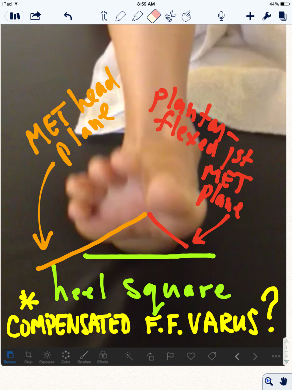
The clients right foot appears to have a dropped 1st met head. (we hate this term, because it is not accurate and is a sloppy clinical description). In this still photo it appears plantarflexed. But in this video, consider the descended 1st met head as due to the disuse or weakness of the EHL muscle (extensor hallucis longus) of the 1st toe. Or, is this in fact a compensated forefoot varus ? Sure looks like it. But with all that anterior compartment weakness (as we discussed in the previous blog post link above) it could just be a mirage. In the photo above, in a normal foot the rearfoot plane (greenline) should parallel the forefoot line (orange line). In this case, in this actively postured foot (thus some inaccuracy here, we are merely making a teaching point from the photo) the upslope of the orange line suggests a forefoot varus. This would be true if the first Metatarsal head also was on this line, but you can see that it has its own idea. This represents, in theory (regarding this photo), a compensated forefoot varus. But remember, this client is holding the foot actively in this posture. A true hands on assessment is needed to truly define a Forefoot varus, and whether it is anatomic, flexible, rigid or in many cases, just a learned functional posturing from weakness of the flexor/extensor pairing of the 1st metatarsal complex or from other weaknesses of the other forefoot evertors. It gets complicated as you can see.
As always, knowledge of the anatomy and functional anatomy allows for observation, and observation leads to understanding, which leads to answers and then remedy implementation. Our thoughts, knowing the case, is that this is a functional appearance illusion of a compensated forefoot varus due to the EHL, EDL and tibialis anterior weakness (anterior compartment) and how they play together with the flexors. One must be sure to assess the EHL when examining the foot. Test all of the muscles one by one. We have been talking about toe extensors for a long time, they can be a paramount steering wheel for the forefoot and arch posture. Podcast 71 talks about this Forefoot varus, and you should care.
In a 2009 study by Reynard et al they concluded:
- “The activity of extensor digitorum longus muscle during the swing phase of gait is important to balance the foot in the frontal plane. The activation of that muscle should be included in rehabilitation programs.” (1)
Have a burning desire to learn more about forefoot varus, here are 25 blog post links from our last few years. And/or you can take our National Shoe Fit program (downloadable links below).
Knowing what you are seeing during your exam and gait analysis can only truly come from coupling your observations with a clinical exam. Anything less is speculation and guess work. It is gambling, and this is not Vegas baby, this is someone’s health.
Shawn and Ivo, The Gait Guys
________________
National Shoe Fit Certification Program:
Gait Guys online /download store (National Shoe Fit Certification and more !) :
http://store.payloadz.com/results/results.aspx?m=80204
1. Foot (Edinb). 2009 Jun;19(2):69-74. Epub 2008 Dec 31. Foot varus in stroke patients: muscular activity of extensor digitorum longus during the swing phase of gait. Reynard F, Dériaz O, Bergeau J.
Other web based Gait Guys lectures:
www.onlinece.com type in Dr. Waerlop or Dr. Allen, ”Biomechanics”Reference


