Can you say Forefoot Adductus?

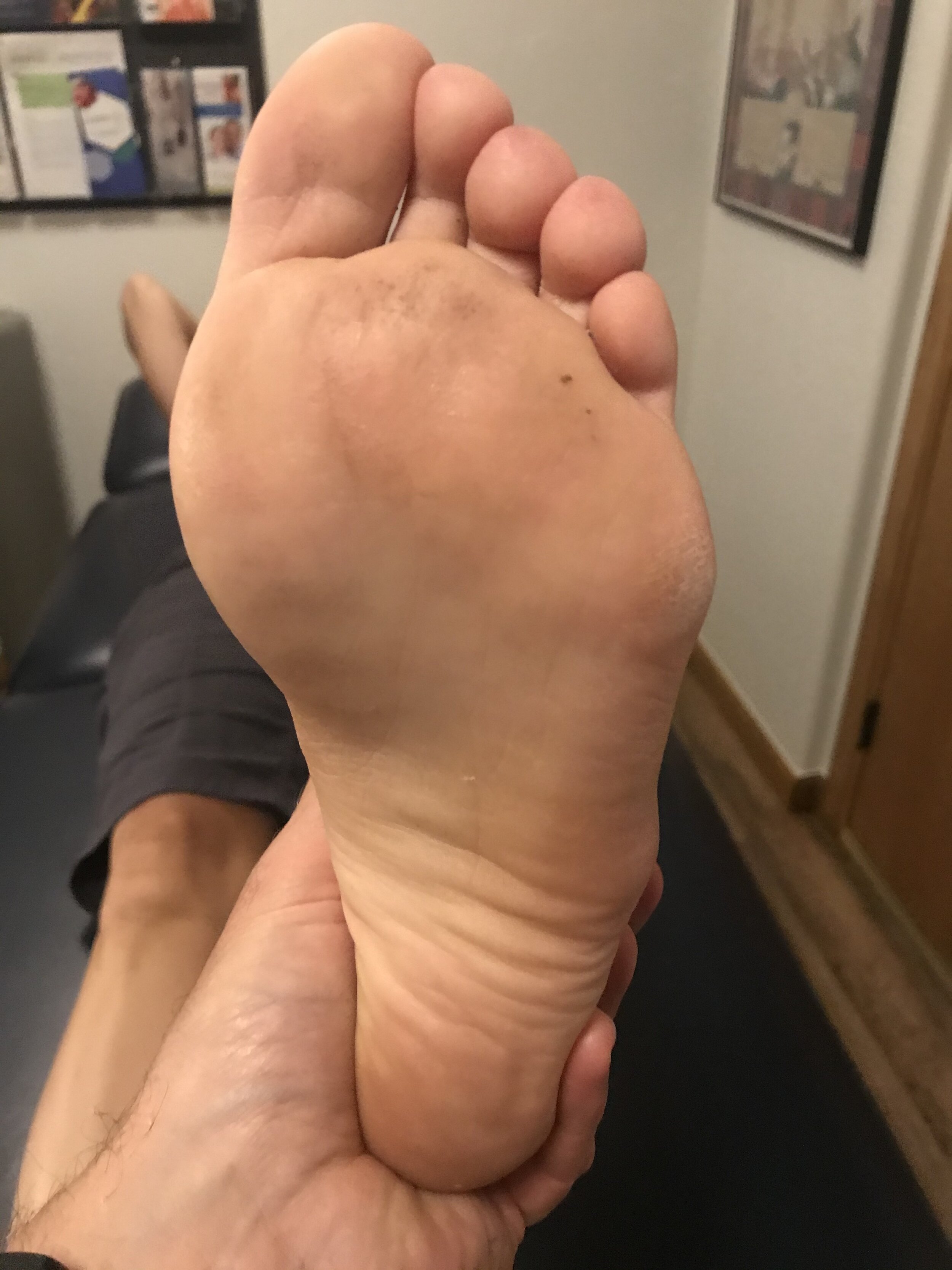
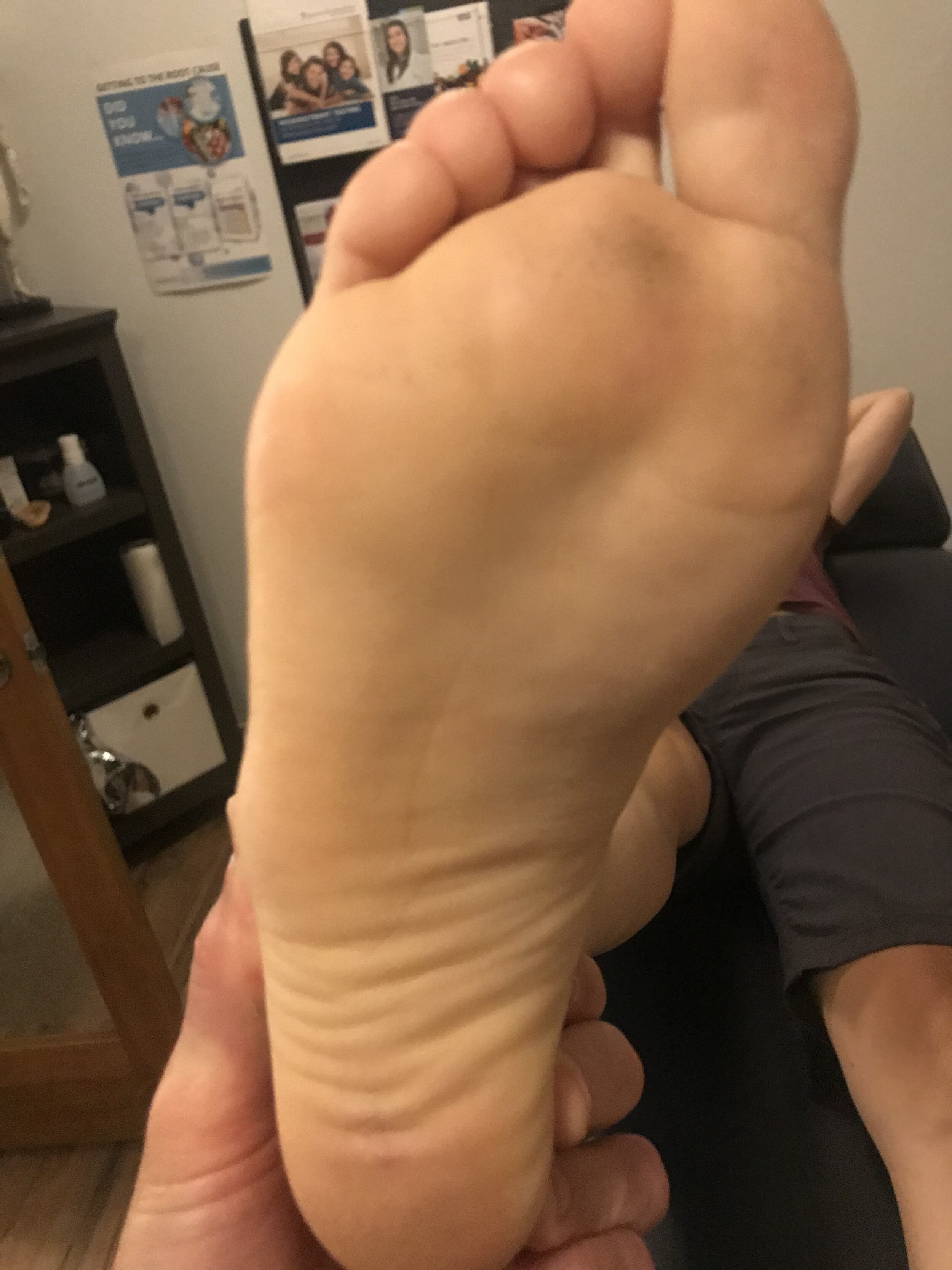
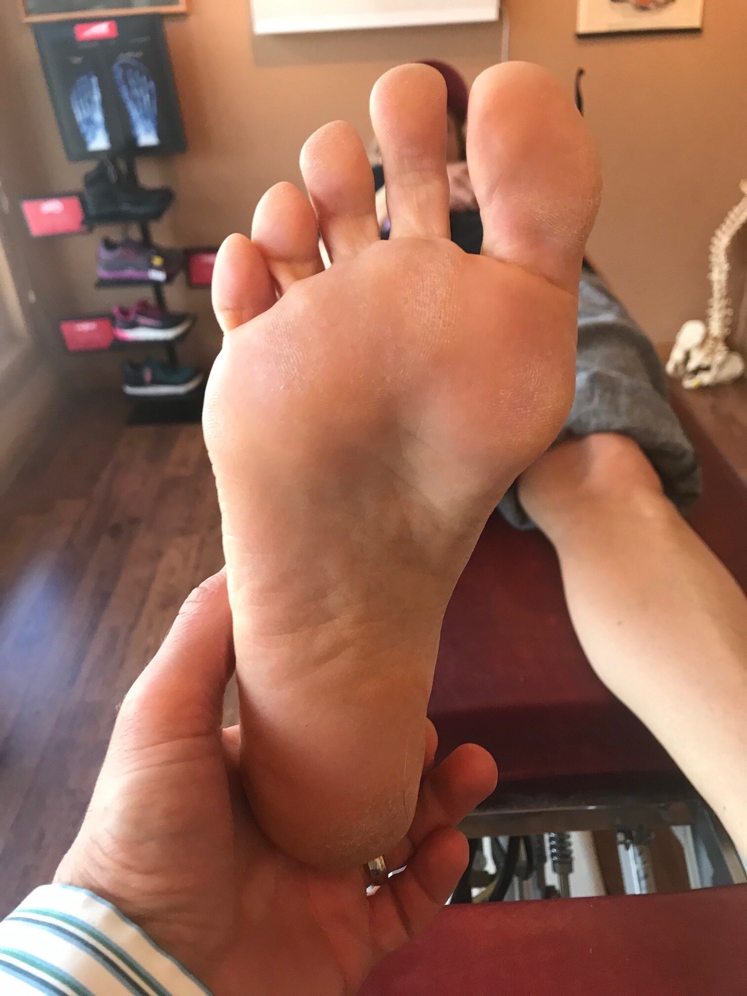
-Take a look at these tootsies. Draw an imaginary lines for the center of the hill: this should normally pass up through the foot either through the second metatarsal or between the second and third. Can you see how the foot is somewhat banana shaped?

IMG_8466.jpg

– This is also called metatarsus adductus. The deformity is at the apex of the tarsal/metatarsal joint, also known as Lisfranc joint. The fifth metatarsal base is often prominent in the foot is convex in shape with a higher arch.

-This is usually caused by intrauterine positioning and if caught early will usually spontaneously resolve. Since this gal is over 34, that’s probably not her reality. It is interesting to note that along with this congenital deformity, hip dysplasia and internal tibial torsion (which she has) are extremely common.

IMG_8468.jpg

IMG_8467.jpg

– Gait abnormalities seen with this include a decreased progression angle, and a propulsive gait often secondary to poor intrinsic function of the foot musculature

#forefoot #forefootadductus #adductus #foot #footproblem #gait #clinicalexam #thegaitguys 

The consequences of an inverted forefoot

A forefoot that is inverted with respect to the rearfoot. Whether it is a forefoot varus, forefoot supinatus or an everted rearfoot ( because the forefoot is still inverted with respect to the rearfoot), what are the biomechanical sequelae?

If we accept the premise that the foot is basically a tripod between the calcaneus, base of the first and base of the fifth metatarsal‘s, we know that all of these parts needs to be on the ground at certain points in the gait cycle. Forces should travel from the calcaneus, up the lateral aspect of the foot, across the metatarsal heads to the first metatarsal head and hopefully out through the hallux.

The foot should hit the ground in slight inversion of the entire foot at initial contact and pronate through the middle of mid stance and then supinate through the remainder of the gait cycle. There’s an intricate balance of biomechanical events that must occur, especially in the latter half of the gait cycle when the rear foot is inverting where the forefoot is everting, so that we can have high gear push off through the distal first ray.

If the forefoot remains inverted then somehow the head of the first metatarsal needs to be brought down to the ground. If there’s not adequate range of motion in the foot, particularly the first ray, then you may pronate through the midfoot, rearfoot or in cases where this is insufficient, bring them immediately over the foot to get it down. This of course shifts center of gravity to midline and the body above must compensate in someway.

Take a look at this video footage and what do you see? She strikes on the outside of her foot but does not have adequate motion in her forefoot and therefore “crashes“ down on the forefoot, forcing a valgus moment into the ankle and the need to shift immediately by the pelvis attempts to dampen it. Notice how this is worse on the right side with more medial knee shift, pelvic shift as well as a lateral bending of the body to the right. Notice also how the upper body twists more to the left than to the right.

So what’s the fix? Well the answer is, “what’s bothering the patient?” We don’t necessarily fix what we see; we correlate what we see with what the patient’s symptoms are because that’s usually why they show up in your office. Yes, we do get people from time to time that come in strictly for “performance enhancement“ but this is pretty rare.

This woman has very little motion and plantar flexion of the first Ray complex so our primary goal was to get her to descend the first ray. We accomplished this by the following:

1. Manipulation in plantar and dorsiflexion of the first ray complex
2. Soft tissue work in the first intermetatarsal interval
3. Exercises of muscles to assist in descending the first ray including the following: extensor hallucis brevis, peroneus longus, flexor digitorum brevis
4. Pelvic stability work to improve the skill, endurance and strength of the gluteus medius complex as well as abdominal endurance work.

Your rehab program should change as the patient has more functional gains, tailoring it to the patient’s deficiencies.


Dr Ivo Waerlop, one of The Gait Guys

#invertedforefoot #forefootsupinatus #forefootvarus #pronation #forefoot #gaitanalysis

Internal tibial torsion puts pressure on the outside of the foot

IMG_7174.jpg
IMG_7175.jpg
IMG_7176.jpg

Take a look at these pictures. This is also a good reason to always look at the insoles. Take a good look. Can you see the increase printing on the lateral aspect of the right foot?

You’ll note that he has internal tibial torsion on the right side. This often presents with a forefoot supinatus and results in pressuring of the lateral column of the foot and an inability to descend the first ray. Note that the footbed on the right shows increased pressure of the lateral column and a lack of pressure under the head of the first.

Stand up and put the weight on the outside of your right foot. Can you feel how the toes on right side pressing more in an attempt to shift the center of gravity medially while it offloading the toes on the left foot? This is also represented in the foot beds.

Yet another great reason to not only look at the wear on the outside, but also on the inside of your clients/patients shoes.

Dr Ivo Waerlop, one of The Gait Guys

#footbeds #internaltibialtorsion #lateralfootpressure #insoles

The “ banana foot”

IMG_7018.jpg

So, you see at foot that looks like this and what do you think? What are some of the biomechanical characteristics of people with the foot that when, you bisect the calcaneus, the line passing forward passes lateral to the second metatarsal or a line between the second and third?

This condition can be congenital, in conditions like forefoot adductus or compensatory.

The first thing that springs to mind when we see deformities like this is “things usually occur in threes“. So we would expect to see other anatomical and/or genetic abnormalities. An adducted forefoot, like you see here, often occurs as the result of lack of internal rotation of the hip on that side so therefore will often be present with conditions like internal tibial torsion and femoral retrotorsion, which we often, but not always, see together. Because of the increased gait and foot progression angle in these individuals, the forefoot compensates and adducts to bring the center of gravity more to midline.

Feet like this are often, but not always, cavus and rigid. If it remains in relative supination (plantarflexion, abduction and inversion) it is an excellent level but poor shock absorber.

Forefoot adduction can also be a compensation pattern if an individual is unable to get the head of their first ray completely down to the ground. It could be a true forefoot varus or more commonly, a forefoot supinatus; either results in an inability to get the first ray down. This often causes the foot to adduct in compensation, and, due to the tarsal articulations, often raises the base of the first metatarsal increasing the inclination angle of the first ray. This frequently leads to limited dorsiflexion of the first metatarsophalangeal articulation.

So what is a clinician to do?

Ensure that the mechanics of the foot are clean through manipulation and mobilization

Make sure there are appropriate flexors/extensor ratios of skill, endurance, and strength of the foot musculature both intrinsically and extrinsically. This means making sure that the long flexors and extensors are in some degree of balance.

Work on balance and coordination of the lower extremity. This can be impeded if they’re unable to get ahead of the first right down to the ground. Exercises for the peroneus longus, extensor hallucis brevis and short flexors of the foot will often help with this.

“Banana foot”. Coming to your clinic, or a clinic near you. Maybe today…

Dr. Ivo Waerlop, one of The Gait Guys.

#forefootadductus #bananafoot #supination #thegaitguys

Unilateral calcaneal valgus: what can it mean?

right calcaneal valgus

right calcaneal valgus

Take a good look at this picture and what do you see? Do you see the calcaneal valgus on the right side. What runs through your mind?

Possibilities for causing this condition, as well as the clinical implications are numerous.

The short list should include:

  • A shorter leg on the contralateral side: often times we will pronate more on the longer leg side to compensate for a short leg on the opposite

  • Increased rear foot and/or fore foot pronation on the valgus side. Laxity of the spring ligament or incompetency of the musculature which helps to maintain your arch (tibialis posterior, foot intrinsics, tibialis anterior to name a few) often causes more collapse on the effected side

  • A lack of available rearfoot eversion on the contralateral side. It may be that the increase calcaneovalgus is normal and the opposite side is more rigid.

  • If you were seeing this in the middle of the gait cycle it could be that that is their strategy to get around a loss of hip extension or ankle rocker

  • External tibial torsion on that side. Go ahead, stand up and spin your right foot into external rotation and keep your left foot with a normal progression angle. Can you see how your arch collapses to a greater degree on the side with the external torsion? Remember that pronation is dorsiflexion, eversion and abduction.

  • Internal tibial torsion on the contralateral side. Internal tibial torsion puts the foot into supination which makes it into more of a rigid lever rather than mobile adapter.

    And the list goes on…

    Next time you see a unilateral deformity like this, hopefully some of these things run through your mind and will help you to pinpoint where the problem actually is.

    Dr Ivo Waerlop, one of The Gait Guys

    #calcanealeversion #rearfootvalgus
    #lowerextremitydeformities

Foot Types? Do they really matter?

IMG_6218 copy.jpg

The answer is " yes, often times".

Did you miss our 3rd Wednesdays presentation last week on foot types and obligate biomechanics (and pathomechanic) that ensue? Here is the video feed that you can watch and get ce credits for:

https://www.chirocredit.com/course/Chiropractic_Doctor/Biomechanics_214

#foottypes #biomechanics #thegaitguys

Determining foot types...In a nutshell

Screen Shot 2019-10-15 at 8.31.27 PM.png

We talked yesterday about how foot types (i.e., the forefoot to rear foot relationship) can often produce predictable pathomechanics. Here's How to do it. Pretty basic, eh? Its the characteristics, along with the other anatomical goodies they may have that helps to clinch the diagnosis and dictate treatment.

To find out about how to apply your newfound knowledge, join us tomorrow night on our 3rd Wednesdays tele seminar: Biomechanics 314 on online.com

5 PST, 6MST, 7CST, 8EST

Foot types: do they really matter?

forefoot varus: note how the forefoot is inverted with respect to the rear foot

forefoot varus: note how the forefoot is inverted with respect to the rear foot

Foot type. You know what we are talking about. The relation in anatomically and in space of the rear foot to the forefoot. We believe that this anatomical relationship holds key clinical insights to predictable biomechanics in that particular foot type.

Simply put, the rear foot can be either inverted, everted or neutral; Same with the forefoot. If the rear foot is inverted we call that a rearfoot varus. If the foot is inverted we call that a forefoot varus. If the rear foot is everted we call that a rear foot valgus and if the forefoot is inverted we call that a forefoot valgus.

Now think about the simple motions of pronation and supination. Pronation is dorsiflexion, eversion and abduction; supination is plantar flexion, inversion and adduction. If it remains in eversion, we say that it is in vslgus and that means they will be qualities of pronation occurring in that foot while it is on the ground. If the foot is inverted, it will have qualities of supination.

We think of pronation as making the foot into a mobile adapter and supination is making the foot into a rigid lever.

During a typical gait cycle the foot is moving from supination at initial contact/loading response to full pronation at mid stance and then into supination from mid stance to terminal stance/pre-swing. I know that if the foot remains and pronation past mid stance that it is a poor lever and if it remains in supination prior to mid stance it will be a poor shock absorbers. Foot type plays into this displaying or amplifying the characteristics of that particular foot type during the gait cycle: if this occurs at a time other than when it supposed to occur, then we can see predictable biomechanics such as too much pronation resulting in increased rear foot eversion, midfoot collapse, abduction of the forefoot and internal rotation of the knee with most often medial knee fall. Now, consider these mechanics along with any torsions or versions in the lower extremity that the patient may have.

This Wednesday night we will be discussing foot types and their biomechanics. Join us on onlinece .com for Biomechanics 314 6:00 MST

Dr Ivo Waerlop, one of The Gait Guys

Yep, these shoes stink for this gal...

IMG_6882.jpg

Look at the left shoe and compare it to the right. See how the upper is canted on the outsole? This “varus cant” can create lots of problems or could actually be beneficial, believe it or not, depending upon the pathology.

In this particular persons story, it was NOT a good thing. They have an anatomical short leg on the left-hand side. If you remember from following us here in the past, generally speaking, the shorter leg tide tends to be more supinated and the forefoot tends to be in more varus. This means more of a “reach” with that foot during the contact phase of gait, Whether that’s running or walking. This generally means that the forefoot will pronate more on the long leg side.

This shoe “defect“ may actually be benefit for someone who has too much rear or mid foot pronation as it would “delay” pronation by starting to rearfoot in an inverted position at heel strike.

The Fix?

You could grind the sole into varus an equal amount to equal the varus cant. In our opinion, not a good idea.

You could return the shoe (that’s what this person is doing) and get another one

In addition, you could…

Give the person a 3 mm sole lift to correct for the leg length discrepancy

Make sure they have adequate range of motion in the first ray on the short leg side to be be able to plantar flex the 1st ray and reach the ground

Make sure they have adequate control of the core musculature as well as foot intrinsic musculature during stance phase.

Dr Ivo Waerlop, one of The Gait Guys

#badshoes #theshoeistheproblem #forefootvarus #leglengthdifference
#gaitproblem

Plantar Plate Gait

This girl has a (healing) plantar plate lesion on the left hand side at the head of the second met. She also has an anatomical short leg on the same side. Her second metatarsal of both feet or longer than the first

A few things I hope you notice about the video:

  • Can you see how she “reaches“ to get to the ground with her left foot?

  • Can you see how her left foot is more inverted that strikes in the right, creating a greater amount of forefoot pronation that needs to be controlled?

  • Can you see how poor her motion control is of her pronation on the left foot with the sudden “crash” at impact?

  • Have you noticed her “crossover“ gait?


Does it make sense that because of her anatomy and running style, that the constant reach, increased forefoot inversion and lack of pronation control (which causes more abduction of the forefoot at toe off); this drives the force to the second metatarsal head which is longer and more prominent and is more than likely what led to her plantar plate lesion in the first place?


Remediation?

  • A 3 mm full length sole lift for the left foot

  • Foot intrinsic strengthening exercises

  • Hip abduction strengthening exercises/drills

  • Moving her more to a “midfoot strike” running gait with toes extended to engage the windlass


Dr Ivo Waerlop, one of The Gait Guys


#plantarplate #gaitanalysis #crossovergait #leglengthdifference #thegaitguys


When you see this, you should be thinking one of 3 possible etiologies...

Cardinal sign of either a forefoot supinatus/forefoot varus or collapsing midfoot

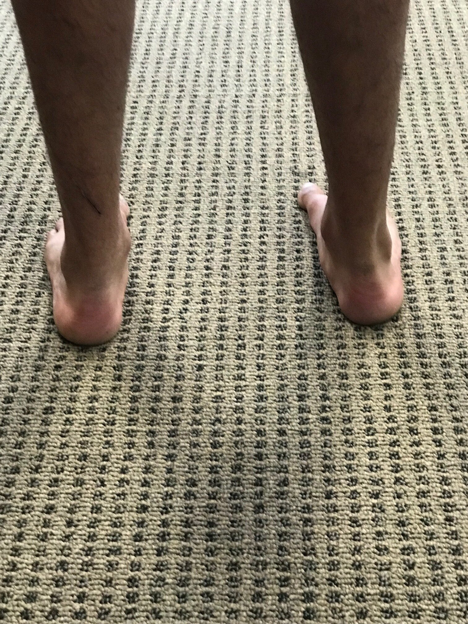
I was hiking behind this young chap over the weekend along with my son and friends. Note the amount of calcaneal eversion present on the right side that is not present on the left. Also note the increased progression angle of the right foot and subtle circumduction of the extremity.

In my experience, you would generally see this much calcaneal diversion and one of three scenarios:

1. Moderate leg length discrepancy with the increased calcaneovalgus occurring on the longer leg side. This would support the amount of circumduction were seeing on the right side.

2. When there is a forefoot supinatus present and and inadequate range of motion available in the midfoot and/or forefoot. This is most likely the case here.

3. In moderate To severe midfoot collapse. This is clearly not the case as the medial aspect of the shoe is usually “blown out”.

Next time you see an everting rearfoot, think about these three possible etiologies.

Dr Ivo Waerlop, on of The Gait Guys

#evertedrrarfoot #calcanealvalgus #shortleg #forefootsupinatus #forefootvarus #gaitanalysis #thegaitguys

The amazing power of compensation. Coming to a patient in your office… Maybe today

This gal has had a right sided knee replacement. She has an anatomical right short leg, a forefoot supinatus, an increased Q angle and a forefoot adductus. So, what’s the backstory?

When we have an anatomical short leg, we will often have a tendency to try to “lengthen“ that extremity and “shorten” the longer extremity. This is often accomplished through pelvic rotation although sometimes can be with knee flexion/extension or change in the Q angle. When the condition is long-standing, the body will often compensate in other ways, such as what we are seeing here.

IMG_6736.jpg

The fore foot can supinate in an attempt to lenthen the extremity. Note how the right extremity forefoot is in varus with respect to the rearfoot, effectively lengthening the extremity. As you can see from the picture, this is becoming a “hard“ deformity resulting in a forefoot varus.

IMG_6740.jpg

Over time, the forefoot has actually “adducted “ as you can see, again in an attempt to lengthen the extremity. Remember that supination is plantar flexion, abduction and inversion, all three which are visible here.


You will also see that the Q angle is less on the right side (se above), effectively lengthening that extremity, but not quite enough as we can see from the picture :-)


Dr Ivo Waerlop, one of The Gait Guys

#forefootadductus #shortleg #kneereplacement #tkr #forefootvarus #gait #thegaitguys

Sixth toe disease...That growth on the outside of your foot… Or on somebody’s foot is coming to see you…

IMG_6704.JPG

You know what we’re talking about. That extra growth on the lateral aspect of the foot that happens way too often and many of your clients. A Taylor’s bunion or sometimes referred to as a “bunionette”. 

What is the usual fix?

Usually in a ski boot or hiking boot, they blow out the lateral side of the shoe. This is usually not a good fix because most of these folks have internal tibial torsion and somewhat of a forefoot supinatus/varus.

IMG_6706.JPG
IMG_6707.JPG

The internal tibial torsion places the knee outside the saggital plane and an arch support without a forefoot valgus post will just push it further out, creating a conflict at the knee. The forefoot supinatus and/or varus places them on the outside of the foot as well. Remember, most of these folks are ALREADY on the outside of the foot and the foot wants to migrate laterally...so creating more space just means it migrates farther. Good thought, doesn’t work that way.

IMG_6710.JPG

So what did we do?

  • We created a valgus post for the forefoot (see picture above) tapering from lateral to medial and to help “push“ the distal aspect of the first ray down (because there was motion available that was not being used)

  • We gave him exercises to help descend the first ray like the extensor hallucis brevis exercise, toe waving as well as peroneus longus exercises

  • We gave him plenty of balance and coordination work

    Dr Ivo Waerlop, one of The Gait Guys


#6thtoe #internaltibialtorsion #forefootvarus # forefootsupinatus #gaitanalysis #thegaitguys


Holy Forefoot Flare, Batman!


Some sources say foot strike pattern does not matter. We disagree.

Look at this gal who midfoot/forefoot strikes. She also has a forefoot supinatus, a plastic condition where the forefoot is inverted with respect to the rearfoot. Take that combination and put it in a shoe with a forefoot flare and what do you get? Can you say AMPLIFICATION?

We’re not saying this is a bad shoe or even the wrong shoe. But, if she is going to run in this shoe, we will need to help her gain more ROM in her forefoot ( and some pelvic and hip stability) dodge doesn’t have to crash into eversion on each landing.

Help your patients with shoe selection. Something with less of a lateral flare in the forefoot would certainly make her life easier.

Need to know more? Consider taking our National Shoe Fit Program: link here:

Dr Ivo Waerlop, one of The Gait Guys

#badshoes #forefootflare #thegaitguys #forefootsupinatus #lateralflare #inversion

The muscle they named wrong?

Why would you name a muscle after its supposed function when its function is actually something totally different? Probably due to what made sense from how it looked, not by how it acted. Of course, we are talking about the abductor hallucis.

tumblr_nn1ca5fx0C1qhko2so1_r1_1280.jpg

Think about all the anatomy you have learned over the years. Think about all the taxonomy and how it was done: sometimes by thename of the discoverer and more often by its anatomical location. The abductor hallucis seems to be the latter. 

The abductor and adductor hallicus function from approximately midstance to pre swing (1-4) (toe off), applying equal and opposite rotational vectors of force (in an ideal world) of the proximal phalynx of the hallux. This should resolve into a purely compressive force (5). In a closed chain environment, the transverse head of the adductor hallicus should act to prevent “splay” of metatarsals, along with the lumbricals and interossei (6), providing stabilzation of the forefoot (7) and rearfoot (8) during preswing, while the oblique head serves to help maintain the medial longitudinal arch. 

The abductor hallicus is actually a misnomer, as it most cases it is not an abductor but rather a plantar flexor of the 1st ray, particularly the proximal hallux, (assisting the peroneus longus) and supinator about the oblique midtarsal joint axis (5).  In the majority of cases, there doesn’t appear to be a separate, distinct insertion of the adductor hallicus to the base of the proximal phalynx, but rather a conjoint insertion with the lateral head of the flexor hallicus bevis into the lateral sesamoid and base of the proximal phalynx (9-11), emphasizing more of its plantar flexion function and stabilizing actions, rather than abduction. 

In one EMG study of 20 people with valgus (12) they looked at activity of adductor and abductor hallucis, as well as flexor hallucis brevis and extensor hallucis longus. They found that the abductor hallucis had less activity than the adductor. No surprise here; think about reciprocal inhibition and increased activity of the adductor when the 1st ray cannot be anchoroed. They also found EMG amplitude greater in the abductor hallucis by nearly two fold in flexion. 

So, the abductor hallucis seems to be important in abduction but more important in flexion. Either way, it is a stance phase stabilizer that we are beginning to know a lot more about. As for the name? You decide...


Dr Ivo Waerlop, one of The Gait Guys


1. Basmajian JV, Deluca CJ . Muscle Alive. Their Functions Revealed by Electromyography Williams and Wilkins. Baltimore, MD 1985, 377

2. Root MC, Orien WP, Weed JH. Normal and Abnormal Function of the Foot. Clinical Biomechanics, Los Angeles, CA 1977

3. Mann RA. Biomechanics of Running. In Pack RP. d. Symposium on the foot and leg in running sports. Mosby. St Louis, MO 1982:26

4. Lyons K, Perry J, Gronley JK. Timing and relative intensity of the hip extensor and abductor muscle action during level and stair ambulation. Phys Ther 1983: 63: 1597-1605

5. Michaud T. Foot Orthoses and Other Forms of Conservative Foot Care. Newton MA 1993: 50-55

6. Fiolkowski P, Brunt D, Bishop et al. Intrinsic pedal musculature support of the medial longitudinal arch: an electromyography study. J Foot & Ankle Surg 42(6) 327-333, 2003

7. Travell JG, Simons DG. Myofascial Pain and Dysfunction: The Trigger Point Manual. Williams and Wilkins, Baltimore 1992; 529

8. Kalin PJ, Hirsch BE. The origin and function of the interosseous muscles of the foot. J Anat 152, 83-91; 1987

9. Owens S, Thordarson DB. The adductor hallucis revisited. Foot Ankle Int. 2001 Mar;22(3):186-91. Am J Phys Med Rehabil. 2003 May;82(5):345-9.

10. Brenner E.Insertion of the abductor hallucis muscle in feet with and without hallux valgus. Anat Rec. 1999 Mar;254(3):429-34.

11. Appel M, Gradinger R. [Morphology of the adductor hallux muscle and its significance for the surgical treatment of hallux valgus][Article in German] Orthop Ihre Grenzgeb. 1989 May-Jun;127(3):326-30.

12. Arinci I, Geng H, Erdem HR, Yorgancioglu ZR Muscle imbalance in hallux valgus: an electromyographic study. Am J Phys Med Rehabil. 2003 May;82(5):345-9.


#halluxvalgus #halluxabductovalgus #bunion #footmuscleactivity #gait #thegaitguys


The Short Foot Exercise

Here it is, in all its glory...Our version of the short foot exercise. Love it or hate it, say it “doesn’t translate”, we find it a useful training tool for both the patient/client as well as the clinician. It awakens and creates awareness of the sometimes dormant muscles in the user and offers a window to monitor progression for them, as well as the observer.

Remember that the foot intrinsics are supposed to be active from midstance through terminal stance/pre swing. Having the person “walk with their toes up” to avoid overusing the long flexors is a cue that works well for us. This can be a useful adjunct to your other exercises on the road to better foot intrinsic function.


Dr Ivo Waerlop, one of The Gait Guys

Sulowska I, Mika A, Oleksy Ł, Stolarczyk A. The Influence of Plantar Short Foot Muscle Exercises on the Lower Extremity Muscle Strength and Power in Proximal Segments of the Kinematic Chain in Long-Distance Runners Biomed Res Int. 2019 Jan 2;2019:6947273. doi: 10.1155/2019/6947273. eCollection 2019

Okamura K, Kanai S, Hasegawa M, Otsuka A, Oki S. Effect of electromyographic biofeedback on learning the short foot exercise. J Back Musculoskelet Rehabil. 2019 Jan 4. doi: 10.3233/BMR-181155. [Epub ahead of print]

McKeon PO, Hertel J, Bramble D, et al. the foot core system: a new paradigm for understanding intrinsic foot muscle function Br J Sports Med March 2014 doi:10.1136/bjsports-2013- 092690

Dugan S, Bhat K: Biomechanics and Analysis of Running Gait Phys Med Rehabil Clin N Am 16 (2005) 603–621

Bahram J: Evaluation and Retraining of the Intrinsic Foot Muscles for Pain Syndromes Related to Abnormal Control of Pronation http://www.aptei.ca/wp-content/uploads/Intrinsic-Muscles-of-the-Foot-Retraining-Jan-29-05.pdf


#shortfootexercise #footexercises #footrehab #thegaitguys #gaitanalysis #gaitrehab #toesupwalking


https://vimeo.com/342800960

Have you seen this?

Patterns. That’s what it’s about a lot of times. Dr Allen and I are always looking for patterns or combinations of muscles which work together and seem to cause what appear to be predictable patterns; like a weak anterior compartment and a weak gluteus maximus, or a weak gluteus medius and contralateral quadratus lumborum.

Here is an interesting story and a new combination that at least I have never seen before

I had a 11-year-old right footed soccer player from my son’s soccer team coming to see me with bilateral posterior knee pain which began during a soccer game while he was “playing up” on his older brothers team. He did need to do a lot of jumping as well as cutting. He is generally a midfielder/Forward. Well experienced player and “soccer is his life“.

My initial thoughts were something like a gastroc dysfunction or a Baker’s cyst. On examination, no masses or definitive swelling noted behind either knee. He did have tenderness to moderate degree over the right plantaris and tenderness as well as 4/5 weakness of the left popliteus. There was a loss of long axis extension of the talo crural articulations bilaterally with the loss of lateral bending to the right and left at L2-L3.

If you think about the mechanics of the right footed kicker (and try this while kicking a soccer ball yourself) it would be approximately as follows: left foot would be planted near the ball and the tibia/femur complex would be internally rotating well the foot is pronating and the popliteus would be eccentrically contracting to slow the rotation of the femur and the tibia. The right foot will be coming through and plantarflexion after a push off from the ball of the foot firing the triceps surae and plantaris complexes. He would be “launching“ off of the right foot and landing on his left just prior to the kick, causing a sudden demand on the plantar flexors; with the plantaris being the weak link. As the kicking leg follows through, the femur of the stance phase leg needs to externally rotate (along with the tibia) at a faster rate than the tibia (otherwise you could injure the meniscus) the popliteus would be contracting concentrically. A cleat, because it increases the coefficient of friction with the ground would keep the foot on the ground solidly planted and The burden of stress would go to the muscles which would be extremely routine leg and close chain which would include the semimembranosus/tendinosis  complex as well as the vastus medialis and possibly gracilis and short adductor, along with the popliteus.

I have to say and all of my years of practice I’ve never seen this combination type of injury before involving these two muscles specifically and am wondering if anyone else has seen this?

Dr Ivo Waerlop, one of The Gait Guys

#footproblem #gait #thegaitguys #soccerinjury #bilateralkneepain #popliteus #plantaris

image credit: https://commons.wikimedia.org/wiki/File:Slide2ACCA.JPG

image credit: https://commons.wikimedia.org/wiki/File:Slide2ACCA.JPG

The LAST word....on Lasts

The last (look inside the shoe on top of the shank) is the surface that the insole of the shoe lays on, where the sole and upper are attached).

Shoes are generally board lasted, slip lasted or combination lasted.

A board lasted shoe is very stiff and has a piece of cardboard or fiber overlying the shank and sole (sometimes the shank is incorporated into the midsole or last) . It can be effective for motion control (pronation) but can be uncomfortable for somebody who does not have this problem.

A slip lasted shoe is made like a slipper and is sewn up the middle. It allows great amounts of flexibility, which is better for people with more rigid feet.

A combination lasted shoe has a board lasted heel and slip lasted front portion, giving you the best of both worlds.

When evaluating a shoe, you want to look at the shape of the last (or sole). Bisecting the heel and drawing an imaginary line along the sole of the shoe determines the last shape. This line should pass between the second and third metatarsal. Drawing this imaginary line, you are looking for equal amounts of shoe to be on either side of this line.

Shoes have a straight, curved or semi curved last. The original idea of a curved last (banana shaped shoe) was to help with pronation. A curved last puts more motion into the foot and may force the foot through mechanics that is not accustomed to. Most people should have a straighter lasted shoe.

The shape of a last will effect the biomechanics of the foot. It should match why shape of the foot as closely as possible. Generally speaking, we recommend straighter lasts for folks that have a tendency to overpronate through the midfoot and curvier lasts for folks that have a more rigid foot.

Because the fore foot abducts during mid stance, if the last is curved, the lateral aspect of the foot can rub against the side of the shoe and create blistering of the little toe and if present long enough, a tailors bunion. A general rule of thumb is: "when in doubt, opt for a straighter one"

Dr Ivo Waerlop, one of The Gait Guys

#last #gait #foot #thegaitguys #lastshape #curvedlast #straightlast #gaitanalysis #pronation

Is your (or your athletes) cleat neutral or in varus?

Cleats are often the athletes primary interface with the ground and are responsible for transmitting the forces from the core and appendicular muscles down to the ground. The construction of the cleat as well as its characteristics (such as a forefoot varus cant in the forefoot, like this one here) can make all the difference in the world in athletic performance.

Dr Ivo Waerlop, one of The Gait Guys

#gait #thegaitguys #forefoot #varus #valgus #gaitanalysis #cleatproblems #cleatconstruction

The problems with some cleats....

Spring is here and Dr Ivo Waerlop of The Gait Guys talks about some common problems seen due to manufacturers defects in cleats and how they can affect athletes. From uppers put on the outsole incorrectly and contributing to and potentiating rearfoot varus and valgus to poor cleat placement affecting the 1st mtp mechanics; they all contribute to athlete performance.