Dragging your tongue ? When the tongue of your shoe keeps getting pulled to the side. Do you know what it means ? It means plenty, if you are sharp.By: Dr. Shawn AllenThis one pisses off most people it happens to. Why does it typically happen only o…

Dragging your tongue ? When the tongue of your shoe keeps getting pulled to the side. Do you know what it means ? It means plenty, if you are sharp.

By: Dr. Shawn Allen

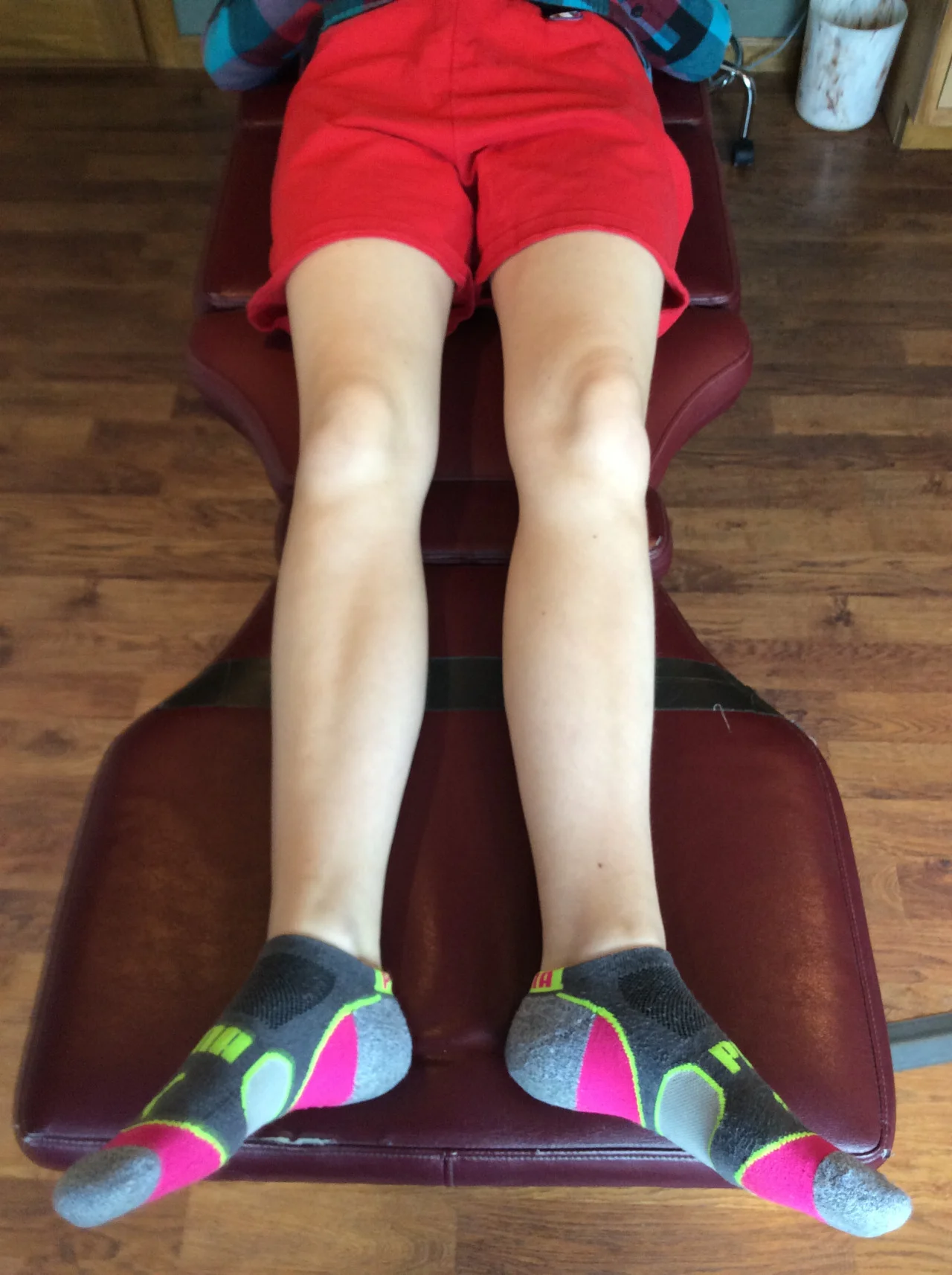
This one pisses off most people it happens to. Why does it typically happen only on one side, on one shoe ? Look at the photo case above. Look closely to the left foot, the tongue of the shoe is pulled laterally compared to the right, or shall I say, dragged.

This is a fairly common phenomenon, and there is a reason for it, several actually. So, no, you do not need to staple the tongue to the shoe upper, or tighten your shoe laces, or stitch the tongue to the medial shoe upper. You need to stop externally spinning your foot in your darn shoe.  What ?!

Yes, you very well may be avoiding normal internal rotation progression of the pelvis over the fixated limb. Loss of internal hip rotation is often a common finding clinically. As one passes the swing leg forward, the forward progressing pelvis eventually meets this loss of internal rotation over the fixated leg and femoral head. The swing leg none the less progresses further forward to get to its’ heel strike and the stance phase leg has to externally spin over the ground (I like to give the analogy of putting out a cigarette butt on the ground or squishing a bug (PETA don’t come after me)). This is called an Abductory or Adductory twist (good video demo here) depending on whether your reference point is the forefoot or rear foot. Regardless, the heel is spinning inward, the forefoot is relatively spinning outward. This spin of the foot inside the shoe (this happens minutely just before the shoe spins on the ground) and pulls the tongue laterally with it.  

This problem can also come from, and often does, a premature heel rise from things like a:

  •  loss of ankle rocker
  • short calf
  • lack of hip extension
  • hallux rigidus / limitus or even a painful big toe
  • etc

There are even several other causes I will not list here today, I could have you waste your whole day on the list and the mental gymnastics of things to consider. Basically, anything that impairs the stance phase mechanics creating a premature heel rise or failure of completing internal hip rotation can cause an Abd/Adductor twist of the foot/heel and drag the tongue laterally. Sure, there are others, but the purpose of my blog post here today was to explain a neat little biomechanical phenomenon that  has huge clinical insight if you know what it means.  You cannot fix this problem if you do not do a physical exam, understand clean and faulty gait biomechanics, and maybe can even find small objects in a dark room.  What I mean is it takes some educated exploration and a curiosity to want to fix things.  

There are clues often right in front of you, all you have to do is pay attention and sometimes ask a simple question. 

“Mr. Jones, when you stick out your tongue, does it drag laterally ?”  

Ok, maybe not that exact question. But, when I see a loss of internal rotation or terminal hip extension in a runner, and when I have time to explain things deeply with a openly receiving client, I might start the conversation with that fun question and then explain what I really meant was the tongue of the shoe on that affected side. 

You can’t swallow bandaids to fix things, as much as you wish it was that easy. Sure, you can avoid all of this fun by buying a shoe that has the tongue of the shoe sewn to the medial upper of the shoe, but then you wouldn’t have to fix anything.  Where would you “get your fun on” then ?  Be brave, go all in, fix the problem dammit.  

These are the things that keep me up at night. Welcome to my nightmares.

Dr. Shawn Allen, one of the gait guys

Photo courtesy of this weartested.org link: http://weartested.org/wp-content/uploads/2015/03/altra-superior-2-top-socks.jpg

Why is that muscle so tight? We often think of neurological reasons (increased facilitation of the agonist, decreased reciprocal inhibition of the antagonist, increase gamma drive, etc), but how about the series elastic element (ie the connective ti…

Why is that muscle so tight?


We often think of neurological reasons (increased facilitation of the agonist, decreased reciprocal inhibition of the antagonist, increase gamma drive, etc), but how about the series elastic element (ie the connective tissue)? Or perhaps the sarcomere (individual contractile unit of the muscle)? How can we fix that? It is easier than you thought!

An oldie but a goodie. A great FREE FULL TEXT paper on sarcomere loss and how to prevent it. Yep, would you have guessed static stretching? Yes, this study was on mice and it seems plausible that it would be applicable to humans as well.

“When muscle is immobilised in a shortened position there is both a reduction in muscle fibre length due to a loss of serial sarcomeres and a remodelling of the intramuscular connective tissue, leading to increased muscle stiffness. Such changes are likely to produce many of the muscle contractures seen by clinicians, who find that such muscles cannot be passively extended to the full length, which normal joint motion should allow, without the production of muscle pain or injury.

…These experiments show that in addition to preventing the remodelling of the intramuscular connective tissue component daily periods of stretch of ½ h or more also prevent the loss ofserial sarcomeres which occurs in mouse soleus muscles immobilised in the shortened position.”

http://www.ncbi.nlm.nih.gov/pmc/articles/PMC1004076/pdf/annrheumd00439-0044.pdf


link to full text: http://www.ncbi.nlm.nih.gov/pmc/articles/PMC1004076/pdf/annrheumd00439-0044.pdf

A great, quick read from one of our fav’s: Dr Tom Michaud. Here is my favorite excerpt. I had not thought of imaging the ankle quite this way“Physical examination reveals pinpoint sensitivity over the anteromedial capsule. When the ankle…

A great, quick read from one of our fav’s: Dr Tom Michaud.

Here is my favorite excerpt. I had not thought of imaging the ankle quite this way

“Physical examination reveals pinpoint sensitivity over the anteromedial capsule. When the ankle is slightly plantarflexed, the osteophytes on the talus and tibia can be readily palpated. Surprisingly, lateral X-rays only identify approximately 40 percent of the talotibial spurs, because the natural torsion of the distal tibia obstructs direct visualization of the anteromedial tibia. To improve radiographic accuracy, van Dijk, et al., recommend oblique radiographs be taken with a 45-degree craniocaudal angle, with the lower extremity externally rotated 30 degrees. The authors demonstrated that oblique radiographs identify 73 percent of the spurs located on the talus and 85 percent of the spurs located on the distal tibia.”

Where your gait might break down.

Gait appears most robust to weakness of hip and knee extensors, which can tolerate weakness well and without a substantial increase in muscle stress. In contrast, gait is most sensitive to weakness of plantarflexors, hip abductors, and hip flexors. - van der Krogt

In the past few weeks I have shared my thoughts on some articles regarding low back paraspinal musculature fatigue and the subsequent effects on motorneuron pools, specifically excitability of the soleus and quadriceps. These shared thoughts are from recent papers in the literature (search the blog over the last week). These effects are suggested to indicate a postural response to preserve lower limb function. In other words, as paraspinal fatigue set in, lower extremity muscle compensation ramped up to sustain postural locomotion demands.  Obviously, one should think this a step further and translate it all into questions of assessment of ankle dorsiflexion (ankle rocker) and control of progressing knee and hip flexion when pertaining to these muscles. The issues of stability and mobility should heighten. The one big problem in these studies, and you have even likely had these thoughts during your clinical examinations, is that one cannot truly fatigue one muscle group alone especially during activity, nor can one assess a single muscle group during manual testing. Luckily we have EMG testing capabilities in this day and age and we can more easily look into the function and reaction of a muscle and its’ direct response reactions. 

Today I have an article by van der Krogt that we read long ago, but that which one of our readers brought back into our wheelhouse.  This is pretty amazing stuff.

“This study examines the extent to which lower limb muscles can be weakened before normal walking is affected. We developed muscle-driven simulations of normal walking and then progressively weakened all major muscle groups, one at the time and simultaneously, to evaluate how much weakness could be tolerated before execution of normal gait became impossible. We further examined the compensations that arose as a result of weakening muscles. Our simulations revealed that normal walking is remarkably robust to weakness of some muscles but sensitive to weakness of others. Gait appears most robust to weakness of hip and knee extensors, which can tolerate weakness well and without a substantial increase in muscle stress. In contrast, gait is most sensitive to weakness of plantarflexors, hip abductors, and hip flexors. Weakness of individual muscles results in increased activation of the weak muscle, and in compensatory activation of other muscles. These compensations are generally inefficient, and generate unbalanced joint moments that require compensatory activation in yet other muscles. As a result, total muscle activation increases with weakness as does the cost of walking.“-van der Krogt

So, if your client comes in with knee, hip or ankle pain and a history of low back pain, you might want to pull out these articles. You may want to consider which muscles are, according to this article, most robust and sensitive to weakness. Remember what I mentioned when i reviewed the soleus article ? I mentioned that the reduced ankle dorsiflexion range may be from a soleus muscle postural compensation reaction to low back pain. Today’s article seemed to confirm that this muscle group is sensitive to weakness. In today’s discussion, not only is the impairment of the hip ranges of motion or control of the knee (from quadriceps adaptive compensation) possibly related to low back pain, in this case, paraspinal fatigue but it may be a muscle group robust to weakness which is a darn good thing when the paraspinals go to nap.

Sometimes the problem is from the bottom up, sometimes it is from the top down. It is what makes this game so challenging and mind numbing at times. If this is all too much for you, in teasing out this quagmire of a system, just throw corrective exercises at your client and hope for the best. What is the worst that can happen if you get it wrong ? Stronger compensations on already present compensations … . . why not, it is good for return business (insert sarcasm emoticon).  But, lets be honest, if it was easy everyone would be doing it the right way. But the truth is that it is a long journey, and we are on the same bus of discovery with you all. 

Dr. Shawn Allen, one of the gait guys.

Reference:

Gait Posture. 2012 May;36(1):113-9. doi: 10.1016/j.gaitpost.2012.01.017. Epub 2012 Mar 3.How robust is human gait to muscle weakness?van der Krogt MM1, Delp SL, Schwartz MH.

Low back pain and quadriceps compensation. A study.

“Neuromuscular changes in the lower extremity occur while resisting knee and hip joint moments following isolated lumbar paraspinal exercise. Persons with a history of LBP seem to rely more heavily on quadriceps activity while jogging.“- Hart et al.

Recently I discussed a paper (link below) about how soleus  motoneuron pool excitability increased following lumbar paraspinal fatigue and how it may indicate a postural response to preserve lower extremity function.
Today I bring you an article of a similar sort.  This paper discusses the plausibility that a relationship exists between lumbar paraspinal muscle fatigue and quadriceps muscle activation and the subsequent changes in hip and knee function when running fatigue ensued. 


"Reduced external knee flexion, knee adduction, knee internal rotation and hip external rotation moments and increased external knee extension moments resulted from repetitive lumbar paraspinal fatiguing exercise. Persons with a self-reported history of LBP had larger knee flexion moments than controls during jogging. Neuromuscular changes in the lower extremity occur while resisting knee and hip joint moments following isolated lumbar paraspinal exercise. Persons with a history of LBP seem to rely more heavily on quadriceps activity while jogging.”- Hart et al.

Whether this or any study was perfectly performed or has validity does not matter in my discussion here today. What does matter pertaining to my dialogue here today is understanding and respecting the value of the clinical examination (and not depending on a gait analysis to determine your corrective exercise prescription and treatment). When an area fatigues and cannot stabilize itself adequately, compensation must occur to adapt. Protective postural control strategies must be attempted and deployed to stay safely upright during locomotion. The system must adapt or pain or injury may ensue, sometimes this may take months or years and the cause is not clear until clinical examination is performed. Your exam must include mobility and stability assessments, motor pattern evaluation, and certainly skill, coordination, ENDURANCE and strength assessments if you are to get a clear picture of what is driving your clients compensation and pain. 

So, if your client comes in with knee, hip or ankle pain and a history of low back pain, you might want to pull out these articles and bash them and other similar ones into your brain. Remember what I mentioned when i reviewed the soleus article ? I mentioned that the reduced ankle dorsiflexion range may be from a soleus muscle postural compensation reaction to low back pain. In today’s discussion, impairment of the hip ranges of motion or control of the knee (from quadriceps adaptive compensation) may also be related to low back pain, in this case, paraspinal fatigue.  

Sometimes the problem is from the bottom up, sometimes it is from the top down. It is what makes this game so challenging and mind numbing at times. If only it were as simple as, “you need to work on abdominal breathing”, or “you need to strengthen your core”.  If only it were that simple. 

Dr. Shawn Allen, one of the gait guys


References:
J Electromyogr Kinesiol. 2011 Jun;21(3):466-70. doi: 10.1016/j.jelekin.2011.02.002. Epub 2011 Mar 8.
Effects of paraspinal fatigue on lower extremity motoneuron excitability in individuals with a history of low back pain. Bunn EA1, Grindstaff TL, Hart JM, Hertel J, Ingersoll CD.

J Electromyogr Kinesiol. 2009 Dec;19(6):e458-64. doi: 10.1016/j.jelekin.2008.09.003. Epub 2008 Dec 16. Jogging gait kinetics following fatiguing lumbar paraspinal exercise.
Hart JM1, Kerrigan DC, Fritz JM, Saliba EN, Gansneder B, Ingersoll CD

tumblr_o471xcV05d1qhko2so1_1280.jpg
tumblr_o471xcV05d1qhko2so3_1280.jpg
tumblr_o471xcV05d1qhko2so2_1280.jpg
tumblr_o471xcV05d1qhko2so4_1280.jpg

Holy twisted tibias Batman! What is going here in this R sided knee pain patient?

In the 1st picture note this patient is in a neutral posture. Note how far externally rotated her right foot is compared to the left. Note that when you drop a plumbline down from the tibial tuberosity it does not pass-through or between the second and third metatarsals. Also note the incident left short leg
In the next picture both of the patients legs are fully externally rotated. Note the large disparity from right to left. Because of the limited extra rotation of the right hip this patient most likely has femoral retro torsion. This means that the angle of her femoral head is at a greater than 12° angle. We would normally expect approximately 40° of external Rotation. 4 to 6° is requisite for normal gait and supination.

In the next picture the patients knees are fully internally rotated you can see that she has an excessive amount of internal rotation on the right compare to left, confirming her femoral antetorsion.

When this patient puts her feet straight (last picture), her knees point to the inside causing the patello femoral dysfunction right greater than left. No wonder she has right-sided knee pain!

Because of the degree of external tibial torsion (14 to 21° considered normal), activity modification is imperative. A foot leveling orthotic with a modified UCB, also inverting the orthotic is helpful to bring her foot somewhat more to the midline (the orthotic pushes the knee further outside the sagittal plane and the patient internally rotate the need to compensate, thus giving a better alignment).

a note on tibial torsion. As the fetus matures, The tibia then rotates externally, and most newborns have an average of 0- 4° of internal tibial torsion. At birth, there should be little to no torsion of the tibia; the proximal and distal portions of the bone have little angular difference (see above: top). Postnatally, the tibia should twist outward (externally) a total of 15 degrees until adult values are reached between ages 8 and 10 years of 23° of external tibial torsion (range, 0° to 40°). more cool stuff on torsions here

Wow, cool stuff, eh?

Your client’s strategic, protective compensation patterns are a power leak and costing them greatly in efficiency and energy. There is a greater cost to compensation other than pain avoidance and protection. The cost is a loss in energy and ef…

Your client’s strategic, protective compensation patterns are a power leak and costing them greatly in efficiency and energy. There is a greater cost to compensation other than pain avoidance and protection. The cost is a loss in energy and efficiency as these numbers elude to. Think of how many people walking amongst us are in protective pain patterns causing a deep energy suck on the system. So, do your client and yourself a favor… . if you are instructing your clients on how to fix their arm swing on a local (arm/upper quarter) level, please stop. Please don’t use your gait analysis software to coach out arm swing changes, you are missing the boat if you are doing that. Arm swing aberrancies are quite often a manifestation of a greater global problem. Arm swing is largely a passive pendulum effect. Changes in the pendulum are often a sign of something bigger. This takes deep knowledge.

The death of meniscal surgery?

Here is a big one when it comes to gait and clients in our offices.

Here is a big one when it comes to gait and clients in our offices.
We tell our clients all the time that cartilage, at best, has a tenuous blood supply, and just in the peripheral red zone at that, and so true healing is not likely, at least not to any significant degree … . we tell them that their so called surgical “repair” is likely nothing more than debridement, a mere clean up most of the time.  We tell our clients that meniscal tears are likely a dime a dozen after 40 years in most people. The cartilage loses some resilience and pliability with age and does not resist loading and shear as well as it used to making it more prone to loading damage.  
“greater than 90% of the surgeries on the structure are not repairs, as most patients believe, but are instead excisions, or cutting out, of the torn part of the meniscus.”
“The most common orthopedic surgery in America had it’s final epitaph written this month with a level-1 study showing that surgery for meniscus locking is no better than placebo. ”

Mind you, some tears are massive and do create obstruction to joint function. Large tears like bucket handle tears and large free fragments often do need surgery because they are just too obtrusive to safe joint function. However, perhaps for the others one should consider the following:Stabilize the joint and return full symmetrical, balanced, coordinated function with endurance and strength. It may just be the best you can do…….and it seems it is often more than sufficient. 

Reference:

http://www.regenexx.com/should-i-have-meniscus-surgery/#

“those with chronic neck pain demonstrated a narrower step width, a shorter step length and slower gait speed during walking with the head movements and at maximum speed” -  Uthaikhup et al. study:Head movement and Gait Parameters:By now you should …

“those with chronic neck pain demonstrated a narrower step width, a shorter step length and slower gait speed during walking with the head movements and at maximum speed” Uthaikhup et al. study:

Head movement and Gait Parameters:

By now you should have a good grasp of the global impact of gait and how it presents and translates in everything we do. It is how we move through this world, and everything we do, and everything that has happened to us, impacts our gait. And, our gait impacts things in turn, from our mental state to how we think and act.  

By now, if you have been with us here on The Gait Guys long enough, you know that with the tremendous proprioceptive impact of the system from the cervical spine, that neck pain can influence sensorimotor function and thus motor function. However, little is known about the effects of head movement and walking speed on gait characteristics in patients with neck pain.

From the Uthaikhup et al. study:

Patient sample:  20 women aged between 18 and 59 years with chronic neck pain (>3 months) and 20 healthy controls of similar age, weight and height

Indexes used: Neck Disability Index and Visual Analogue Pain Scale.

“The experiment consisted of two walking sessions. The first session included walking with head straight, head up-down, and head turns from side to side. The second session included walking at comfortable and maximum speeds. Each trial was performed twice. Gait parameters measured using GAITRite walkway system were step length, stride length, step time, stride time, step width, cadence and gait speed.”

According to this study, the clients with chronic neck pain showed several changes in how they implemented their gait. They displayed step width narrowing, a reduction in step length and speed of gait, and even an overall reduction in gait speed when neck movements were induced or encouraged or when there was pain..

As Uthaikhup et al. summarized, “The results suggest that patients with chronic neck pain have gait disturbances. This supports the notion that assessment of gait should be addressed in patients with persistent neck pain.”

You have to know your gait norms to understand abnormals. We have written about other parameters that affect gait speed, step length and width here on the blog. Here is one more parameter for you to store in your noggin. It is all connected. So, when you goto your gait analysis guru, ask them if they are going to clinically assess your painful neck as part of the gait analysis (be prepared for the “deer in the headlights” look).

Dr. Shawn Allen, one of the gait guys

Reference:

Man Ther. 2014 Apr;19(2):137-41. doi: 10.1016/j.math.2013.09.004. Epub 2013 Sep 27.The effects of head movement and walking speed on gait parameters in patients with chronic neck pain. Uthaikhup S, Sunkarat S, Khamsaen K, Meeyan K, Treleaven J.

What are we listening to this week? The Physio edge podcast with David pope. This week they interview Kurt Lisle about anterior knee pain. Here is our synopsis:One of the things they empahasized right off the bat was that patellofemoral pain not onl…

What are we listening to this week? 

The Physio edge podcast with David pope. This week they interview Kurt Lisle about anterior knee pain. Here is our synopsis:

One of the things they empahasized right off the bat was that patellofemoral pain not only refers about the knee but also below or most importantly posterior to the knee. The fat pad had a tendency to refer more locally where is other structures can refer to other areas.

Aggravating factors for patello femoral dysfunctional pain tends to be flexion or activities involving flexion as well as compression of the knee and rest is in alleviating factor.

The fat pad pain tends to be to either side of the patellar tendon and sometimes directly under it. This can be aggravated by standing, particularly with the knee and hyperextension, which compresses the fat pad.

Patellar tendon pain tends to remain at the inferior pole of the patella on the tendon whereas patellofemoral pain has a tendency to refer more.

Physical examination pearls:

  • Patellar tendonopathy alone generally does not have effusion present where as the patellofemoral or fat pad injury may.
  • Is there pain in passive hyperextension? This generally can mean fat pad injury or potential he ligamentous injury.
  • Visually you may palpate a thickened fat pad, particularly in females.
  • Pain with passive motions generally points away from patellar tendon.
  • Dialing in as to where and when they are having their pain is an important part of the functional evaluation.

Kurt likes to do a table top examination first to ensure functional integrity of the knee before jumping right to functional tasks. His concerns are (which are valid) is the knee up to the task you’re about to ask it to do? Good advice here.
He emphasizes the need to be systematic and consistent in your examination, no matter how you examine them. Develop a routine that you follow each and every time. He recommends passively looking at the knee in extension and 90° flexion.

There is a discussion on functional movement about the hip and pelvis, knee, and foot and ankle. Emphasis is made, for example at the knee, as to “is the knee moving medially and laterally or are the femur and tibia rotating mediately or laterally” in which is precipitating the pain?

“Catching” of the patella is often due to patellofemoral pathology such as a subchondral defect, slap tear of the chondral surface, or abnormalities of the trochlea of the femur.

Advanced imaging strategies are also discussed with a brief overview of some of the things to look for.

Finally treatment strategies were discussed. It is emphasized that identifying the specific activity or change activities that’s causing any pain he’s made as well as activity modification. We were happy to hear that footwear and its role in knee as well as hepatology was discussed as well as looking at occupational contributions to the pain.

There was emphasis on exercise specificity particularly with respect to if the problem was unilateral not giving “blanket” exercises for both knees but rather concentrating on the symptomatic side.

A discussion on the use of EMG and activation patterns was also entertained with some good clinical pearls here. More marked rather than subtle changes and activation side to side seem to be more clinically significant. In other words, with respect training, can they achieve similar levels of activation on each side with a similar activity (for example isometric knee extension with the leg bent 60°).

The judicious use of tape from a functional testing standpoint was interesting. Emphasis was made that tape is not a cure and will merely a tool.

All in all and informative, concise podcast with some great clinical pearls and a nice review of the knee and patellofemoral pain.


link to PODcast: http://physioedge.com.au/pe-029-acute-knee-injuries-with-kurt-lisle/

tumblr_nx0apl3SVJ1qhko2so2_1280.jpg
tumblr_nx0apl3SVJ1qhko2so3_1280.jpg
tumblr_nx0apl3SVJ1qhko2so4_1280.jpg
tumblr_nx0apl3SVJ1qhko2so5_r1_1280.jpg
tumblr_nx0apl3SVJ1qhko2so6_r1_1280.jpg

Congenital clubfoot anyone?

This gentleman, a longtime patient came in for new orthotics, as his old ones were 10 years old. From the pedographs above, you can see it is his LEFT foot. 

Note the following:

  • shortened apparent foot length left compared to right (the foot is merely deformed and plantarflexed)
  • The increased plantar pressures laterally, from the foot being supinated 
  • increased arch height L > R
  • clawing of digits 2-4 to provide stability

This case made me think about some common issues that you may be wondering about if you see these folks. 

There are several things you should think about:

  • People with clubfoot generally have a high arched, rigid, cavus foot. 
  • These folks generally are fixed in some degree of plantar flexion.
  • Because of the plantar flexed posture of their foot, they generally have a loss of a ankle rocker
  • If you utilize an orthotic with these patients, you need to make sure that there is significant ramp delta (heel higher than the 1st metatarsal)
  • Clubfoot can often be unilateral.
  • Clubfoot is usually not congenital
  • Gait training and balance (proprioceptive) work can be especially helpful in these cases. 

Which sports burn the most calories?

Photo: Gallo Images/iStockphoto  

Look at this photo. Do you see it ? How much posterior rotation (left rotation) is being driven through that left shoulder/torso rotation. That is nuts! We have a hard time believing that is not a compensation. We would be assessing for stability and mobility issues elsewhere. Heck, the elbow practically crosses the spine posteriorly ! Sheesh ! When you cannot put the movement where it should be, or control it (stabilize) where it should be controlled, sometimes you try to get it or put it elsewhere. We would love to see this lady run, we bet there is a host of clean gait problems down below. We would bet some cross over gait is present as well, after all, that left arm swing is largely predicated off of the right leg swing. Arm swing is far less independent than people think, we have written about that here on our blog numerous times. Just search “arm swing” over on our Tumblr blog.

Remember this, and if you need to go back to read about phasic and anti-phasic gaits head over and search our blog, but the amount of shoulder “girdle” (essentially thoracic rotation) is typically met by the same amount of pelvis rotation. These should be symmetrical. And, when they are not, we can drive it through various means, even as in this case, through more arm swing unilaterally. We wish we could see some axial photos from above to see how much pelvis rotation is noted  here.

Just some brief thoughts from Dr. Allen

Photo link attributed to owner: http://africanspotlight.com/wp-content/uploads/2016/03/CdBVhTzUkAELVp6.jpg

tumblr_o39jesCIny1qhko2so1_1280.jpg
tumblr_o39jesCIny1qhko2so2_1280.jpg
tumblr_o39jesCIny1qhko2so3_1280.jpg
tumblr_o39jesCIny1qhko2so4_1280.jpg
tumblr_o39jesCIny1qhko2so5_1280.jpg

Got big toe pain? Think it’s gout? Think again!   Things are not always what they appear to be. 

This gent came in with first metatarsophalangeal pain which had begun a few months previous. His uric acid levels were borderline high (6) so he was diagnosed with gout.  It should be noted his other inflammatory markers (SED rate and CRP) were low. Medication did not make the symptoms better, rest was the only thing that helped. 

The backstory is a few months ago he was running in the snow and “punched through"the snow, hitting the bottom of his foot on the ground. Pain developed over the next few days and then subsided. The pain would come on whenever he try to run or walk along distances and he noticed a difficult time extending his big toe.

 Examination revealed some redness mild swelling over the 1st metatarsophalangeal joint (see pictures above) and hallux dorsiflexion of 10°.   If we raised the base of the first metatarsal and pushed down on the head of the 1st, he was able to dorsiflex the 1st MTP approximately 50°. He had point tenderness over the medial sesamoid. We shot the x-rays you see above. The films revealed a fracture of the medial sesamoid with some resorption of the bone.

The  sesamoid fracture caused the head of the 1st metatarsal to descend on one side, and remain higher on the other, altering the axis of rotation of the joint and restricting extension. We have talked about the importance of the axis of this joint in may other posts (see here and here).

 He was given exercises to assist in descending the first ray (EHB, toe waving, tripod standing).  He will be reevaluated in a week and if not significantly improved we will consider a wedge under the medial sesamoid. 

A pretty straight forward case of “you need to be looking in the right place to make the diagnosis”. Take the time to examine folks and get a good history.

Podcast 103: Effects of Cold on Physiology/Athletes

Using Cold adaptation to your advantage, Walking Rehab “Carries”, Walking and Proprioception.

Show Sponsors:
newbalancechicago.com
Softscience.com

Other Gait Guys stuff

A. Podcast links:

direct download URL: http://traffic.libsyn.com/thegaitguys/pod_103f.mp3

permalink URL: http://thegaitguys.libsyn.com/podcast-103-effects-of-cold-on-physiologyathletes

B. iTunes link:
https://itunes.apple.com/us/podcast/the-gait-guys-podcast/id559864138


C. Gait Guys online /download store (National Shoe Fit Certification & more !)
http://store.payloadz.com/results/results.aspx?m=80204

D. other web based Gait Guys lectures:
Monthly lectures at : www.onlinece.com type in Dr. Waerlop or Dr. Allen, ”Biomechanics”

-Our Book: Pedographs and Gait Analysis and Clinical Case Studies
Electronic copies available here:

-Amazon/Kindle:
http://www.amazon.com/Pedographs-Gait-Analysis-Clinical-Studies-ebook/dp/B00AC18M3E

-Barnes and Noble / Nook Reader:
http://www.barnesandnoble.com/w/pedographs-and-gait-analysis-ivo-waerlop-and-shawn-allen/1112754833?ean=9781466953895

https://itunes.apple.com/us/book/pedographs-and-gait-analysis/id554516085?mt=11

-Hardcopy available from our publisher:
http://bookstore.trafford.com/Products/SKU-000155825/Pedographs-and-Gait-Analysis.aspx

Show Notes:
Cold
Switching on a cold-shock protein may restore lost connections between brain cells & memory function in aging brain.  
http://www.bbc.com/news/health-30812438

-“Connections between brain cells - called synapses - are lost early on in several neurodegenerative conditions, and this exciting study has shown for the first time that switching on a cold-shock protein called RBM3 can prevent these losses.
http://www.pnas.org/content/111/20/7379.abstract

New study in mice in the inaugural issue of Brain Plasticity reports that new brain cell formation is enhanced by running.
http://neurosciencenews.com/neurogenesis-exercise-memory-3165/

Walking changes our mental state, and our mental state changes our walking.  60 sec audio clip.
http://www.scientificamerican.com/podcast/episode/bouncy-gait-improves-mood/

http://www.sciencedaily.com/releases/2015/…/151119122246.htm

Walking. You don’t have to have the pedal to the metal.
"Those who walked an average of seven blocks per day or more had a 36%, 54% and 47% lower risk of CHD, stroke and total CVD, respectively, compared to those who walked up to five blocks per week.”

http://www.sciencedaily.com/releases/2015/…/151119122246.htm
New proprio study:
http://www.nature.com/neuro/journal/v18/n12/abs/nn.4162.html
Piezo2 is the principal mechanotransduction channel for proprioception
Seung-Hyun Woo et al,
Nature Neuroscience 18, 1756–1762 (2015) doi:10.1038/nn.4162Received 14 July 2015 Accepted 13 October 2015 Published online 09 November 2015

Magnesium intake higher than 250 mg/day associated with a 24% increase in leg power & 2.7% increase in muscle mass.
http://onlinelibrary.wiley.com/doi/10.1002/jbmr.2692/full

Dietary Magnesium Is Positively Associated With Skeletal Muscle Power and Indices of Muscle Mass and May Attenuate the Association Between Circulating C-Reactive Protein and Muscle Mass in Women

Ailsa A Welch et al.
http://www.readcube.com/articles/10.1002%2Fjbmr.2692?r3_referer=wol&tracking_action=preview_click&show_checkout=1&purchase_referrer=t.co&purchase_site_license=LICENSE_DENIED

Gray Cook
https://duker2p.wordpress.com/2015/11/16/illuminating-insights-gray-cook-part-1/

Carries, lots of carries
https://www.facebook.com/otpbooks/videos/1004044686323688/

And what have we been saying? parallel processing seems to be OK (balancing and reading), but dual or multitasking has its hazards…decreased speed of movement. not surprising because of the dual taskingincreased ankle dorsiflexion (not necess…

And what have we been saying? parallel processing seems to be OK (balancing and reading), but dual or multitasking has its hazards…

  • decreased speed of movement. not surprising because of the dual tasking
  • increased ankle dorsiflexion (not necessarily a bad thing. This is probably to create a wider and more stable base through pronation
  • reduced cadence
  • decreased stride length

we were surprised there was not a increased “base of gait”, as balance requirements increase, gait usually decomposes (see here for a cool post and video we did on this a while ago)

“Numerous studies have analyzed the impact of dual tasks—specifically, tasks that cause cognitive distraction—on gait. With regard to texting as a dual task, many studies have consistently found that it does have an effect on gait, and that’s mostly to slow a person down.


For instance, Italian researchers in the Journal of NeuroEngineering and Rehabilitation assessed 18 healthy young adults who did not have problems with vision, or neurological or musculoskeletal disorders that could affect their gait.3 Barefoot participants walked a straight path of 15 meters (about 50 feet) for three minutes under two conditions: walking alone and walking while texting.


They found that texting while walking differed from walking alone in terms of muscle activation, kinematics, and spatiotemporal variables. Texting was associated with delayed activation of the gastrocnemius lateralis muscle and slightly increased ankle dorsiflexion followed by slightly reduced plantar flexion. It was also associated with a slower gait speed, reduced cadence and stride length, increased flat-foot contact, and decreased push-off. The researchers also found increased co-contraction of the ankle antagonist muscles during what they called the “critical” gait phase—from load response to midstance, corresponding to the transfer of body weight from one leg to the other.”


its a short one. Take the time to check it out…


link to article: http://lermagazine.com/cover_story/texting-while-walking-gait-adaptations-and-injury-implications

Using a boot to heal a bone, tendon, post-op ?  Think deeper please.Please please, please ! If you are going to put your client in a CAM rocker boot/shoe for a fracture, or post-op can you please try to level out the leg length discrepancy caused by…

Using a boot to heal a bone, tendon, post-op ?  Think deeper please.

Please please, please ! If you are going to put your client in a CAM rocker boot/shoe for a fracture, or post-op can you please try to level out the leg length discrepancy caused by the thickness of the boot’s sole ? Please ? Pretty please with sugar on top?

Some boot brands have a huge midsole thickness. This leads to a functionally longer leg length. If they are barefoot much of the day, there will be a huge leg length discrepancy. If in shoes all day, you can offset this with a sole lift in the healthy foot’s shoe or you can add something like this to the outsole. Use common sense. IF someone is in a CAM boot for 6 weeks and thus a longer leg, this is going to promote a knee flexed posture on the boot side (ie. shortens the leg) and/or hyperextension of the healthy leg’s knee, supination of the foot, more forefoot habitus (sustained calf loads) and even frontal plane lurch pelvis gait mechanics (this is why many folks will get opposite hip pain). These embedded gait flaws must be addressed and remedied after they are out of the boot to reset normal gait. We have seen enough problems come to our offices that are suspect as a result of prolonged boot use and failure to reteach normal gait patterns, meaning, to reduce the learned gait behaviors of being in a boot for prolonged periods. Gait retraining is just as important as the rehab post-boot removal.  Of course, this is rarely done.  Using logic is never a bad thing.   

Dr. Shawn Allen, one of the gait guys

Here is a neat device we found to help.http://www.braceshop.com/procare-evenup-shoe-balancer-walker-system.htm?gdftrk=gdfV28018_a_7c2568_a_7c10961_a_7c32290&gclid=Cj0KEQiA37CnBRChp7e-pM2Mzp0BEiQAlSxQCCeL74AvCkYXbQX_jV1jEP27mfocB87f8pSfbo2PZMIaAsOV8P8HAQ

Medial longitudinal Arch age stablization.

It seems to depend on what source you read as to when the MLA (medial longitudinal arch) stabilizes, but here is a number, between 7-9 years old. 

Conclusions: The MLA of children remained stable from 7 to 9 years old. Gender and the type of footwear worn during childhood may influence MLA development.

Reference:

Medial Longitudinal Arch Development of Children Aged 7 to 9 Years: A Longitudinal Investigation.Jasper W.K. Tong, Pui W. Kong Journal of American Physical Therapy Assoc.   DOI: 10.2522/ptj.20150192 Published February 18, 2016

http://ptjournal.apta.org/content/early/2016/02/17/ptj.20150192