Calf Size Matters

Calf size matters. Truly, and here is why.

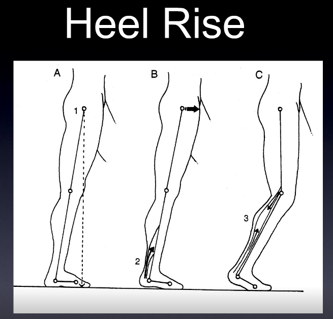
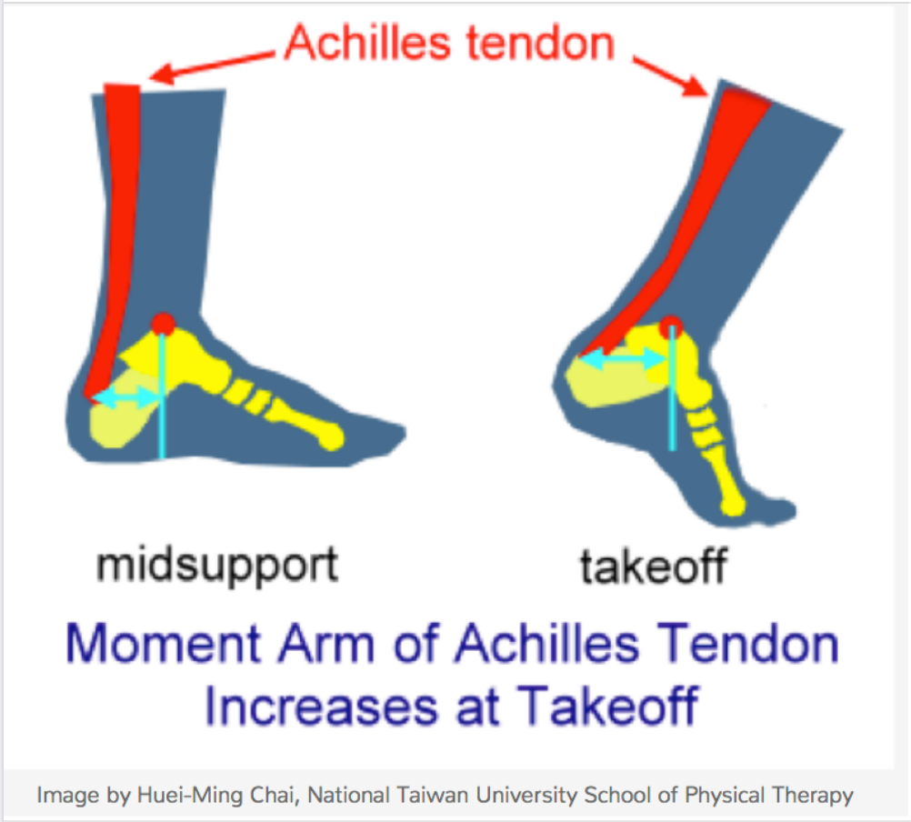
The Achilles tendon (AT) moment arm transforms triceps surae muscle forces into a moment about the ankle which is critical for functional activities like walking and running. The achilles tendon moment arm changes continuously during walking. It changes as movement occurs and as the triceps surae contracts. But, it also changes as the muscle grows in size, or shrinks. This has relevance to exercise, strengthening, and atrophy of the calf compartment. When the muscle contracts, its cross sectional area changes, and this can change the pull angle on the achilles. One might think of the achilles tendon as being "tented" away from the tibia slightly, and one can feel this on their own foot when palpating the soft tissue space between the tendon and the tibia on plantarflexion even when sitting. Better yet, sit down, your foot on the ground. Now, palpate the soft tissue space between the achilles and tibia. Now raise your heel, thus forefoot loading. You will feel the space gap open, the moment arm has increased because of this, thus changing its moment arm. On dorsiflexion the moment arm shortens, on plantarflexion it lengthens. Sure, this does not create a monstrous line of pull change, but by the time we get down to the pivot point at the ankle mortise, a small change in moment arm can translate to significant changes in torque and force production. This is why a foot that does not supinate in time for heel off, or supinate sufficiently, meaning the rear foot isn't inverted optimally, means that the ankle mortise (talus position) might change/shorten that moment arm. This is not efficient mechanics. Want to jump higher ? You have to get that excessive pronation in ankle dorsiflexion under control and convert it to supination, and rearfoot inversion. Jumping from a collapsed foot tripod is a power leak and you will not optimize the triceps surae-achilles complex and their lever arm. This also goes for toe off in walking and especially running, particularly sprinting where you are up on that forefoot. Said another way, when the arch is more collapsed and the talus is thus more plantarflexed the moment arm is sorter for the achilles. Strength, power, torque all suffer. One does not want to engage heel rise and calf contraction from this ineffective position of pronation taken too far, or heel rise while still pronated. This can also put undue load, and angle of pull, through the achilles. Meaning, the linear pull one desires through the achilles, can be through a calcaneal insertion that is not oriented optimally. One might postulate, rightly so we believe, that the lateral bundles/fascicles of the achilles tendon might see more loading than the medial. At the very least, we might postulate that the medial and lateral achilles tensile loads are offset and unequal. This could create problems over time, meaning changes in tendon morphology.

Screen Shot 2020-06-18 at 8.25.41 AM.png

In the below Rasske & Franz article, they posit that aging negatively effects the architecturally complex AT moment arm during walking, which thereby contributes to well-documented reductions in ankle moment generation during push-off. They looked at the "AT moment arms of young (23.9 ± 4.3 years) and older (69.9 ± 2.6 years) adults during walking, their dependence on triceps surae muscle loading, and their association with ankle moment generation during push-off. Older adults walked with 11% smaller AT moment arms and 11% smaller peak ankle moments during push-off than young adults. Moreover, as hypothesized, these unfavourable changes were significantly and positively correlated (r2 = 0.38, p < 0.01). More surprisingly, aging attenuated load-dependent increases in the AT moment arm (i.e., those between heel-strike and push-off at the same ankle angle); only young adults exhibited a significant increase in their AT moment arm due to triceps surae muscle-loading. Age-associated reductions in triceps surae volume or activation, and thus muscle bulging during force generation, may compromise the mechanical advantage of the AT during the critical push-off phase of walking in older adults. Thus, strategies to restore and/or improve locomotor performance in our aging population should consider these functionally important changes in musculoskeletal behavior."

Great article spawning deeper thoughts, here at The Gait Guys blog.
More to come on this most likely.

Aging effects on the Achilles tendon moment arm during walking. Kristen Rasske, Jason R.Franz
Journal of Biomechanics
Volume 77, 22 August 2018, Pages 34-39

Photo credit: Image by Huei-Ming Chai, National Taiwan University School of Physical Therapy as found on www.runsmartproject.com

The 4 Factors of Heel Rise.

Screen Shot 2019-01-14 at 12.48.19 PM.png
Screen Shot 2019-01-14 at 12.48.08 PM.png

These SHOULD all happen to have appropriate heel rise and forward progression

1. active contraction of the posterior compartment of the calf

2. passive tension in the posterior compartment of the calf

3. knee flexion and anterior translation of the tibia ankle rocker

4. the windlass mechanism

a problem with any one of these (or more collectively) can effect heel rise, usually causing premature heel rise.

ask yourself:

  • Do you think the posterior compartment is actively contracting? not enough or too much? Remember the medial gastrocnemius adducts the heel at the end of terminal stance to assist in supination. Don't forget about the tibialis posterior as well as the flexor digitorum longs and flexor hallucinate longus.

  • Does there appear to be increased passive tension in the posterior compartment? How visible and prominent are their calf muscles?

  • Do they have forward progression of the body mass?

  • How is his windlass mechanism? Good but not good enough.

Dr Ivo Waerlop. One of The Gait Guys…

#gait, #gaitanalysis, #continuingeducation, #limp, #casestudy, #gaitparameters, #heelrise, #prematureheelrise, #windlassmechanism

The gastroc can causse ankle dorsi and plantarflexion ? Yup. What ?

The gastroc, does it cause ankle dorsiflexion and ankle plantarflexion ? Yup. What ?

You may think you know the answer, the gastrocs are ankle plantarflexors, because that is the easy one we all recognize. But I stew on things when unique cases come in and do not fit the "normal" models and it got me reviewing principles I need to always keep in mind.

Think about it, the gastroc cross the knee, so it causes knee flexion. And when the knee flexes, the proximal tibia is progressing forward in the sagittal plane. Now remember, the foot is on the ground, so the distal tibia is (relatively) fixated in relation to the upper tibia. So, as this proximal top tibial moves forward, because of gastroc contraction, the muscle is actually causing ankle dorsiflexion !

So, it is it important to know your normal gait cycle events ? Yes, Ivo and i harp on that all the time ! One has to know the normal cycles to know when abnormal gait cycles are presenting clues.
So, am I saying that the gastroc are helpers of ankle rocker and ankle dorsiflexion ? Yes, they can be. It is a timing thing. So, we have to again get out of our model of open chain events, and thinking that only the anterior compartment muscles are ankle dorsiflexors. We also have to remember that a bent knee heel raise is not the same as a straight leg (knee extension) heel raise. One can stimulate and assist in ankle dorsiflexion and the other cannot so much. So, in clients with loss of ankle dorsiflexion/ankle rocker should you be assessing the function of the gastroc at the proximal knee, for its effects of dorsiflexion at the ankle ? Yes. Go ahead and try it, bend knee and straight knee heel raises, they are different beasts. This gets more complicated, and i will go into that next week ! I have had some deeper epiphanies i wish to share.
Also, remember, single and biarticular muscles have varied and vast capabilities. Thus it is always vital to consider whole body movements where muscles have abilities to accelerate, decelerate, and control and stablize joints they span, and do not span, via dynamic coupling.
Dr. Allen

What do you know about the Ia Afferents?

This is a nice study looking at lateral gastroc activity and changing firing patterns with speed of movement. Great if you treat anyone or anything that walks...

Ia afferents

You remember them, large diameter afferent (sensory) fibers coming from muscle spindles and appraising the nervous system of vital information like length and rate of change of length of muscle fibers, so we can be coordinated. They act like volume controls for muscle sensitivity. Turn them up and the muscle becomes more sensitive to ANY input, especially stretch (so they become touchy…maybe like you get if you are hungry and tired and someone asks you to do something); turn them down and they become less or unresponsive.

Their excitability is governed by the sum total (excitatory and inhibitory) of all neurons (like interneuron’s) acting on them (their cell bodies reside in the anterior horn of the spinal cord).

If we slow things down, the rate of change of length slows as well and excitability decreases, like we see in this study (3-6% slower). We also notice that the length of contraction increases; hmmm, why doesn’t it decrease?

Remember these folks are on a treadmill. The treadmill is constantly moving, opposite the direction of travel. With the foot on the ground, this provides a constant rate of change of length of the gastroc/soleus (ie, it is putting it through a slow stretch); so , once the muscle is activated, it contracts for a longer period of time because of the treadmill putting a slow stretch on the gastroc (and soleus).

This article also talks about people with upper motor neuron lesions. An important set of inhibitory neurons come from higher centers of the brain, in the motor cortex. These tend to attenuate the signals affecting the Ia afferents, and keep us stable. When we have an upper motor neuron lesion (like a brain lesion or stroke), we lose this “attenuation” and the stretch reflexes (and muscle tone) becomes much more active (actually hyperactive), making the muscle more sensitive to stretch. This loss of attenuation, along with differing firing patterns of the gastroc are important to remember in gait rehab.

The soleus and medial gastroc begin firing in the first 10% of the gait cycle (at the beginning of loading response) and fire continuously until pre swing (peaking just after midstance). The lateral head begins firing at midstance; both heads (along with soleus) decelerate the forward momentum of the tibia, flex the knee at midstance, and the medial head assists in adducting the calcaneus to assist in supination.

Making sure these muscles fire appropriately is important and needling is just one way of helping them to function better. Don’t overlook the tricep surae on your next patient that has a “hitch in their giddyup”.


Effects of treadmill walking speed on lateral gastrocnemius muscle firing.

by Edward A Clancy, Kevin D Cairns, Patrick O Riley, Melvin Meister, D Casey Kerrigan

American journal of physical medicine rehabilitation Association of Academic Physiatrists (2004) Volume: 83, Issue: 7, Pages: 507-51 PubMed: 15213474

Abstract

OBJECTIVE: To study the electromyographic profile-including ON, OFF, and peak timing locations-of the lateral gastrocnemius muscle over a wide range of walking speeds (0.5-2.1 m/sec) in healthy young adults. DESIGN: We studied gastrocnemius muscle-firing patterns using an electromyographic surface electrode in 15 healthy subjects ambulating on a treadmill at their normal walking speed and at three paced walking speeds (0.5, 1.8, and 2.1 m/sec). Initial heel contact was determined from a force-sensitive switch secured to the skin over the calcaneous. RESULTS: For all speeds, the gastrocnemius firing pattern was characterized by a main peak, occurring 40-45% into the gait cycle, that increased in amplitude with walking speed. Speeds of > or =1.3 m/sec produced a common electromyographic timing profile, when the profile is expressed relative to the stride duration. However, at 0.5 m/sec (a speed typical of individuals with upper-motor neuron lesions), the onset of gastrocnemius firing was significantly delayed by 3-6% of the gait cycle and was prolonged by 8-11% of the gait cycle. CONCLUSION: Many patients with upper motor neuron lesions (e.g., stroke and traumatic brain injury) walk at speeds much slower than those commonly described in the literature for normal gait. At the slow walking speed of 0.5 m/sec, we have measured noticeable changes in the electromyographic timing profile of the gastrocnemius muscle. Given the importance of appropriate plantar flexor firing patterns to maximize walking efficiency, understanding the speed-related changes in gastrocnemius firing patterns may be essential to gait restoration.