Pod #124: Gluteal gripping, Runner's dystonia. Are leg length differences real ?

Key tag words:
running, gait, injuries, kidney, kidneydamage, marathoners, foot, feet, dehydration, heatstroke, elon musk, neural lace, hip pain, crossfit, squats, deadlifts,  LLD, short leg, dystonia, runner's dystonia, posture, 

Summary:  Today we hit some very important topics on how to examine a client and how asymmetries play into gait, running, posture and pathomechanics. We hope you enjoy today's show, it is our first one back in 6 weeks. We are back strong after a brief early summer sabbatical. Back to the "podcast every 2 weeks" again. Thanks for being patient while Ivo recharged for the second half of the year.   Plus, on today's show, we also dive into Runner's kidney, dehydration, gluteal gripping, runner's dystonia, functional leg length differences due to asymmetries, and more !

Show links:

http://traffic.libsyn.com/thegaitguys/pod_124final.mp3

http://thegaitguys.libsyn.com/pod-124

Our Websites:
www.thegaitguys.com

summitchiroandrehab.com   doctorallen.co     shawnallen.net
Our website is all you need to remember. Everything you want, need and wish for is right there on the site.
Interested in our stuff ? Want to buy some of our lectures or our National Shoe Fit program? Click here (thegaitguys.com or thegaitguys.tumblr.com) and you will come to our websites. In the tabs, you will find tabs for STORE, SEMINARS, BOOK etc. We also lecture every 3rd Wednesday of the month on onlineCE.com. We have an extensive catalogued library of our courses there, you can take them any time for a nominal fee (~$20).
 
Our podcast is on iTunes, Soundcloud, and just about every other podcast harbor site, just google "the gait guys podcast", you will find us.
 
Show Notes:

Kidney Damage in Runners. 82%  !?
http://www.newsweek.com/running-bad-you-marathons-damage-kidneys-runners-bodies-575829
 
Kidney nephropathy in mesoamericans.
http://www.ajkd.org/article/S0272-6386(15)01257-3/fulltext  
 
Elon Musk's Neural Lace.
https://www.scoopwhoop.com/elon-musk-launches-neuralink-which-hopes-to-combine-your-brain-with-artificial-intelligence/
 
Leg length discrepancies,do they really even exist ?
Dystonia ?
The Gluteal gripping phenomenon.

What is the deal with "toe separators"?

Less pain through better mechanics?

We have been using toe separators for various foot problems like hallux valgus, hammer toes and flexor dominance/extensor weakness. Our reasoning is that through changing the angle of attachment of the muscle, you alter the mechanical advantage of that muscle and help it to work more efficiently. This seems implied in the literature with respect to foot orthoses (1-3) but we could not find any data regarding toe separators. Toe separators DO seem to reduce pain and increase function (4-6). Perhaps this is through better biomechanics, mechanical deformation, proprioceptive changes, or most likely a combination of all these factors and more.  We think clinical results speak volumes. It is nice to see more data coming out on these easy to implement clinical tools.

What is you clinical reasoning or rationale for using these devices? We would love to hear and if you have an article for reference you could share, that would be great.

TGG

1. Scherer PR, Sanders J, Eldredge DE, Duffy SJ, Lee RY. Effect of functional foot orthoses on first metatarsophalangeal joint dorsiflexion in stance and gait. J Am Podiatr Med Assoc. 2006 Nov-Dec;96(6):474-81.

2. Halstead J, Chapman GJ, Gray JC, Grainger AJ, Brown S, Wilkins RA, Roddy E, Helliwell PS, Keenan AM, Redmond ACFoot orthoses in the treatment of symptomatic midfoot osteoarthritis using clinical and biomechanical outcomes: a randomised feasibility study. Clin Rheumatol. 2016 Apr;35(4):987-96. doi: 10.1007/s10067-015-2946-6. Epub 2015 Apr 28.

3. Bishop C, Arnold JB, May T. Effects of Taping and Orthoses on Foot Biomechanics in Adults with Flat-Arched Feet. Med Sci Sports Exerc. 2016 Apr;48(4):689-96. doi: 10.1249/MSS.0000000000000807.

4. Chadchavalpanichaya N, Prakotmongkol V, Polhan N, Rayothee P, Seng-Iad S. Effectiveness of the custom-mold room temperature vulcanizing silicone toe separator on hallux valgus: A prospective, randomized single-blinded controlled trial. Prosthet Orthot Int. 2017 Mar 1:309364617698518. doi: 10.1177/0309364617698518. [Epub ahead of print]

5. Tehraninasr A, Saeedi H, Forogh B, Bahramizadeh M, Keyhani MR. Effects of insole with toe-separator and night splint on patients with painful hallux valgus: a comparative study. Prosthet Orthot Int. 2008 Mar;32(1):79-83. doi: 10.1080/03093640701669074.

6. Tang SF, Chen CP, Pan JL, Chen JL, Leong CP, Chu NK. The effects of a new foot-toe orthosis in treating painful hallux valgus. Arch Phys Med Rehabil. 2002 Dec;83(12):1792-5.

Ankle Rocker Revisited....

How many times have we talked about ankle rocker and its importance? So how are YOU measuring ankle rocker? Are you looking at it on the table? On the ground? Weight bearing? Knee flexed or extended (or both?). The knee is extended at initial contact, flexes through midstance, extends at terminal stance and pre swing and flexes again during swing phase until extending at the end of terminal swing for initial contact again.

What you see on the table may not (and many times doesn't)  translate to real life. Someone with limited ankle dorsiflexion non weight bearing may have normal amounts during gait and vice versa. With gravity in place and a functioning (or malfunctioning) vestibular system, things can change rapidly. Remember that the vestibular system drives the extensors and if inhibited, you will often have flexor dominance. Talk about a tight gastroc/soleus group!


"These findings indicate that nonweightbearing and weightbearing measurements of ankle DF PROM with knee extension should not be used interchangeably and that weightbearing ankle DF PROM with the knee extended is more appropriate for estimating ankle DF during gait."

Kang MH, Oh JS. Relationship Between Weightbearing Ankle Dorsiflexion Passive Range of Motion and Ankle Kinematics During Gait. J Am Podiatr Med Assoc. 2017 Jan;107(1):39-45. doi: 10.7547/14-112.


"There is no relationship between a static diagnosis of ankle dorsiflexion at 0° with dorsiflexion during gait. On the other hand, those subjects with less than -5° of dorsiflexion during static examination did exhibit reduced ankle range of motion during gait."


Gatt A, De Giorgio S, Chockalingam N, Formosa C. A pilot investigation into the relationship between static diagnosis of ankle equinus and dynamic ankle and foot dorsiflexion during stance phase of gait: Time to revisit theory? Foot (Edinb). 2017 Mar;30:47-52. doi: 10.1016/j.foot.2017.01.002. Epub 2017 Feb 6.


Slow Down already...

"too much tripping, soles worn thin.... too much trippin and my souls worn thin"
Scott Weiland: Stone Temple Pilots

Nothing earth shattering here. Tripping (no, not THAT kind) can be due to many variables including biomechanical as well as alterations in surfaces and/or terrain. For probable proprioceptive reasons and less brain "interpolation", walking slower decreases your trip risk. This study looked at minimal clearance of the great toe (requiring adequate ankle dorsiflexion as well as great toe dorsiflexion). This was classically used to determine trip risk. This new measure called the "trip risk integral" calculates stability throughout the gait cycle rather than just a single point in time.

The results?
 "... slower gait is both an important covariate and potential intervention for trip-related falls."

our conclusion: Look at the WHOLE gait cycle, not just at one point intime...


Schulz BW A new measure of trip risk integrating minimum foot clearance and dynamic stability across the swing phase of gait. J Biomech. 2017 Apr 11;55:107-112. doi: 10.1016/j.jbiomech.2017.02.024. Epub 2017 Feb 27.

An often overlooked culprit in hip pain...

We often find clinically that the quadratus femoris as becoming the 1st dysfunctional muscle of the deep 6 external rotators (1) and its pain referral pattern can mimic the piriformis (2) and piriformis syndrome (3) as well as hamstring insertional tendinitis.  It has also been implicated in some cases of femoroacetabular impingement (4)  as well as ishiofemoral impingement (5). It is active during walking stance phase, and moreso during stance while running as well as with a clamshell exercise with external rotation (6). It appears to be maximally lengthened with flexion and adduction or abduction, with internal rotation ( a great position of you need to stretch this muscle), and is deducted to be strongest going from a 60-90 degree flexed position into extension (ie: it has the with the largest moment arms observed for extension in the deduced force-length efficient range of 60-90° flexion)(7).

Needling this muscle can sometimes pose a challenge. Here is a demo of one way to accomplish it I often employ while needling some of the other surrounding hip musculature.

Consider the QF the next time you have someone with hamstring insertional pain, or diffuse hip pain that you are having a difficult time localizing.

  1. Personal observation
  2. Janet G. Travell , M.D., and David G. Simons, M.D., Myofascial Pain and Dysfunction: The Trigger Point Manual, The Lower Extremities vol. 2 (Baltimore: Williams & Wilkins, 1992) pp. 186-193.
  3. Dalmau-Carolà J Myofascial pain syndrome affecting the quadratus femoris Pain Pract. 2010 May-Jun;10(3):257-60. doi: 10.1111/j.1533-2500.2009.00347.x. Epub 2010 Feb 11

  4.  Diamond LEVan den Hoorn WBennell KLWrigley TVHinman RSO'Donnell JHodges PW. Coordination of deep hip muscle activity is altered in symptomatic femoroacetabular impingement.  J Orthop Res. 2016 Aug 11. doi: 10.1002/jor.23391. [Epub ahead of print]

  5. http://radsource.us/ischiofemoral-impingement-syndrome/
  6. Semciw, Adam I. et al. Quadratus femoris: An EMG investigation during walking and running Journal of Biomechanics , Volume 48 , Issue 12 , 3433 - 3439

  7. Vaarbakken KSteen HSamuelsen GDahl HALeergaard TBStuge B .Primary functions of the quadratus femoris and obturator externus muscles indicated from lengths and moment arms measured in mobilized cadavers. Clin Biomech (Bristol, Avon). 2015 Mar;30(3):231-7. doi: 10.1016/j.clinbiomech.2015.02.004. Epub 2015 Feb 11.

More on Rockered footwear and Hallux Limitus

Rockered footwear is for more than Hallux Limitus..

To go along with yesterdays post on Hallux Limitus... In case you missed it, click here

In other words, footwear with more "drop" in the front lessens the need for forefoot rocker (otherwise known as 1st metatarsophalangeal joint extension, or "the ability to bend your big toe backward)

“Most people have to wear MBTs a little at a time until they gain strength and stamina, so we recommend wearing them an hour a day for the first few days and to increase gradually until they feel strong enough to wear them for a full day,”

...sounds an awful lot like our mantra "skill, endurance, strength"...

A great read here. Keep this one around for reference...

http://lermagazine.com/article/rocker-bottom-footwear-effects-on-balance-gait

#rockeredshoe #rocker #footwear

The Q angle and Kids: The Basics

Screen Shot 2017-05-01 at 3.37.08 PM.png

Genu valgum in kids: What you need to know

We have all seen this. The kid with the awful “knock knees”.  It is a Latin word “which means “bent” or “knock kneed”. It appears to have 1st been used in 1884.

This condition, where the Q angle angle exceeds 15 degrees, usually presents maximally at age 3 and should resolve by age 9. It is usually physiologic in development due to obliquity of the femur, when the medial condyle is lower than the lateral. Normal development and weight bearing lead to an overgrowth of the medial condyle of the femur. This, combined with varying development of the medial and lateral epiphysies of the tibial plateau leads to the valgus development. Gradually, with increased weight bearing, the lateral femoral condyle (and thus the tibial epiphysis) bear more weight and this appears to slow, and eventually reverse the valgum.

Normal knee angulation usually progresses from 10-15 degrees varus at birth to a maximal valgus angle of 10-15 degreesat 3-3.5 years (see picture).  The valgus usually decreases to an adult angle of 5-7 degrees.  Remember that in women, the Q angle should be less than 22 degrees with the knee in extension and in men, less than 18 degrees. It is measured by measuring the angle between the line drawn from the ASIS to the center of the patella and one from the center of the patella through the tibial tuberosty, while the leg is extended.

Further evaluation of a child is probably indicated if:

  • The angle is greater than 2 standard deviations for their age (see chart) 
  • If their height is > 25th percentile 
  • If it is increasing in severity 
  • If it is developing asymmetrically

Management is by serial measurement of the intermalleolar distance (the distance between ankles when the child’s knee are placed together) to document gradual spontaneous resolution (hopefully). If physiologic genu valgum persists beyond 7-8 years of age, an orthopaedic referral would be indicated but certainly intervention with attempts at corrective exercises and gait therapy should be employed. Persistence in the adult can cause a myriad of gait, foot, patello femoral and hip disorders, and that is the topic on another post.

Promotion of good foot biomechanics through the use of minimally supportive shoes, encouraging walking on sand (time to take that trip to the beach!), walking on uneven surfaces (like rocks, dirt and gravel), gentle massage (to promote muscle facilitation for those muscles which test weak (origin/insertion work) and circulation), gait therapeutic exercises and acupuncture when indicated, can all be helpful.

To drop or not to drop...That is the question...

Like to run in Zero Drop shoes? Good... we do too... but look at this:

"Barefoot running induced higher loading rates during overground running than the highest drop condition, while it was the opposite during treadmill running. Ankle plantar flexion and knee flexion angles at touchdown were higher during treadmill than overground running for all conditions, except for barefoot which did not show any difference between the tasks."

So, if you want to reduce vertical loading rates, run barefoot on a treadmill.

Does this mean if we want to decrease vertical loading rates when running overground (NOT on a treadmill) we should run in shoes with a large drop?

It seems, according to this study, that kinematics are the same with barefoot but not with shoes.

Which is best for you? You decide...


http://link.springer.com/article/10.1007%2Fs00421-014-3072-x

Medial tibial stress syndrome (MTSS) and the long flexor of the big toe.

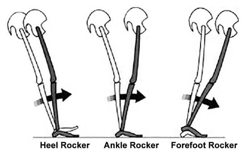
There are several mechanisms in place to stabilize the medial foot tripod. Some, we would hope many in fact, would argue that the process starts up in the glutes in controlling the rate of internal rotation of the limb during the weight bearing response. The glute has to help slow down that spin, hopefully to the point that when the knee reaches its sagittal tracking plane, the spin stops. And, if the knee stops its inward course, the tibia will stop spinning and thus excessive pronation loads into the foot tripod.
Jumping past a bunch of other variables here, for the sake of a shorter article, many typically feel that the tibialis posterior and peroneal sling is additionally important and cannot be ignored.  Then there is a concept we pound hard here on TGG, that one must have adequate control of the rate, speed, degree of pronation.  Again, that comes in part from the sling we just mentioned and of course the skill, endurance and strength of the anterior compartment components, mostly the toe extensors and tibialis anterior.  Their eccentric control slows the forefoot loading and thus controls the rate and degree of foot tripod splay/collapse. If too weak /too fatigued will render too much splay and too much pronation for too long.  All this, and of course other components (too many to get into here today) when insufficient will create a differential in rotation and bending/flexing loads into the tibia, classically presenting at the distal third of the tibia where Medial Tibial Stress Syndrome (MTSS) classically takes root.
We have discussed many times, here on the blog and on our 100+ podcasts about having a competent foot tripod for a given loading response. Specifically addressing the medial tripod, because this is classically where the foot tripod fails, this anchoring of the medial foot tripod around the 1st metatarsophalangeal joint can only occur if there is a harmonious activation of the long and short flexors and extensors of the hallux, and predicated on a proper plantarflexion of the 1st metatarsal to procure proper orientation and purchase of the metatarsal head on the surface/ground not to mention a competent foot tripod.

This study, investigated and determined some curious findings of linking a history of MTSS with increased isometric FHL (flexor hallucis longus) strength.  Namely, they felt that when there was a history of MTSS, it resulted in increased FHL isometric strength.
Why might this be pre-predictive in our mind ? Well, hammering the big toe into aggressive flexion will act to help synergistically stabilize the medial foot tripod, and thus assist in pronation control even though it is not a primary optimal strategy.  This scenario, thought clearly not an optimal strategy, rather a compensation, often sets up global toe gripping strategies, and as we have discussed long ago, a strong link to subungal hematomas (black toenail, bleeding under the toe nails. (link:https://thegaitguys.tumblr.com/post/6355488304/the-black-plague-ok-kinda-sort-ofsubungal). However, to be fair, as mentioned at the end of this article, the authors of the study in question felt that the increased FHL  might be from an attempt to make up for weakened FDL to the lesser toes.  If you are looking for it, someone who has an incompetent medial foot tripod, will often hammer the big toe down while sometime completely disabling the 1st metatarsal purchase on the ground. I have termed these clients as "knuckle poppers" because they cannot find the medial tripod, and their attempts are from over-recruitment of the long hallux flexor (FHL).  The problem here in lies that one cannot properly toe off the big toe, which should be in extension, when they were still just in a FHL life or death strategy. This can set up a functional hallux limitus.  Often, someone with such a functionally troubled medial tripod strategy, will toe off more laterally, choosing not to drive through the medial tripod and hallux, merely because they cannot do so well.


"In this study, the MVIC torque of the 1st MTPJ plantar flexion was significantly higher in runners with a history of MTSS than without it." 

"Our results suggest that runners with a history of MTSS adopt a strategy of reducing the load to the medial tibia because of their history of MTSS."

This may be the case in some, but i am not sure we entirely agree this is the case. We are trying to postulate that the failed tripod is more likely.  Meaning, more uncontrolled medial loading of the foot and thus increased load to the medial tibia. This leans towards this articles comment that, 

"A previous study reported that excessive pronation during motion is a risk factor of developing MTSS [9, 11]. The results also suggest that the FDL and TP muscles that act to support the arch of the foot tend to be stressed in runners who potentially have a risk for developing MTSS. On the other hand, although the FHL, which is an agonist of 1st MTPJ plantar flexion, has a function similar to those of the FDL and TP as the inversion muscle of the ankle, FHL is not likely to be related to development of MTSS because the FHL does not connect to tibial fascia [22]. "

We would suggest that, it does not have to connect to the tibial fascia to be a relevant issue. It is about the loads directly and indirectly across the tibia, not the actual connections. 

However, we do not argue with their statement,  

"Thus, this characteristic increase in MVIC torque of the 1st MTPJ in runners with a history of MTSS could be considered to be a result of increased activity of the FHL to avoid pain caused by contraction stress of the FDL, which could be a possible cause of MTSS." But, what we are suggesting is that perhaps the increased FHL is not to avoid the pain, but an attempt to help stabilize the medial foot tripod. 

Two interesting side notes/thoughts from this article are,

"Collectively, muscle strength characteristics in runners with a history of MTSS could be considered to be a result of increasing activity of the FHL to reduce the load on FDL and avoid pain caused by contraction stress of the FDL."

 "A previous study reported that the FHL tendon branches to the 2nd and 3rd toe in most cases [24]. Therefore, it could be considered that the absence of a significant difference in the MVIC torques of the 2nd–5th MTPJ between runners with and without a history of MTSS could be due to the effects of the branching of FHL tendon to the 2nd and 3rd toe."

This investigators did admit some possible limitations in this study, one of which was "we investigated the relationships between MTSS and muscle strength as a static assessment. Therefore, it is unclear whether the results directly reflect muscle endurance or activity during running. " 

- the gait guys

Reference:

Ankle and toe muscle strength characteristics in runners with a history of medial tibial stress syndrome

Junya Saeki
Masatoshi Nakamura,
Sayaka Nakao,
Kosuke Fujita,
Ko Yanase,
Katsuyuki Morishita and
Noriaki Ichihashi

https://jfootankleres.biomedcentral.com/articles/10.1186/s13047-017-0197-2

Tibial Torsion and Genu Valgum

Join us in this brief video about tibial torsion and genu valgum in a 6-year-old

Mooney JF 3rd Lower extremity rotational and angular issues in children. Pediatr Clin North Am. 2014 Dec;61(6):1175-83. doi: 10.1016/j.pcl.2014.08.006. Epub 2014 Sep 18.

Killam PE. Orthopedic assessment of young children: developmental variations. Nurse Pract. 1989 Jul;14(7):27-30, 32-4, 36.

Kling TF Jr, Hensinger RN. Angular and torsional deformities of the lower limbs in children. Clin Orthop Relat Res. 1983 Jun;(176):136-47.

Podcast 123: The Rear foot: Understanding your RearFoot type

Key tag words:
foot types, rearfoot, forefoot, pronation, supination, shoe fit, forefoot varus, forefoot supinatus, rearfoot inversion, ankle rocker, injuries, rehab, corrective exercises

Rearfoot varus and Rearfoot valgus. Knowing the anatomy of your rear foot and its anatomic and functional posturing can lead to many problems in anyone. If you do not know the rearfoot type and posturing, you will not understand the rest of the foot mechanics. Without this knowledge, you will not know the reason for midfoot or forefoot problems, not understand what shoe you are in, or even why the shoe, footbed, orthotic you have chosen is either not fixing your problems, or causing them.  Join us on a journey down the rearfoot rabbit hole over the next hour.  Plus a few funny stories to lighten the biomechanics-heavy dialogue.
 

Show links:
http://traffic.libsyn.com/thegaitguys/pod_123final_cut.mp3

http://thegaitguys.libsyn.com/podcast-123-the-rear-foot-understanding-your-rearfoot-type

Show sponsors:
www.newbalancechicago.com

www.thegaitguys.com
That is our website, and it is all you need to remember. Everything you want, need and wish for is right there on the site.
Interested in our stuff ? Want to buy some of our lectures or our National Shoe Fit program? Click here (thegaitguys.com or thegaitguys.tumblr.com) and you will come to our websites. In the tabs, you will find tabs for STORE, SEMINARS, BOOK etc. We also lecture every 3rd Wednesday of the month on onlineCE.com. We have an extensive catalogued library of our courses there, you can take them any time for a nominal fee (~$20).
 
Our podcast is on iTunes, Soundcloud, and just about every other podcast harbor site, just google "the gait guys podcast", you will find us.
 
Show Notes:

https://www.ncbi.nlm.nih.gov/pubmed/27134364

https://www.ncbi.nlm.nih.gov/pubmed/25364132

RearFoot positions:

https://www.ncbi.nlm.nih.gov/pmc/articles/PMC3588658/

https://www.ncbi.nlm.nih.gov/pmc/articles/PMC3990938/

Powers CM, Maffucci R, Hampton S. Rearfoot posture in subjects with patellofemoral pain. J Orthop Sports Phys Ther. 1995 Oct;22(4):155-60.

Power V, Clifford AM. The Effects of Rearfoot Position on Lower Limb Kinematics during Bilateral Squatting in Asymptomatic Individuals with a Pronated Foot Type. J Hum Kinet. 2012 Mar;31:5-15. doi: 10.2478/v10078-012-0001-0. Epub 2012 Apr 3.

Shultz SP, Song J, Kraszewski AP, Hafer JF, Rao S, Backus , Mootanah R, Hillstrom HJ. An Investigation of Structure, Flexibility and Function Variables that Discriminate Asymptomatic Foot Types. J Appl Biomech. 2016 Dec 19:1-25. [Epub ahead of print]


You better keep that Hallux Dorsiflexion

Geee....Looks like forefoot rocker really IS IMPORTANT, eh?

...and what have we been saying about being able to dorsiflex your big toe? Watch how well you or your client can descend the 1st ray (remember that if the head of the 1st does not go down and anchor, its axis of rotation moves dorsally and posterior, limiting dorsiflexion of the joint)

This article should make you look at the "toe break" in a shoe...

"They found that increasing bending stiffness assisted with propulsion during running, reducing the metabolic cost of running by about 1%. However, at a certain level, the increased elasticity began to interfere with the natural flexion of the first MTP joint, reducing the contribution of ankle joint torque to push-off and counteracting the metabolic benefits. Within the study population, the threshold of bending stiffness for optimal energetics varied significantly from one runner to the next, suggesting running shoe design may need to be tuned to an individual runner’s needs." 

http://lermagazine.com/news/in-the-moment-sports-medicine/stiffer-shoes-improve-running-energetics-as-long-as-first-mtp-flexion-is-preserved

Metatarsus Adductus: The Basics

Metatarsus Adductus: The Basics

A few points to remember:

  • Metatarsus adductus deformity is a forefoot which is adducted in the transverse plane with the apex of the deformity at LisFranc’s (tarso-metatarsal) joint. The fifth metatarsal base will be prominent and the lateral border of the foot which is convex in shape . The medial foot border is concave with a deep vertical skin crease located at the first metatarso cuneiform joint level. The hallux (great toe) may be widely separated from the second digit and the lesser digits will usually be adducted at their bases (se below). ln some cases the abductor hallucis tendon may be palpably taut just proximal to its insertion into the inferomedial aspect of the proximal phalanx (1)
  • To measure the deviation of the metatarsals, the midline of the foot correspondingto bisecting the heel is used as a reference. This is the line that divides the heel pad into equal parts and, when extended, runs through the second toe or the second web space. In mild deformities, the midline of the foot runs through the third toe. In moderate adductus deformities, it falls between the third and fourth toes. In severe deformities the line is lateral to the third web space.(2)
  • If detected early, stretching is a common and effective treatment for mild and some moderate cases. The heel is steadied with one hand while the forefoot is abducted in relation to the hind foot. This is done for 5 reps, 5-7 times per day. (2)
  • 85% will resolve spontaneously, is caused by intrauterine position, is flexible & resolves spontaneously in more than 90 % of children. (3)
  • Though often used interchangeably, the term "metatarsus adductus" is usually reserved for milder cases, where the forefoot is adducted on the hindfoot at the tarso-metatarsal articulation. Metatarsus varus is often reserved for conditions where the matatrsals are actually curved AND the forefoot is adducted on the hindfoot. (4) The term "Metatarsus primus varus" is reserved for feet which have the same neutral or valgus hindfoot and varus forefoot but, in addition, increased divergence of the first and second metatarsals. (5)
  • It is interesting to note that along with forefoot adductus, hip dysplasia and internal tibial torsion are common (6) and this patient has the latter
  • Gait abnormalities seen with this deformity include a decreased progression angle, in toed gait, excessive supination of the feet with low gear push off from the lesser metatarsals. 


1.  Bleck E: Metatarsus adductus: classification and relationship to outcomes of treatment. J Pediatric Orthop 3:2-9,1983.

2. Bohne W. Metatarsus adductus. Bulletin of the New York Academy of Medicine. 1987;63(9):835-838.  link to FREE full text: https://www.ncbi.nlm.nih.gov/pmc/articles/PMC1629274/

3. http://www.wheelessonline.com/ortho/metatarsus_adductus

4. Peabody, C.W. and Muro, F.: Congenital metatarsus varus. J. Bone Joint Surg. 15:171-89, 1933.

5. Truslow, W.: Metatarsus primus varus or hallux valgus? J. Bone Joint Surg.23:98-108, 1925.

6. Jacobs J: Metatarsus varus and hip dysplasia. C/inO rth o p 16:203-212, 1960


additional references:

Kane R. Metatarsus varus. Bulletin of the New York Academy of Medicine. 1987;63(9):828-834. link to FREE full text: https://www.ncbi.nlm.nih.gov/pmc/articles/PMC1629282/

Wynne-Davies R, Littlejohn A, Gormley J. Aetiology and interrelationship of some common skeletal deformities. (Talipes equinovarus and calcaneovalgus, metatarsus varus, congenital dislocation of the hip, and infantile idiopathic scoliosis). Journal of Medical Genetics. 1982;19(5):321-328. link to FREE full text: https://www.ncbi.nlm.nih.gov/pmc/articles/PMC1048914/


Take the minute to tie your shoes properly.

Plantar thermal stress response (TSR) leads to skin shear forces.

"Fifteen eligible subjects were enrolled. The left foot was used as a reference and fitted to a self-adjusted and habitual lace-tightening method by each subject. The right foot was used as a test closure and fitted into three lace closure conditions: loose, tight, and preset optimal closure (reel clutch, BOA technology). Thermal images were taken after 5 minutes of acclimatization (pre-trial) and immediately after 200 walking steps in each shoe closure condition (post-trial)."

Their conclusions:
"The results from this study suggest that shoelace closure technique can have a profound effect on TSR. It therefore stands to reason that optimal lace closure may have an impact in reducing risk of plantar ulcers in people with Diabetic peripheral neuropathy (DPN). Interestingly, results revealed that even a self-adjusted lace closure may not be necessarily optimal and a preset closure setting like reel clutch might ultimately be recommended to minimize risk."


Lace Up for Healthy Feet: The Impact of Shoe Closure on Plantar Stress Response

Hadi Rahemi, PhD, David G. Armstrong, DPM, MD, PhD, Ana Enriquez, BSc, Joshua Owl,BSc, Talal K. Talal, DPM, Bijan Najafi, PhD, MSc
First Published April 18, 2017


http://journals.sagepub.com/doi/abs/10.1177/1932296817703669?platform=hootsuite&journalCode=dsta

Wool socks ?

Blisters, wet feet, smell, skin maceration, who needs that. 
Here at The Gait Guys, we swear by wool socks. We wear them everyday, every season, and even in our running and hiking or athletic shoes. Cotton and synthetics are not in our life, and this is a personal choice. Ivo likes the Darn Tough brand, so do I, but I also love the REI thinner black dress socks for every day wear. I could wear a single pair all week if i wanted, and admittedly often do, without any smell. Yes, you read that right, no odor.  That is the genius of wool. But, there are other benefits as well.
Wool is also king for moving moisture, the fatty acids in the wool fibers eat up the bacteria and microbes that can possibly cause infection. 

Darn Tough ran a test where they soaked their socks in a bacteria that hikers might encounter on the trail; within an hour, 50 to 80 percent of the bacteria ws gone. 

Wool is also great in all temperatures. They insulate when its cold and assist in evaporation when warm. Plus, these things are durable, it is why we love them, and hence the name, Darn Tough Socks. The will get you through a 1000 mile hike, as long as you have shoes on over them :)

https://www.outsideonline.com/2173601/do-i-really-need-25-merino-hiking-socks

https://darntough.com/


The "Bad Guys" are watching your gait, too.

It is cold coffee and stale donut Thursday here on The Gait Guys, poorly palatable stuff, but stuff we need to eat anyways.  This is gait related. Read on.

"The cues used by prisoners were: gait, body posture (body movements not related to gait), age, gender, attractiveness, build, clothing, attention, fitness, environment (e.g., lack of lighting), and whether target was alone."

Yes, "Bad Guys" are looking at your gait.
___________

"Bad guys" are relentless and patient, opportunistic and flexible — and they pay attention, to us. And so, what does this mean to us ? And to our gait, how we move about our communities ?

It is in the MSM everyday, so we might as well pay attention to it. It is happening all around us, all the time. Opportunists. You should up your awareness game when you are out an about. We are not talking about becoming paranoid, but we should all be more aware. And, this article proves it.  

Are you a soft target ? "Bad Guys" like easy soft targets. So, read this and don't be a softie.  

"In a classic study conducted by a prison psychologist where dozens of convicted felons were interviewed, the psychologist played videos or presented several snapshots of random crowds of people in shopping malls, subway stations, and busy city streets and asked each of the inmates to point out for him the softest target in the pictures–that is the individual that they would most likely attack if given the opportunity. All responses were recorded. The study concluded that nearly every inmate selected the same people in each segment for similar reasons—these reasons form the baseline definition of Soft Target Indicators."
"Examples that help describe a Soft Target Indicator come from snippets of those recorded study responses, and include “looks sheepish,” “eyes were looking down,” ”obviously lost,” “an easy mark,” “looks out of place,” “not paying attention,” “distracted,”  and the most common—“unaware of their surroundings.”

These are all gait related. We have talked about dual tasking and many other gait and locomotion distractors that can make us vulnerable to falling and stepping off of cliffs and into ponds at the local mall. But now we should all be aware that there are other people watching our gait for reasons other than capturing the next viral video for YouTube. They are opportunists. 

The top vulnerable cues for these opportunists were: 

- Walk/gait – walking with confidence versus walking like a soft target. 

- Gender – females appear as softer targets than males. 

- Body type – In good physical shape versus heavy set or slow - - - - Apparent Fitness level – 

- Attention to surroundings versus Not paying attention; Appears to be cautious versus appears to be clueless.

So, our advice, especially if you are in a new environment or travelling?  Plan ahead, so you can pay attention. Keep your head up and look around, walk with confidence and be observant. If your head is down in your phone and you are dual tasking, this articles suggests that the "Bad Guys" are looking for you. 


http://www.recoilweb.com/adversarial-attraction-predators-optic-90271.html

Podcast 122: Achilles problems, glutes, & feet.

Key tag words:
neuroscience, elon musk, achilles, tendonitis, tendonopathy, eccentric loading, tendon loading, gluteus maximus, gmax, glutes, abductor hallucis, foot pain, hip biomechanics, navicular drop, BEAR, ACL tear, ACL reconstruction, plantar fascitis
 

Show links:

http://traffic.libsyn.com/thegaitguys/pod_122f.mp3

http://thegaitguys.libsyn.com/podcast-122-achilles-problems-glutes-the-feet
Show sponsors:
 www.newbalancechicago.com


www.thegaitguys.com
That is our website, and it is all you need to remember. Everything you want, need and wish for is right there on the site.
Interested in our stuff ? Want to buy some of our lectures or our National Shoe Fit program? Click here (thegaitguys.com or thegaitguys.tumblr.com) and you will come to our websites. In the tabs, you will find tabs for STORE, SEMINARS, BOOK etc. We also lecture every 3rd Wednesday of the month on onlineCE.com. We have an extensive catalogued library of our courses there, you can take them any time for a nominal fee (~$20).
 
Our podcast is on iTunes, Soundcloud, and just about every other podcast harbor site, just google "the gait guys podcast", you will find us.
 
Show Notes:

Stanford Develops Computer That Literally Plugs Into People's Brains

https://www.entrepreneur.com/article/289645


Elon Musk says humans must become cyborgs to stay relevant. 

https://www.theguardian.com/technology/2017/feb/15/elon-musk-cyborgs-robots-artificial-intelligence-is-he-right

1. achilles tendonopathy:

http://www.jospt.org/doi/abs/10.2519/jospt.2016.6462?platform=hootsuite&code=jospt-site

2. achilles tendinitis and tendonosis.

Ohberg L, Lorentzon R, Alfredson H, Maffulli N. Eccentric training in patients with chronic Achilles tendinosis: normalised tendon structure and decreased thickness at follow up. British Journal of Sports Medicine. 2004;38(1):8-11. doi:10.1136/bjsm.2001.000284.

link to abstract: http://www.ncbi.nlm.nih.gov/pmc/articles/PMC1724744/

3. Is Achilles tendon blood flow related to foot pronation?
 E. Wezenbeek,T. M. Willems,N. Mahieu,I. Van Caekenberghe,E. Witvrouw,D. De Clercq

http://onlinelibrary.wiley.com/doi/10.1111/sms.12834/full

4.  The effects of gluteus maximus and abductor hallucis strengthening exercises for four weeks on navicular drop and lower extremity muscle activity during gait with flatfoot

Young-Mi Goo, MS, PT,1 Tae-Ho Kim, PhD, PT,1,* and Jin-Yong Lim, MS, PT1  J Phys Ther Sci. 2016 Mar; 28(3): 911–915.
https://www.ncbi.nlm.nih.gov/pmc/articles/PMC4842464/

5. BEAR
https://www.youtube.com/watch?v=k3g-CagCrZM

Bridge-Enhanced Anterior Cruciate Ligament Repair (BEAR) procedure uses stitches and a bridging scaffold (a sponge injected with the patient’s blood) to stimulate healing of the torn ACL eliminating the need tendon graft.

References:
Murray, M., Flutie, B., Kalish, L., Ecklund, K., Fleming, B., Proffen, B. and Micheli, L. (2016). The Bridge-Enhanced Anterior Cruciate Ligament Repair (BEAR) Procedure: An Early Feasibility Cohort Study. Orthopaedic Journal of Sports Medicine, 4(11).

L. Proffen, B., S. Perrone, G., Roberts, G. and M. Murray, M. (2015). Bridge-Enhanced ACL Repair: A Review of the Science and the Pathway Through FDA Investigational Device Approval. Annals of Biomedical Engineering, 43(3), pp.805-818.

Distracted Kids Walk Slower

kid-walking-on-road-2592x3408_47863.jpg

Do Gait rehab on Kids?

Distracted kids walk slower and spend more time in double leg stance.

We know it can effect adults, usually resulting a mild decomposition of gait, by decreasing step length in many and slowing of gait in older folks (becoming more primitive).

Watch where you do your gait rehab in the younger set; keep them focused and engaged for better outcomes.

"Significant main effects of walking experience groupand visual distraction condition were found. Visual environmental distraction significantly affected gait performance in children regardless of walking experience. Velocity decreased from 110.04 to 97.73 cm/sec (p = 0.003) while double limb stance % of gait cycle increased from 18.29% to 20.39% (p = 0.025)."

Phys Occup Ther Pediatr. 2017 Apr 10:1-10. doi: 10.1080/01942638.2017.1297987. [Epub ahead of print]
The Effect of Visual Environmental Distraction on Gait Performance in Children.
Bizama F, Medley A, Trudelle-Jackson E, Csiza L.

J Neuroeng Rehabil. 2014 Apr 28;11:74. doi: 10.1186/1743-0003-11-74.
Effect of explicit visual feedback distortion on human gait.
Kim SJ, Mugisha D.

Curr Gerontol Geriatr Res. 2011;2011:651718. doi: 10.1155/2011/651718. Epub 2011 Jun 16.
Effects of a Visual Distracter Task on the Gait of Elderly versus Young Persons.
Bock O, Beurskens R.