Metatarsalgia happens...
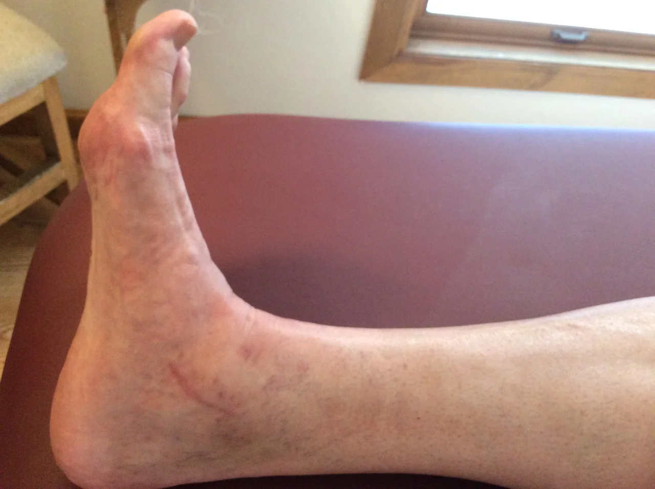
So a patient presents with forefoot pain, worse in the am upon awakening, with 1st weight bearing that would improve somewhat during the day, but would again get worse toward the end of the day and with increased activity. It began insidiously a few months ago (like so many problems do) and is getting progressively worse. Rest, ice and ibuprofen can offer some relief. You may see a dropped metatarsal head and puffiness and prominence in that area on the plantar surface of the foot, maybe not. Maybe you do a diagnostic ultrasound and see a lesion of the plantar plate as well? How did it get there? 

image courtesy of Tom Michaud: with permission

image courtesy of Tom Michaud: with permission

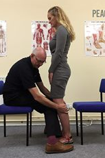
Lets look at the anatomy of the short flexors of the foot, as well as some biomechanics of the foot, ankle and hip. 

The flexor digitorum brevis (FDB) is innervated by the medial plantar nerve and arises from the medial aspect of the calcaneal tuberosity, the plantar aponeurosis (ie: plantar fascia) and the areas bewteen the plantar muscles. It travels distally, splitting at the metatarsal phalangeal articulation (this allows the long flexors to travel forward and insert on the distal phalanges); the ends come together to divide yet another time and each of the 2 portions of that tendon insert onto the middle of the middle phalanyx (1) 

As a result, in conjunction with the lumbricals, the FDB is a flexor of the metatarsophalangeal and proximal interphalangeal joints. In addition, it moves the axis of rotation of the metatasophalangeal joints dorsally, to counter act the function of the long flexors, which, when tight or overactive, have a tendency to drive this articulation anteriorly .Do you see any subtle extension of the metatarsophalangeal joint and flexion of the proximal interphalangeal joints on your exam?

We know that the FDB contracts faster than the other intrinsic muscles (2), playing a role in postural stability (3) and that the flexors temporally should contract earlier than the extensors (4), assumedly to move this joint axis posteriorly and allow proper joint centration. When this DOES NOT occur, the metatarsal heads are driven into the ground, causing irritation and pain.

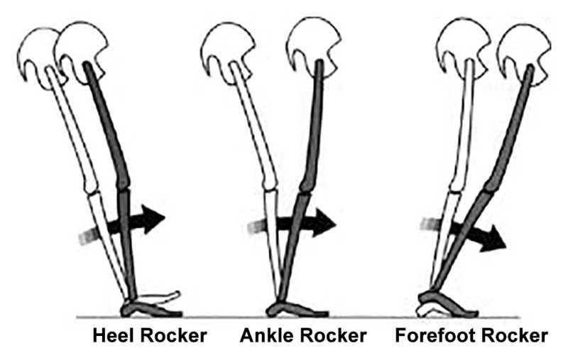
If there is also a loss of ankle rocker this problem is made (much) worse. Why? Because, with the loss of one rocker, another must make up for the loss: ankle rocker decreases, forefoot rocker has to increase; this equals increased metatarsal head pressure. 

If you have been with us for any length of time, you know that ankle rocker and hip extension are intimately related, as one should equal the other, something we call “The “Z” angle”, that you have probably (hopefully?) read about here before. 

So what is the fix? Getting the FDB back on line for one. 

  • How about the toe waving exercise? 

  • How about the lift spread reach exercise? 

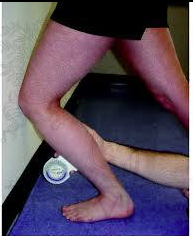
  • How about retraining ankle rocker and improving hip extension?

  • How about an orthotic with a metatarsal pad in the short term? 

  • How about some inflammation reducing modalities, like acupuncture, ice laser and pulsed ultrasound. 

  • Maybe some herbal or enzymatic anti inflammatories?


Dr Ivo Waerlop, one of The Gait Guys.

#gait #footpain #metatarsalgia #metatarsalpain #anklerocker #hipextension #thegaitguys


1. http://en.wikipedia.org/wiki/Flexor_digitorum_brevis_muscle

2. Tosovic D1, Ghebremedhin E, Glen C, Gorelick M, Mark Brown J.The architecture and contraction time of intrinsic foot muscles.J Electromyogr Kinesiol. 2012 Dec;22(6):930-8. doi: 10.1016/j.jelekin.2012.05.002. Epub 2012 Jun 27

3.Okai LA1, Kohn AF. Quantifying the Contributions of a Flexor Digitorum Brevis Muscle on Postural Stability.Motor Control. 2014 Jul 15. [Epub ahead of print]

4. Zelik KE1, La Scaleia V, Ivanenko YP, Lacquaniti F.Coordination of intrinsic and extrinsic foot muscles during walking.Eur J Appl Physiol. 2014 Nov 25. [Epub ahead of print]


Got hip extension?

Because she sure could use some...

we have see this gal before… yesterday in fact

  • left plantar plate lesion (yes, conformed on ultrasound)

  • left sided anatomical leg length discrepany

  • bilateral internal tibial torsion

  • incompetent L quadratus lumborum

  • adequate hip extension and ankle dorsiflexion available to her

  • lack of endurance in her abs

yep, lots more, but that is enough for now


note that she has plenty of ankle dorsiflexion, more on the right. this is due to her right leg being anatomically longer and has to travel through a greater range of motion

look at the knee and the hip articulations to assess hip extension. It should match ankle dorsiflexion, no?


Dr Ivo Waerlop, one of The Gait Guys


#gait #gaitguys #thegaitguys #hipextension #LLD #quadratuslumborum #internaltibialtorsion #anklerocker #ankledorsiflexion

Two out of Three ain't Bad...But sometimes it is

image credit: https://commons.wikimedia.org/wiki/File:Meatloaf_(1).jpg

image credit: https://commons.wikimedia.org/wiki/File:Meatloaf_(1).jpg

“What do you mean my plantar fasciitis is due to my hip?”

I recently saw a 60 YO male patient with right-sided plantar fasciitis of approximately 1-1/2 months duration. It began insidiously with pain located at the medial calcaneal facet on the right hand side. He had localized tenderness in this area with some spread distally towards the metatarsal heads. He has ankle dorsiflexion was relatively symmetrical with mild impairment on the right compared to left but only approximately 2 degrees. He had hip extension is 0 degrees on the affected side and 10 degrees on the affected side. Sacroiliac pathomechanics were present as well with the loss of flexion and extension. He had a slight leg length discrepancy, short on the symptomatic side.

So what is going on?

Moving forward in the sagittal plane requires a few things:

Adequate hip extension

Adequate ankle dorsiflexion

Adequate hallux dorsiflexion with an intact Windlass mechanism

He has a diminished step length going from right to left. Because of the lack of hip extension, the motion needs to occur somewhere. His ankle dorsiflexion is almost sufficient but less sufficient on the right (symptomatic) side than it is on the left. He has adequate hallux dorsiflexion but lacks adequate hip extension. Like the song goes, begin "Two of of three ain’t bad". However in this case, it is bad. He has an intact windlass mechanism. In fact, a little too intact. This is causing a tug at the medial calcaneal facet, creating an insertional tendinitis that we know as "plantar fasciitis".

So we did we do?

  • Manipulated the right sacroiliac joint

  • Gave him lift she/spread/reach exercises

  • Gave him shuffle walk exercises

  • Worked on hip flexor lengthening

  • Treated the plantar fascial insertion locally with acupuncture and laser therapy

Dr Ivo Waerlop, one of The Gait Guys

#gait, #gaitanalysis,#thegaitguys, #anklerocker#halluxdorsiflexion, #plantarfascitis

Flip Flops not so bad? We still think they suck and here's why...

journal.pone_.0193653.g001-374x500.jpg

We have talked about the dangers of open back shoes (Including flip-flops)  and loss of ankle rocker as well as changes in forefoot rocker and great toe dorsiflexion on our blog many times.

The findings of this study, with slower cadence and shortened stance are not surprising (especially since you need to fire your long flexors to keep them on!) nor are ankle joint kinematics (flip flops have no heel counter and are not torsionally rigid, so naturally there would be increseased subtalar motion), however we really question the interpretation.

 "Many have long suspected the answer, but a new study would appear to resolve the question: Are flip flops really that bad for your feet? According to Chen and colleagues from the Department of Biomedical Engineering at The Hong Kong Polytechnic University, flip flops are most likely no better than barefoot when it comes to lower-limb co-contraction and joint contact force in the ankle. The authors had hypothesized that the popular rubber footwear would increase co-contraction of the muscles between the knee and ankle joints in what they thought was a compensatory mechanism for the unstable foot–sole interface and would affect gait kinematics and kinetics.

In the study, the researchers had 10 healthy males perform 6 walking trials under 3 conditions: barefoot, sports shoes, and thong-type flip flops. Participants, who reported they were not “regular flip flop wearers,” were fitted with numerous markers that were monitored while they walked on a 10-meter pathway. The study looked at several muscle pairings that stabilize the knee, ankle, and subtalar joints, including vastus lateralis and gastrocnemius medialis; vastus lateralis and biceps femoris; and peroneus longus and tibialis anterior.

In pairwise comparisons, the walking velocity of flip flops was lower than that of sports shoes (p<0.01) but comparable to barefoot (p>0.05), findings that were consistent with the published literature. Although not significant, the minimalist footwear produced a slower cadence and shortened stance phase in walking trials compared to the other 2 types of footwear. Joint kinematics differed significantly in the ankle joint (F[2,18]=6.73, P<.05) and subtalar joint (F[2,18]=4.45; P<.05); Furthermore, ankle and subtalar range of motion was higher for flip flops than for sports shoes. However, co-contraction was not enhanced. The authors propose that walking speed does not need to be consistent for real-world activities and the slower speed could be a natural approach to avoid injury.

The authors conclude that the slowed walking speed of flip flop users could account for the comparable joint biomechanics between flip flop use and barefoot. They note, however, that, for injury prevention, the closed-toe design of the sports shoe would provide better support for joint motion and loading compared to the other 2 options."

Source:

Chen TL, Wong DW, Xu Z, Tan Q, Wang Y, Luximon A, Zhang M. Lower limb muscle co-contraction and joint loading of flip-flops walking in male wearers. PLoS One. 2018;13(3):e0193653."

image and article source: http://lermagazine.com/issues/may/flip-flops-bare-feet-or-sports-shoes-which-are-best-and-which-are-worst

Try THIS at home...

Screen Shot 2018-02-07 at 11.23.24 AM.png

Cool test, results you can see and some literature to back it up. If you are interested at all in proprioception, this is an interesting read.

So, the question for us is: "Does ankle dorsiflexion actually create more stability, like is purported?"

“The point is that if I make their ankle rigid this way, then they can more effectively use the balance mechanisms at their knees, hips, and proximally, because they’re on a stable base. My proposition is that their balance is actually normal, apart from that distal segment. When their ankle is stabilized, they use their knees more effectively, and they become less dependent on their eyesight to maintain their balance.”

http://lermagazine.com/article/afos-and-balance-issues-in-peripheral-neuropathy

Your Gait Changes when you text....

GettyImages-534572749web-57a3087d3df78c3276b9fc80-2.jpg

Does texting alter your gait? It sure seems to slow you down, and according to this study, alter firing patterns of muscles about your ankle. Perhaps you are trying to preserve ankle rocker and maintain stability? It is interesting that ankle dorsiflexion actually increased and plantar flexion decreased.

"Young adults showed, overall, small gait modifications that could be mainly ascribable to gait speed reduction and a modified body posture due to phone handling. We found no significant alterations of ankle and knee kinematics and a slightly delayed activation onset of the left gastrocnemius lateralis. However, we found an increased co-contraction of tibialis anterior and gastrocnemius lateralis, especially during mid-stance. Conversely, we found a reduced co-contraction during terminal stance."


link to FREE FULL TEXT: https://www.ncbi.nlm.nih.gov/pmc/articles/PMC4579642/

Ankle Rocker Revisited....

How many times have we talked about ankle rocker and its importance? So how are YOU measuring ankle rocker? Are you looking at it on the table? On the ground? Weight bearing? Knee flexed or extended (or both?). The knee is extended at initial contact, flexes through midstance, extends at terminal stance and pre swing and flexes again during swing phase until extending at the end of terminal swing for initial contact again.

What you see on the table may not (and many times doesn't)  translate to real life. Someone with limited ankle dorsiflexion non weight bearing may have normal amounts during gait and vice versa. With gravity in place and a functioning (or malfunctioning) vestibular system, things can change rapidly. Remember that the vestibular system drives the extensors and if inhibited, you will often have flexor dominance. Talk about a tight gastroc/soleus group!


"These findings indicate that nonweightbearing and weightbearing measurements of ankle DF PROM with knee extension should not be used interchangeably and that weightbearing ankle DF PROM with the knee extended is more appropriate for estimating ankle DF during gait."

Kang MH, Oh JS. Relationship Between Weightbearing Ankle Dorsiflexion Passive Range of Motion and Ankle Kinematics During Gait. J Am Podiatr Med Assoc. 2017 Jan;107(1):39-45. doi: 10.7547/14-112.


"There is no relationship between a static diagnosis of ankle dorsiflexion at 0° with dorsiflexion during gait. On the other hand, those subjects with less than -5° of dorsiflexion during static examination did exhibit reduced ankle range of motion during gait."


Gatt A, De Giorgio S, Chockalingam N, Formosa C. A pilot investigation into the relationship between static diagnosis of ankle equinus and dynamic ankle and foot dorsiflexion during stance phase of gait: Time to revisit theory? Foot (Edinb). 2017 Mar;30:47-52. doi: 10.1016/j.foot.2017.01.002. Epub 2017 Feb 6.


You better keep that Hallux Dorsiflexion

Geee....Looks like forefoot rocker really IS IMPORTANT, eh?

...and what have we been saying about being able to dorsiflex your big toe? Watch how well you or your client can descend the 1st ray (remember that if the head of the 1st does not go down and anchor, its axis of rotation moves dorsally and posterior, limiting dorsiflexion of the joint)

This article should make you look at the "toe break" in a shoe...

"They found that increasing bending stiffness assisted with propulsion during running, reducing the metabolic cost of running by about 1%. However, at a certain level, the increased elasticity began to interfere with the natural flexion of the first MTP joint, reducing the contribution of ankle joint torque to push-off and counteracting the metabolic benefits. Within the study population, the threshold of bending stiffness for optimal energetics varied significantly from one runner to the next, suggesting running shoe design may need to be tuned to an individual runner’s needs." 

http://lermagazine.com/news/in-the-moment-sports-medicine/stiffer-shoes-improve-running-energetics-as-long-as-first-mtp-flexion-is-preserved

and what have we been saying about loss of ankle rocker and achilles tendon problems for years now?Here is a FREE, FULL TEXT article talking all about it“A more limited ankle Dorsi Flexion ROM as measured in Non Weight Bearing with the knee bent inc…

and what have we been saying about loss of ankle rocker and achilles tendon problems for years now?

Here is a FREE, FULL TEXT article talking all about it

“A more limited ankle Dorsi Flexion ROM as measured in Non Weight Bearing with the knee bent increases the risk of developing Achilles Tendinopathy among military recruits taking part in intensive physical training.”


J Foot Ankle Res. 2014 Nov 18;7(1):48. doi: 10.1186/s13047-014-0048-3. eCollection 2014.Limited ankle dorsiflexion increases the risk for mid-portion Achilles tendinopathy in infantry recruits: a prospective cohort study.Rabin A1, Kozol Z, Finestone AS.

link to full text: http://www.ncbi.nlm.nih.gov/pmc/articles/PMC4243387/

tumblr_o8tlm8zPXq1qhko2so1_1280.jpg
tumblr_o8tlm8zPXq1qhko2so2_1280.jpg
tumblr_o8tlm8zPXq1qhko2so3_1280.jpg
tumblr_o8tlm8zPXq1qhko2so4_r1_1280.jpg

Wow!  Can you figure out why this person at the distal end of her first metatarsal under her medial sesamoid.

She recently underwent surgery for a broken fibula (distal with plate fixation) and microfracrure of the medial malleolus. You are looking at her full range of dorsiflexion which is improved from approximately 20° plantarflexion. She is now at just under 5°.

She has just begun weight-bearing and developed pain over the medial sesamoid.

The three rockers, depicted above from Thomas Michauds book, or necessary for normal gait.  This patient clearly has a loss of ankle rocker. Because of this loss her foot will cantilever forward and put pressure on the head of the first metatarsal.  This is resulting in excessive forefoot rocker.  Her other option would have been to pronate through the midfoot. Hers is relatively rigid so, as Dr. Allen likes to say, the “buck was passed to the next joint. ”

There needs to be harmony in the foot in that includes each rocker working independently and with in its normal range. Ankle rocker should be at least 10° with 15° been preferable and for footlocker at least 50° with 65 been preferable.

 If you need to know more about rockers, click here.

Achilles TendonitisThe motion needs to occur somewhere&hellip;Make sure you look at the whole pictureSince the knee was bent, perhaps we should be looking at the soleus? And the talo crural articulation?&ldquo;A more limited ankle Dorsi Flexion Rang…

Achilles Tendonitis

The motion needs to occur somewhere…Make sure you look at the whole picture

Since the knee was bent, perhaps we should be looking at the soleus? And the talo crural articulation?

“A more limited ankle Dorsi Flexion Range Of Motion as measured in Non Weight Bearing with the knee bent increases the risk of developing Achilles Tendonitis among military recruits taking part in intensive physical training.”

J Foot Ankle Res. 2014 Nov 18;7(1):48. doi: 10.1186/s13047-014-0048-3. eCollection 2014.
Limited ankle dorsiflexion increases the risk for mid-portion Achilles tendinopathy in infantry recruits: a prospective cohort study.
Rabin A1, Kozol Z1, Finestone AS2.

tumblr_nahfwdDSeS1qhko2so1_1280.jpg
tumblr_nahfwdDSeS1qhko2so2_1280.jpg

Proprioception trumps Biomechanics

As I sit here on a rare Friday afternoon, not working (OK, I am writing this, so sort of working) and looking out at the lake (picture above), while on a family camping trip, I think about a walk on the rocks this morning with my kids. I was watching my very skilled 7 year old jump from rock to rock while my 3 ½ year old, that thinks he is seven, tried to follow his older brother.

I had my foot on a rock which lowered the front of my foot in plantar flexion and stood on that leg. I noticed that my balance was not as great as it was when my foot was in dorsiflexion. This made me think about pronation and supination. Yes, it is not uncommon for me to think about such things, especially when I have some spare time. That is one of the things about being a foot and gait nerd; these sorts of things are always on our minds.

So, why was my balance off? Did I need more proprioceptive work? Were my foot intrinsics having issues? No, it was something much more mundane.

Pronation consists of dorsiflexion, eversion and abduction. This places the foot in a  “mobile adaptor” posture, reminiscent of our hunter/gatherer ancestors, who needed to adapt to uneven surfaces while walking over terra firma barefoot. Supination, on the other hand (which is the position my foot was in), consists of plantarflexion, inversion and adduction. It places the foot (particularly the midtarsals) in a locked position for propulsion (think of the foot position during toe off).

So why when my foot was plantar flexed and adducted while standing on this rock so much more unstable in this supposedly more stable, supinated position? I would encourage you, at this point, to try this so you can see what I mean. When I placed my foot in dorsiflexion on the rock, I was much more stable. A most interesting conundrum for a biomechanist.

Experimenting for a few minutes, alternating plantar flexion and dorsi flexion, gave me the answer. When we are walking on the flats, our foot is (usually) not pushed to the extremes of dorsiflexion; with the front of the foot up on a rock, it is much more so. This “extra” upward force on the front of the foot, provides much more sensory input (and thus proprioception) from the ball of the feet. Take a look at the sensory homunculus and you can see how much brain real estate is dedicated to your foot, especially the front portion. With this information, we are able to apply more  force through the posterior compartment of the leg,which is stronger than my anterior compartment (as it is with most folks).

When the front of your foot is in plantar flexion (ie, your heel is on the rock), we have less sensory input to the balls of the feet, and rely more on the anterior compartment (weaker in many folks, including myself) to counterbalance the weight of our body.

Mystery solved: proprioception trumps biomechanics; more proof that the brain is smarter than we are.

The Gait Guys. Solving the worlds great gait questions, one at a time.

tumblr_n3vbw7hW5t1qhko2so1_1280.jpg
tumblr_n3vbw7hW5t1qhko2so2_400.jpg

Welcome to Rewind Friday, Folks. Today we review the importance of the great toe extensor. Enjoy!

Gait Topic: The Mighty EHB (The Short extensor of the big toe, do not dismiss it !)

Look at this beautiful muscle in a foot that has not yet been exposed to hard planar surfaces and shoes that limit or alter motion! (2 pics above, toggle back and forth)

The Extensor Hallicus Brevis, or EHB as we fondly call it (beautifully pictured above causing the  extension (dorsiflexion) of the child’s proximal big toe) is an important muscle for descending the distal aspect of the 1st ray complex (1st metatarsal and medial cunieform) as well as extending the 1st metatarsophalangeal joint. It is in part responsible for affixing the medial tripod of the foot to the ground.  Its motion is generally triplanar, with the position being 45 degrees from the saggital (midline) plane and 45 degrees from the frontal (coronal) plane, angled medially, which places it almost parallel with the transverse plane. With pronation, it is believed to favor adduction (reference). Did you ever watch our video from 2 years ago ? If not, here it is, you will see good EHB demo and function in this video. click here

It arises from the anterior calcaneus and inserts on the dorsal aspect of the proximal phalynx. It is that quarter dollar sized fleshy protruding, mass on the lateral aspect of the dorsal foot.  The EHB is the upper part of that mass. It is innervated by the lateral portion of one of the terminal branches of the deep peronel nerve (S1, S2), which happens to be the same as the extensor digitorum brevis (EDB), which is why some sources believe it is actually the medial part of that muscle. It appears to fire from loading response to nearly toe off, just like the EDB; another reason it may phylogenetically represent an extension of the same muscle.

*The EDB and EHB are quite frequently damaged during inversion sprains but few seem to ever look to assess it, largely out of ignorance. We had a young runner this past year who had clearly torn just the EHB and could not engage it at all. He was being treated for lateral ankle ligament injury when clearly the problem was the EHB, the lateral ligamentous system had healed fine and this residual was his chief problem.  Thankfully we got the case on film so we will present this one soon for you !  In chronic cases we have been known to take xrays on a non-standard tangential view (local radiographic clinics hate us, but learn alot from our creativity) to demonstrate small bony avulsion fragments proving its damage in unresolving chronic ankle sprains not to mention small myositis ossificans deposits within the muscle mass proper.

Because the tendon travels behind the axis of rotation of the 1st metatarsal phalangeal joint, in addition to providing extension of the proximal phalynx of the hallux (as seen in the child above), it can also provide a downward moment on the distal 1st metatarsal (when properly coupled to and temporally sequenced with the flexor hallicus brevis and longus), assisting in formation of the foot tripod we have all come to love (the head of the 1st met, the head of the 5th met and the calcaneus).

Wow, all that from a little muscle on the dorsum of the foot.

The Gait Guys. Definitive Foot Geeks. We are the kind of people your podiatrist warned you about…

Being a gait geek offers you a unique perspective in many situations.

Perhaps you have been with us for some time now and would like to check your gait acumen. If you are new, or these terms are foreign to you; search here on our blog through hundreds of posts to become more comfortable with some of the vocabulary.

Watch this video a few times (we slowed it down for you) and write down what you see.

Did you see all of these in this brief video?

  • bilateral loss of hip extension
  • bilateral loss of ankle rocker
  • less ankle rocker on right
  • bilateral increased progression angle  
  • dip in right pelvis at right heel strike
  • arm swing increased on R

The Gait Guys. Increasing your gait competency each and every day.

special thanks to NL for allowing us to use this video footage.

Podcast #31: Walking Straight, Mastalgia & Shoes


podcast link:

http://thegaitguys.libsyn.com/podcast-31-walking-straight-matalgia-queen

iTunes link:

https://itunes.apple.com/us/podcast/the-gait-guys-podcast/id559864138

Gait Guys online /download store:

http://store.payloadz.com/results/results.aspx?m=80204

other web based Gait Guys lectures:

www.onlinece.com   type in Dr. Waerlop or Dr. Allen  Biomechanics

Today’s show notes:

1. Neuroscience Piece:

http://www.cell.com/current-biology/abstract/S0960-9822(09)01479-1

http://www.npr.org/blogs/krulwich/2011/06/01/131050832/a-mystery-why-can-t-we-walk-straight

Today we have a neuroscience piece on “turning”, in a matter of speaking. So why, when blindfolded, can’t we walk straight?

These “Turning” field studies appear in Chris McManus’ book, Right Hand, Left Hand, The Origins of Asymmetry in Brains, Bodies, Atoms and Cultures (Phoenix, 2002). 

NPR Story Produced by Jessica Goldstein, Maggie Starbard.

2. neuroscience 2 at the end of the show.
The myth of the 8 hour sleep
3. Blog reader asks:
Any shoe recommendations for an uncompensated forefoot varus?

4. and another from the Blog:
Hi The Gait Guys, what can I do to regain medial tripod? I have a forefoot varus and when I am standing it compensates and my rearfoot everts and gets valgus. I have been having some pain lately and it is annoying me a lot. Please help. Thank you.

5. FACEBOOK readers asks:

Bringing the Foot Back To Life: Restoring the Extensor Hallucis Brevis Muscle.

http://youtu.be/1iZg_e4veWk
6. PUBMED

Foot loading patterns can be changed by deliberately walking with in-toeing or out-toeing gait modifications.

Gait Posture. 2013 Apr 25. pii: S0966-6362(13)00190-2.

7. The Gait Guys are always talking about ankle rocker, dorsiflexion strength and the importance of the anterior compartment of the lower leg. Here is another study to add fuel to our fire.

Ankle dorsiflexor strength relates to the ability to restore balance during a backward support surface translation

Gait & Posture

———-
8. Shoes:

NB new Minimus 10V2

The Minimus 10 is back - and better than ever. The MR10v2 is the latest version of the previous Minimus Road 10,


9.
http://www.runnersworld.com/health/study-one-third-female-marathoners-report-breast-pain

Study: One-Third of Female Marathoners Report Breast Pain

10. Painkiller meds taken before marathons

http://www.labspaces.net/127827/Painkillers_taken_before_marathons_linked_to_potentially_serious_side_effects

from the British Medical Journal

11. The myth of the 8 hour sleep

http://www.bbc.co.uk/news/magazine-16964783
By Stephanie Hegarty BBC World Service