Happy Holidays from The Gait Guys

gnomes.jpg

Twas the night before Christmas, and all through the land, and the Gait Guys were there to give St Nick a hand. 

This poor fellows knees had been in pain as of late. He had taken up running to help lose some weight. 

To his clinician he went, who prescibed an orthotic for pronation, without a look or thought, or a very methodic examination.

So across the country, Dillon, Chicago and the nation, He went to see the Gait Guys for a comprehensive evaluation.

They watched him run on the treadmill and analyzed his stride and they saw he had a heavy foot strike on one side

And his knees fell outside of center, left side more than the right and an adductory twist, from a heel cord wound too tight.

They looked at each other and at the same time said they thought that they knew what the problem was with the man who wore red.

Then they placed him on the table, with the highest efficiency, they found that he had a left sided leg length deficiency.

When his knees were straight, his feet pointed to the middle; internal tibial torsion they thought, and that solves the riddle.

An orthotic for internal torsion, without a valgus post is sure to macerate the meniscus, and turn it to toast. 

That orthotic they took, from his shoe in a jiffy and knees were more midline, now wasn’t that spiffy

and a sole lift for his shoe, to correct the difference, even though it was small, it had a significance…

And exercises they gave, to be done three times each day to anchor the medial tripod, and push off through the 1st ray. 

“Thanks Gents”, he said, as he took off running with a smile, His knees were much better, even after running a mile. 

Shawn and Ivo looked at each other feeling fulfilled, Having helped this poor fellow, and they hope they instilled

In each and every reader and follower and student, the desire to look closer and do what is prudent

Happy Holidays we wish to all our sisters and brothers, We hope we have inspired you to continue to learn and teach one another. 


The Cross Over Running Technique: A Quick Case Study

Walk on a piece of string or along a seam in the concrete or walk on the lane dividing lines on your local high school or college track. What happens ? If you walk on a single line you will find yourself more unstable as compared to walking with a foot fall directly under your hips and knees the way it is supposed to occur. The limbs are a pendulum and economy and biomechanical efficiency as well as injury reduction will occur when the parts operate in the most effective manner.

We have all of our cross over runners, as you see her doing in the first half of this video before she corrects to anti-cross over (ie. natural), first walk on a line. In our case we use the metal drainage grate outside our office that you see in the video for just that purpose, they walk the grate. Then they run the grate. We ask them to feel how unstable they are in the frontal plane walking the grate. Then we have them walk with their feet only touching the outer edges of the grate, now not crossing over. They can feel the difference, the increased stability. They all say it is easier to walk with the thighs, knees and feet all barely scuffing past one another but after they feel the other most will comment that they can see and feel how lazy their gait and running gait have become. They can feel the better posture, more gluteals and more power that an anti-cross over gait affords them. Then they run the grate again. Then they run the edges of the grate. You see this skill builder in the video above.

In this video clip, after 60 seconds of coaching, this top NCAA distance track athlete (often injured) was able to make the change immediately. You can see after just a few strides the immediate and dramatic change in her gait. We then had her drift back and forth between lazy cross over and the corrected anti-cross over gait. We do this so that on her long runs, when she notices the inside shoes scuff past one another, when they notice the feet begin to run on a line, when the thighs begin brushing past each other that she can immediately make the correction. It will happen often during the beginning stages of developing the new neurologic skill pattern. Motor pattern learning takes up to 12 weeks before the neuroplasticity becomes more worthy of the dominant pattern of choice.

We have all of our athletes head over to the oval track and run not in the lanes, but on the line. To be precise, they run with their feet on either side of the line, making sure they have that visual feedback for the correction. They run over the line. We drove past a local high school the other day and saw the entire girls cross country team on the track running not in the lanes, but over the lines. We smiled big, and long. We know the coach, he follows our stuff, and he will prevent so many injuries this year in his runners. They have a 15 minute pre-run warm up and skill building for their runners. They will be competitive at the State level once again because they will show up with everyone healthy and free of injury, we can only hope. They will have a better chance than others who keep doing what they did last year, and the year before that, and the year before that.

If you are doing what you did last year in your training, expect last years results.

Want to know more? Join Dr Allen this Wednesday evening on onlineCE.com, Biomechanics 316

Barefoot running is Barefoot running. There is no substitute

umage source: https://commons.wikimedia.org/wiki/File:06patriotsrun5.jpg

umage source: https://commons.wikimedia.org/wiki/File:06patriotsrun5.jpg

There is nothing quite like running barefoot .. literally ..

There are few studies which examined barefoot versus simulated barefoot versus shod running and this is one of them (1). The forefoot strike pattern and shorter stride length (or increased cadence, provided velocity is constant) often associated with barefoot running, as well as simulated barefoot running seems, to decrease vertical impact loading rates, depending upon the angle of the foot on landing and seem desirable for decreasing injury risk (2-4).

Running barefoot has the greatest amount of ankle dorsiflexion, plantar flexion and thus total range of motion with the knee flexion angle being the least when comparing it to shod and stimulated barefoot running. stride length was shorter and cadence increased, as was suspected and has been reported in many other studies. It is surprising that and stimulated barefoot running, the forefoot strike was there however cadence and stride length did not really change.

In short, the runners were able to simulate some elements of barefoot running, but they did not completely mimic it.

Want to know more? Join us this Wednesday on onlinece.com: Biomechanics 303 for a lively discussion of barefoot running and more. 8 EST, 7 CST, 6 MST, 5PST

  1. Leblanc M, Ferkranus H. Lower Extremity Joint Kinematics of Shod, Barefoot, and Simulated Barefoot Treadmill Running. Int J Exerc Sci. 2018;11(1):717-729.

    link to FREE FULL TEXT: https://www.ncbi.nlm.nih.gov/pmc/articles/PMC6033505/#b31-ijes-11-1-717

  2. Shih Y, Lin KL, Shiang TY. Is the foot striking pattern more important than barefoot or shod conditions in running? Gait Posture. 2013;88(4):116–120. [PubMed]

  3. Hobara H, Sato T, Sakaguchi M, Nakazawa K. Step frequency and lower extremity loading during running. Int J Sports Med. 2012;2012;33:310–313. [PubMed]

  4. Thompson MA, Lee SS, Seegmiller J, McGowan CP. Kinematic and kinetic comparison of barefoot and shod running in mid/forefoot and rearfoot strike runners. Gait Posture. 2015;41:957–959. [PubMed]

Barefoot vs Shoes...It's about the strike pattern


Footnotes 7 - Black and Red.jpg

“The influence of strike patterns on running is more significant than shoe conditions, which was observed in plantar pressure characteristics. Heel-toe running caused a significant impact force on the heel, but cushioned shoes significantly reduced the maximum loading rate. Meanwhile, although forefoot running can prevent impact, peak plantar pressure was centered at the forefoot for a long period, inducing a potential risk of injury in the metatarsus/phalanx. Plantar pressure on the forefoot with RFS was lesser and push-off force was greater when cushioned shoes were used than when running barefoot.”


takeaways from the study?

  • forefoot strike reduces heel impact

  • rear foot strike reduces forefoot impact

  • forefoot strike increases and prolongs pressures (in shoes) on the forefoot which could potentially cause forefoot problems

  • cushioned shoes do not really change impact force but change (reduce) the rate of loading

  • in a forefoot strike, pressures are shifted more to the mid foot

want to know more? Join us this Wednesday, December 19th on online.com: Biomechanics 303


Sun XYang YWang LZhang XFu W. Do Strike Patterns or Shoe Conditions have a Predominant Influence on Foot Loading? J Hum Kinet. 2018 Oct 15;64:13-23. doi: 10.1515/hukin-2017-0205. eCollection 2018 Sep.

link to FREE FULL TEXT: https://www.ncbi.nlm.nih.gov/pmc/articles/PMC6231350/


Chronic achilles tendonitis? Transferring a tendon ?

Screen Shot 2018-11-11 at 9.06.57 AM.png

On occasion we understand extreme measures are necessary, some things just do not heal sometimes. But tendon transfers have always boggled our mind. We can see the value in someone of senior age who is not all that active anymore and won't express some of the more extreme side effects of a tendon transfer, and benefit more from the positive effects of the outcome.
But, this article outlines just a portion of the concerns we discuss with our clients who have had these types of issues proposed.
First of all, what is crazy to us, is that these were just chronic achilles cases. We wonder if these cases had undergone some similar work following Jill Cooks tendon reloading paradigm. One has to wonder if ever outlet was attempted, surgery should always be the last option.

But, if you are tendon transferring the FHL (long hallux flexor) or the FDL (long digit/toe flexors) to the achilles to piggyback load, there is gonna be changes in biomechanics obviously.
- the balance between long and short hallux flexors and extensors will be disrupted, possibly leading to windlass mechanism impairments, leading to 1st MTP joint impairment and thus toe off impairment. This could also create complications in hallux extenson (dorsiflexion) which we know can change the foot's arch dynamics.
- the balance between long and short toe flexors and extensors will be disrupted, possibly leading to hammer toes, swan neck deformities of the toes and thus impairment of the lumbricals and thus proper metatarsal loading at late stance phases of gait, there might be fat pad migration issues, gripping attempts for foot stability, more or less pronation-supination events etc. The possible lists are long and winding.

This study also mentioned some changes in "single leg heel rise test in the operated extremity, although there was significant difference when comparing operated and uninvolved sides. One patient reported weakness for plantar flexion of the lesser toes, without balance or gait disturbances."
There are going to be biomechanical changes, and knowing the normal mechanics and gait cycles will help you determine what the post-operative impairments mean, are, and what things you might do to help your client in terms of coming up with some new forced compensations to teach your client.
However, we say, exhaust all of your options. For these chronic achilles problems, safe, pain free, progressive isometric loading does really well for these cases. It just takes time and lots of work for the client. Progressive loading seems to be a huge key though.

Foot Ankle Surg. 2017 Dec 19. pii: S1268-7731(17)31364-4. doi: 10.1016/j.fas.2017.12.003. [Epub ahead of print]
Outcomes of flexor digitorum longus (FDL) tendon transfer in the treatment of Achilles tendon disorders.
de Cesar Netto C1, Chinanuvathana A2, Fonseca LFD2, Dein EJ3, Tan EW4, Schon LC2.

Podcast 142: Running in Circles? How your brain manipulates the body parts to get us to run straight.

Visual targeting during running. How we manage to run in a straight line (when we clearly should be running in circles).

Topics:
gait, running, eyes, visual queues, gait problems, gait analysis, running in circles, head tilt, Vaporfly, foot mechanics

Links to find the podcast:

Look for us on iTunes, Google Play, Podbean, PlayerFM and more.
Just Google "the gait guys podcast".

Our Websites:

www.thegaitguys.com
Find Exclusive content at: https://www.patreon.com/thegaitguys
doctorallen.co
summitchiroandrehab.com
shawnallen.net

Our website is all you need to remember. Everything you want, need and wish for is right there on the site.
Interested in our stuff ? Want to buy some of our lectures or our National Shoe Fit program? Click here (thegaitguys.com or thegaitguys.tumblr.com) and you will come to our websites. In the tabs, you will find tabs for STORE, SEMINARS, BOOK etc. We also lecture every 3rd Wednesday of the month on onlineCE.com. We have an extensive catalogued library of our courses there, you can take them any time for a nominal fee (~$20).

Our podcast is on iTunes and just about every other podcast harbor site, just google "the gait guys podcast", you will find us.

Where to find us, the podcast Links:


iTunes page:
https://itunes.apple.com/us/podcast/the-gait-guys-podcast/id559864138?mt=2

Google Play:
https://play.google.com/music/m/Icdfyphojzy3drj2tsxaxuadiue?t=The_Gait_Guys_Podcast

Direct download URL: http://traffic.libsyn.com/thegaitguys/pod_143final_121418.mp3

Permalink URL: http://thegaitguys.libsyn.com/podcast-142

Libsyn Directory URL: http://directory.libsyn.com/episode/index/id/7927373


1. How exercise reprograms the brain:
https://www.the-scientist.com/features/this-is-your-brain-on-exercise-64934

2. The influence of prolonged running and footwear on lower extremity biomechanics

Gillian Weir , Carl Jewell, Hannah Wyatt , Matthieu B. Trudeau, Eric Rohr, Gert-Peter Brüggemann & show all
Received 20 Aug 2018, Accepted 18 Oct 2018, Published online: 20 Nov 2018

https://www.tandfonline.com/doi/abs/10.1080/19424280.2018.1539127?tokenDomain=eprints&tokenAccess=mWfmhgSBXzETDAAhnAbk&forwardService=showFullText&doi=10.1080%2F19424280.2018.1539127&doi=10.1080%2F19424280.2018.1539127&journalCode=tfws20

3. New study: the thick foam midsole in Nike's Vaporfly 4% shoe stores and returns ~45 times more energy per stride than the bending of the carbon fiber plate.
https://www.outsideonline.com/2367961/how-do-nikes-vaporfly-4-shoes-actually-work?utm_medium=social&utm_source=twitter&utm_campaign=onsiteshare

Balance..

Footnotes 7 - Black and Red.jpg

Did you know that the posterior spinocerebellar tract is essential for normal gait? It receives information from ALL muscle spindles, Golgi tendon organs and joint mechanoreceptors and coordinates them not only with the cerebellum but also with the vestibular system. Abnormalities within this system are present (but perhaps not apparent) all gait pathologies.

Do you know your Torsions? If so, then you here is what you need to know about twisted people...

Are you twisted? Are your patients/clients twisted? You know about tibial torsions from yesterday but do you know about femoral torsions?

To go along with yesterdays post, here is some more info on femoral torsions. If you missed it, click here

The degree of version is the angle between an imaginary line drawn through the condyles of the femur and an imaginary line drawn through the head and neck of the femur. This is often referred to as the femoral neck angle or FNA.

IMAGE SOURCE: Michael T Cibulka; Determination and Significance of Femoral Neck Anteversion, Physical Therapy, Volume 84, Issue 6, 1 June 2004, Pages 550–558, https://doi.org/10.1093/ptj/84.6.550

IMAGE SOURCE: Michael T Cibulka; Determination and Significance of Femoral Neck Anteversion, Physical Therapy, Volume 84, Issue 6, 1 June 2004, Pages 550–558, https://doi.org/10.1093/ptj/84.6.550

Beginning about the 3rd month of embryological development (Lanz and Mayet 1953) the femoral neck angle reaches 60 degrees and decreases, with growth, to about 40 degrees (with an average of 30-60 degrees) at birth. It then decreases 25-30 degrees by adulthood to 8-20 degrees with males being at the lower and females at the upper end of the range.

The FNA angle, therefore, diminishes about 1.5 degrees a year until about 15 years of age. Femoral neck anteversion angle is typically symmetrical from the left side to the right side.

What causes torsion in the first place? By the sixth month in utero, the lumbar spine and hips of the fetus are fully flexed, so perhaps it is positional. Other sources say it coincides with the degree of osteogenesis. There is a growing consensus that muscular forces are responsible, particularly the iliopsoas or possibly the medial and lateral hip rotators.

Additional changes can occur after birth, particularly with sitting postures. “W” sitting or “cross legged” sitting have been associated with altering the available range of motion and thus the FNA, with the range increased in the direction the hip was held in; W sitting causing increased internal rotation and antetorsion and cross legged causing external rotation and retro torsion.

image source: T Michaud, with permission

image source: T Michaud, with permission

As discussed previously, there are at least 3 reasons we need to understand torsions and versions, They can alter the progression angle of gait, they usually affect the available ranges of motion of the limb and they can alter the coronal plane orientation of the limb.

  1. fermoral torsions often alter the progression angle of gait. In femoral antetorsion torsion, the knees often face inward, resulting in an intoed gait and a decreased progression angle of the foot. This can be differentiated from internal tibial torsion (ITT) by looking at the tibia and studying the position of the tibial tuberosity with respect to the foot, particularly the 2nd metatarsal. In ITT, the foot points inward while the tibial tuberosity points straight ahead. In an individual with no torsion, the tibial tuberosity lines up with the 2nd metatarsal. If the tibial tuerosity and 2nd met are lined up, and the knees still point inward, the individual probably has femoral ante torsion. Remember that a decreased progression angle is often associated with a decreased step width whereas an increased angle is often associated with an increased step width.

  2. Femoral torsions affect available ranges of motion of the limb. We remember that the thigh leg needs to internally rotate the requisite 4-6 degrees from initial contact to midstance (most folks have 40 degrees) If it is already fully internally rotated (as it may be with femoral retro torsion), that range of motion must be created or compensated for elsewhere. This, much like internal tibial torsion, can result in external rotation of the affected lower limb to create the range of motion needed.

  • Femoral retro torsion results in less internal rotation of the limb, and increased external rotation.

  • Femoral ante torsion results in less external rotation of the limb, and increased internal rotation.

          3. femoral torsions usually do not effect the coronal plane orientation of the lower limb,      since the “spin” is in the transverse or horizontal plane.

The take home message here about femoral torsions is that no matter what the cause:

  • FNA values that exist one to two standard deviations outside the range are considered “torsions”

  • Decreased values (ie, less than 8 degrees) are called “retro torsion” and increased values (greater than 20 degrees) are called “ante torsion”

  • Retro torsion causes a limitation of available internal rotation of the hip and an increase in external rotation

  • Ante torsion causes an increase in available internal rotation of the hip and decrease in external rotation

  • Femoral ante torsion will be perpetuated by “W” sitting (sitting on knees with the feet outside the thighs, promoting internal rotation of the femur)

  • Femoral antetorsion will be perpetuated by sitting cross legged, which forces the thigh into external rotation.

Michael T Cibulka; Determination and Significance of Femoral Neck Anteversion, Physical Therapy, Volume 84, Issue 6, 1 June 2004, Pages 550–558, https://doi.org/10.1093/ptj/84.6.550

http://www.clinicalgaitanalysis.com/faq/torsion.html

Souza AD, Ankolekar VH, Padmashali S, Das A, Souza A, Hosapatna M. Femoral Neck Anteversion and Neck Shaft Angles: Determination and their Clinical Implications in Fetuses of Different Gestational Ages. Malays Orthop J. 2015;9(2):33-36.

A primer on tibal torsions and versions....

We get tired of reading posts on squats, lifting, lunges and the whole “have your toes in”, “Have your tires pointing out”, “keep your feet straight” sort of advice for best performance. The truth of the matter is, when the knee is in the saggital plane, you will have the best results and cause the least amount of damage to the knee and menisci. In our opinion, if you are not paying attention to femoral and tibial torsions and versions, you are missing the boat.

This is not a post for the faint of heart, but hopefully will help clear up some questions you may (or may not) have had. Grab a cup of your favorite beverage and enjoy...

The tibia and femur are more prone to torsional defects, as they are longer lamellar (layered) bones as opposed to the cancellous bone that makes up the talus. These often present as an “in toeing” or “out toeing” of the foot with respect to the leg; changing the progression angle of gait.

Tibial versions and torsions can be measured by the “thigh foot angle” (the angulation of the foot to the thigh with the leg bent 90 degrees: above right) or the “transmalleolar angle” (the angle that a line drawn between the medial and lateral malleoli of the ankle makes with the tibial plateau).

At a gestational age of 5 months, the fetus has approximately 20° of internal tibial torsion. As the fetus matures, The tibia then rotates externally, and most newborns have an average of 0- 4° of internal tibial torsion. At birth, there should be little to no torsion of the tibia; the proximal and distal portions of the bone have little angular difference (see above: top). Postnatally, the tibia should twist outward (externally) a total of 1.5 degrees per year until adult values are reached between ages 8 and 10 years of 23° of external tibial torsion (range, 0° to 40°).

Sometimes the rotation at birth is excessive. This is called a torsion. Five in 10,000 children born will have rotational deformities of the legs. The most common cause is position and pressure (on the lower legs) in the uterus (an unstretched uterus in a first pregnancy causes greater pressuremaking the first-born child more prone to rotational deformities. Growth of the unborn child accelerates during the last 10 weeks and the compression from the uterus thus increases. As you would guess, premature infants have less rotational deformities than full-term infants. This is probably due to decreased pressure in the uterus. Twins take up more space in the uterus and are more likely to have rotational deformities.

Of interesting note, there is a 2:1 preponderance of left sided deformities believed to be due to most babies being carried on their backs on the left side of the mother in utero, causing the left leg to overlie the right in an externally rotated and abducted position.

Normal ranges of versions and torsions are highly variable. Ranges less than 2 standard deviations are considered internal tibial torsion and greater external tibial torsion.

Internal tibial torsion (ITT) usually corrects 1 to 2 years after physiological bowing of the tibia (ie tibial varum) resolves. External tibial torsion (TT) is less common in infancy than ITT but is more likely to persist in later childhood and NOT resolve with growth because the natural progression of development is toward increasing external torsion.

Males and females seem to be affected equally, with about two thirds of patients are affected bilaterally and the differences in normal tibial version values are often expected to be cultural, lifestyle and posture related.

The ability to compensate for a tibial torsion depends on the amount of inversion and eversion present in the foot and on the amount of rotation possible at the hip. Internal torsion causes the foot to adduct, and the patient tries to compensate by everting the foot and/or by externally rotating at the hip. Similarly, persons with external tibial torsion invert at the foot and internally rotate at the hip. Both can decrease walking agility and speed if severe. With an external tibial torsion deformity of 30 degrees , the capacities of soleus, posterior gluteus medius, and gluteus maximus to extend both the hip and knee were all reduced by over 10%.

So, there you have it. Ina nutshell, the basics that will take you far and wide on your journey to better performance and biomechanics for yourself and your patients/clients.


The “Dodgy Foot”, a UK runner’s dilemma.

We get “help me” emails from all over the world on a regular basis. Recently we received this photo from a runner in Oxford, UK, The runner was frustrated, explaining a “dodgy foot”. We like the word.

We can guarantee you that the solution here to this runner’s form issue is not wholly at the foot which appears “in toed” and slanted and appears ready to kick the back of the right heel, not to mention the knees that are about to brush together. Thus, merely working on their foot strike would be so remedial and corrupt that it would a crime.

Screen Shot 2018-11-11 at 9.03.59 AM.png

Ivo and I do not take on cases via the internet because we cannot give all the information because we cannot examine the client, many do offer such services but people are not being given the whole story and we pledged long ago not to be part of the problem. Anyone who recommends exercises from things they see on a video gait analysis are basically doing the same disservice in our opinion. But sometimes, as in this case, their inquiry is simple, there is a photo or video and it allows us to highlight an important component of an individuals gait which can lead them on a road to appropriate discovery. This is one of those cases. I will not be presenting a solution, because I do not have the examination information I need, but I will propose a solid thought process that further investigation may afford progress towards resolution.

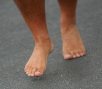
This is a non-pathologic cross over gait in my mind until proven otherwise, there may be other sources, causes and components, but when it quacks like a duck you’d be silly not to check for webbed feet. This runner even confirmed upon questioning that the left foot scuffs the inside of the right ankle/shin often, both sides scuff in fact but more left shoe on right shin. No Einsteinian epiphany there. After all, the thigh adduction on the left is what gives the foot posturing appearance, but it is likely driven by poor stabilization on the stance side leg (the right):

This means a narrow swing through (adducting) left limb.
This means stance and swing phase gluteus medius communication problems.
This means swing leg foot targeting problems.
This often suggests right, but sometimes both right and left, frontal plane pelvis sway problems which means pelvis control is challenged which means core lumbar stability control is challenged.
This means adaptive arm swing changes from the clean norm.
This does NOT mean this runner has pain, or pain yet, or maybe never will have pain but there are many determinants of that which I will discuss below.

But, make no mistake, this is flawed gait mechanics. The left swing leg is clearly targeting a more medial placement, meaning limb adduction (active or passive or both is to be determined) and this is a product of the cross over gait (unfamiliar with the cross over gait ? SEARCH our blog for the term, you will need a few hours of free time to get through it all). Some would call the cross over gait a lazy gait, but I would rather term it an efficient gait taken too far that it has now become a liability, a liability in which they can no longer stabilize frontal plane sway/drift. A wider gait on the other hand, as in most sprinters, is less efficient but may procure more power and the wider base is more stable affording less frontal plane drift. Just go walk around your home and move from a very narrow line walking gait to a wide gait and you will feel a more powerful engagement of the glutes. Mind you, this is not a fix for cross over gaits, gosh, if it was only that simple !

This runner must investigate whether there is right frontal plane drift, and if it is in fact occurring, find the source of the drift. It can come from many places on either limb. (This client says they are scuffing both inside ankles, which is not atypical and so we likely have drift on both right and left). We have discussed many of them here in various places on the blog over the years. Now as for “Why” the foot looks in toed, well that can also come from many places. Quite simply the adducted limb once it leaves toe off can look like this. But, perhaps it is also a product of insufficient external rotation maintenance occurred during that left stance phase, affording more internal rotation which is being unchecked and observed here during early swing. Remember though, if this is in fact a cross over gait result, in this gait the limb approaches the ground unstacked (foot is too far inside a left hip joint plumb line) the foot will greet the ground at a far lateral strike and in supination. Pronation will thus be magnified and accelerated, if there is enough time before toe off. However, and you can try this on your own by walking around your home, put yourself in terminal stance at toe off. Make sure you have the foot inverted so you are toeing off the lateral toes (low gear toe off). Does this foot not look like the one in the photo ? Yes it does, now just lift the foot off the ground and you have reproduced this photo. And when combined with a right pelvis drift, the foot will sneak further medially appearing postured behind the right foot.

Keep this in mind as well, final pronation and efficient hallux (big toe) toe off does often not occur in someone who strikes the ground on a far lateral foot. I am sure this runner will now be aware of how poorly they toe off of the big toe, the hallux. They will tend to progress towards low gear toe off, off the lesser toes. This leaves the foot inverted and this is what you are seeing in her the photo above. That is a foot that is inverted and supinated and it carried through all the way through toe off and into early swing. It is a frequently component of the cross over gait, look for it, you will find it, often.

Final thoughts, certainly this can be an isolated left swing phase gluteus medius weakness enabling an adducted swing limb thus procuring a faulty medial foot placement, but it is still part of the cross over phenomenon. Most things when it comes to a linked human frame do not work in isolation. But i will leave you with a complicating factor and hopefully you will realize that gait analysis truly does require a physical exam, and without it you could be missing the big picture problem. What if she has a notable fixed anatomic internal tibia torsion on that left side. Yup, it could all be that simple, and that is not something you can fix, you learn to manage that one as a runner.

* Side bar rant: Look at any google search of runners photos and you will see this type of swing limb foot posturing often, far too often. And yes, you can take the stance that “I do it as well and i have no injuries or problems so what is the big deal?”. Our response is often “you do have an issue, it may be anatomic or functional, but you do have an asymmetrical gait and you think it is not a problem, YET”. And maybe you will run till you are 6 feet under and not have a problem because you have accomodated over many years and you are a great compensator, yes, some people get lucky. Some people also do not run enough miles that these issues express themselves clinically so lets be fair. But some of these people are reality deniers and spend their life buying the newest brace or gadget, trying a different shoe insert, orthotic or new shoe of the month and shop over and over again for another video gait analysis expert who can actually fix their pain or problem. And then there are those who have a 45 minute home exercise program that they need to do to keep their problems at bay, managing, not fixing anything. Or, they spend an hour a week on the web reading article after article on what are the top 4 exercises for iliotibial band syndrome for example. They shop for the newest Graston practitioner, the newest kinesio taping pattern, Voodoo bands, breathing patterns, compression socks etc. And sometimes they are the ones that say they still dont have a problem.You get the drift. Gosh darn it, find someone who knows what the hell they are doing and can help you fix the issues that are causing the problem. And yes, some of the above accoutrements may be assistive in that journey.

I have dealt with this unique toe off issue way too many times not to roll my eyes at it any longer. It is to the point that it is an automated evaluation and solution program that begins to run in my head. Once you see something enough times, you learn all of the variations and subtle nuiances that a problem can take on. But, trying to fit everyone into a similar solution model is where the novice coach, trainer or clinician will get into trouble. Trust us, it all starts with an examination, a true clinical physical examination. If one leaves the investigatory process to a series of screens or functional movement patterns, “activation” attempts, digital gait analysis or strength tests one is juggling chainsaws and the outcome you want is often not likely to occur. There is nothing wrong with making these components part of the investigation process, but on their own, they are not enough to get the honest answer many times. Of course, Ivo and i were not able to jump the pond and examine this runner with our own eyes and hands so today’s dialogue was merely to offer this runner some food for thought to open their mind to our thought process, in the hopes that they can find someone to help them solve the underlying problem and not merely make the gait look cleaner. Making someone’s walking or running gait look cleaner is not hard, but making it subconsciously competent and clean (without thought or effort) requires a fix to the underlying problem. We can ALMOST guarantee you that the solution here to this runner’s form issue is not wholly at the foot that looks in toed and slanted. Merely working on their foot strike would be so remedial and corrupt that it would a crime.

Dr. Shawn Allen, one of the gait guys

This is a Patreon subscriber VIDEO post.

Patreon members, click the link below to access your video/audio/article.
https://www.patreon.com/thegaitguys

Screen Shot 2018-12-04 at 2.52.16 PM.png

Do you also want access to this content, PLUS MUCH MORE?
When you make even the smallest (1$/month) donation to our Patreon Page, you will get access to some of these private content posts.

Any tier ($1, $5, $10, $20, $50) will receive exclusive content not shared anywhere else. The higher your tier, the juicier the content.
Help support The Gait Guys mission to continue to produce high level content that you have grown accustomed to here.

Click the link to get over to the PATREON page.

https://www.patreon.com/thegaitguys

Patrons........ please comment here or on patreon and let us know if this was helpful !

Whoa! Dangerous shoes ahead....

IMG_5734.jpg
IMG_5736.jpg
IMG_5735.jpg

Holy smokes ! Can you believe this?

Take a look at these BRAND NEW, just out of the box pair of Brooks Cadence shoes. We do not usually see many manufacturer defects from this brand. Looks like someone might have been asleep at the “upper goes on the midsole” machine

Check out the varus cant to the rearfoot of the right shoe. Now look at the forefoot valgus cant to the left shoe. This would not be a great shoe for someone who has too much rear foot eversion and midfoot pronation on the right and and uncompensated forefoot valgus on the left, but we do not think it was designed for that specific, small niche market.

Think of the biomechanical implications on a "neutral" foot. Placing the right rear foot in varus would effectively halt or slow pronation in the rear foot and midfoot of that foot. This could be a good thing for an over pronator but, in a neutral foot, this would cause them to toe off in supination on that side resulting in low gear push off and biomechanical insufficiency, not to mention the increased external rotation of the lower extremity and lack of shock absorption from 1 of the 4 mechanisms of shock absorption left (mid foot pronation, ankle dorsiflexion, knee flexion, thumb flexion, contralateral drop of the pelvis). Now, imagine if that same person had internal tibial torsion. Talk about placing the knee outside of the sagittal plane ! Can you say macerated meniscus?

And now the left shoe. Look at the valgus cant! If you had and uncompensated forefoot valgus, where the forefoot is everted with respect to the rear foot or a forefoot varus, where they had adequate range of motion to allow the first ray to descend, then this could be a good thing, otherwise they are toeing off in too much pronation. This could be a real problem for a midfoot pronator or someone with large amounts of external tibial torsion, because they commonly toe off in too much pronation and low gear to begin with, as this shoe would accelerate pronation from midfoot to the forefoot

The bottom line? Look at your patients/clients shoes, as well as your own before purchasing them and examined for manufacturer defects. The upper should sit squarely on the midsole and the shoe should not rock or tip from side to side.

TGG

The Sartorius: insertional tendinitis and medial knee pain?

We all see folks with medial knee pain, many times women, with the pain located just below the medial tibial plateau. It often results from running, but sometimes with jumping sports like basketball as well. It has been our experience that these people are often diagnosed with an MCL type injury, but when you examine them further, they do not really fit the bill. All the ligaments are stable and there is no tenderness at the joint line. The is often tenderness at the pes anserine, but who is driving the bus here?

image source: https://commons.wikimedia.org/wiki/File:Muscles_and_tendons_of_the_legs_and_feet;_écorch_́figur_Wellcome_V0008276.jpg

image source: https://commons.wikimedia.org/wiki/File:Muscles_and_tendons_of_the_legs_and_feet;_écorch_́figur_Wellcome_V0008276.jpg

The sartorius originates from the anterior compartment of the thigh. During an ideal gait cycle, the sartorius fires from toe off through nearly terminal swing (1)

We remember that the abdominals should initiate thigh flexion with the iliopsoas, rectus femoris, tensor fascia lata and sartorius perpetuating the motion. Sometimes, when the abdominals are insufficient, we will substitute other thigh flexors, often the psoas and/or rectus femoris, but sometimes sartorius, especially in people with excessive midfoot pronation. Think about all of the medial rotation occurring at the knee during excessive midfoot pronation and when overpronation occurs, the extra compensatory external rotation that must occur to try and bring the knee back into the sagittal plane. The sartorius is positioned perfectly for this function, along with the semitendinosus which assists and external rotation in closed chain. This is why it is often implicated as the culprit in many cases of pes anserine bursitis (or as we like to say “sartorius insertional tendinitis” (2-3)

Some other things you may find interesting is that it is utilized more in crossing or cutting maneuvers while changing directions while running (4). This makes sense, given its anatomical course and origin/insertion. It can often be overlooked in adductor strains. It can also be avulsed during sprints, particularly in adolescents (5) and because of the course of the lateral femoral cutaneus nerve beneath it, can be the cause of meralgia paresthetica (6). It is proprotionally smaller in females (along with the gracilis and short head of the biceps femoris) (7). And during vertical jumping, is considered an internal rotator, along with the semimembranosis, semitendinosis, gracilis, and popliteus (8).

The sartorius is superficial in the anterior thigh, just under the skin, running from the ASIS, coursing lateral to medial and inserting at the pes anserine at its most superior aspect, just overlying the gracilis. Since it is an external rotator, knee flexor and assists in thigh abduction, you can easliy locate it by placing the patient in a "figure 4" position and having them resist as you pull downward on the leg. Be careful if you are needling this muscle because of the subsartorial canal (ie Hunters canal) lying just beneath it in the middle 1/3 of the thigh, from the apex of the femoral triangle to the adductor hiatus in the adductor magnus. It houses the femoral artery and vein, as well as the saphenous nerve and nerve to the vastus medialis.


  1. Michaud T: in Human Locomotion: The Conservative Management of Gait-Related Disorders 2011

  2. Imani F, Rahimzadeh P, Abolhasan Gharehdag F, Faiz SH. Sonoanatomic variation of pes anserine bursa. Korean J Pain. 2013;26(3):249-54. 

  3. Gupta, Aman & Saraf, Abhinesh & Yadav, Chandrajeet. (2013). ISSN 2347-954X (Print) High-Resolution Ultrasonography in PesAnserinus Bursitis: Case Report and Literature Review. 1. 753-757. 

  4. Rand MK, Ohtsuki T. EMG analysis of lower limb muscles in humans during quick change in running directions. Gait Posture. 2000 Oct;12(2):169-83.

  5. Manning CJ, Singhai S, Marshall P. Synchronised sartorius avulsions in adolescent sprinter. BMJ Case Rep. 2016 Jul 13;2016.

  6. Hsu CY, Wu CM, Lin SW, Cheng KL. Anterior superior iliac spine avulsion fracture presenting as meralgia paraesthetica in an adolescent sprinter. J Rehabil Med. 2014 Feb;46(2):188-90. doi: 10.2340/16501977-1247.

  7. Behan FP, Maden-Wilkinson TM, Pain MTG, Folland JP. Sex differences in muscle morphology of the knee flexors and knee extensors. PLoS One. 2018 Jan 23;13(1):e0190903.

  8. Cleather DJ. An important role of the biarticular hamstrings is to exert internal/external rotation moments on the tibia during vertical jumping. J Theor Biol. 2018 Oct 14;455:101-108

The gastroc can causse ankle dorsi and plantarflexion ? Yup. What ?

The gastroc, does it cause ankle dorsiflexion and ankle plantarflexion ? Yup. What ?

You may think you know the answer, the gastrocs are ankle plantarflexors, because that is the easy one we all recognize. But I stew on things when unique cases come in and do not fit the "normal" models and it got me reviewing principles I need to always keep in mind.

Think about it, the gastroc cross the knee, so it causes knee flexion. And when the knee flexes, the proximal tibia is progressing forward in the sagittal plane. Now remember, the foot is on the ground, so the distal tibia is (relatively) fixated in relation to the upper tibia. So, as this proximal top tibial moves forward, because of gastroc contraction, the muscle is actually causing ankle dorsiflexion !

So, it is it important to know your normal gait cycle events ? Yes, Ivo and i harp on that all the time ! One has to know the normal cycles to know when abnormal gait cycles are presenting clues.
So, am I saying that the gastroc are helpers of ankle rocker and ankle dorsiflexion ? Yes, they can be. It is a timing thing. So, we have to again get out of our model of open chain events, and thinking that only the anterior compartment muscles are ankle dorsiflexors. We also have to remember that a bent knee heel raise is not the same as a straight leg (knee extension) heel raise. One can stimulate and assist in ankle dorsiflexion and the other cannot so much. So, in clients with loss of ankle dorsiflexion/ankle rocker should you be assessing the function of the gastroc at the proximal knee, for its effects of dorsiflexion at the ankle ? Yes. Go ahead and try it, bend knee and straight knee heel raises, they are different beasts. This gets more complicated, and i will go into that next week ! I have had some deeper epiphanies i wish to share.
Also, remember, single and biarticular muscles have varied and vast capabilities. Thus it is always vital to consider whole body movements where muscles have abilities to accelerate, decelerate, and control and stablize joints they span, and do not span, via dynamic coupling.
Dr. Allen

Why is that joint range of motion absent? Here are some thoughts.

Photo courtesy of Pixabay.com

Photo courtesy of Pixabay.com

Is this how you think ? It is how we approach puzzles. . . .

Said client has a loss of internal hip rotation (pick any joint for that matter). . . . .

-is the loss of rotation present because they cannot get the rotation range because there is weakness of the internal rotators . . .

- or perhaps external rotators more dominant, combined with the weakness of the internal rotators

-or, is the loss there because of neuro-protective shortness/tightness because the brain feels that the said internal rotation is a vulnerable range (pain, instability), a range where it cannot protect the joint ?

-or, is it a combination of the above? (not to dismiss other processes of course, such as pelvis, knee or foot mechanical issues, OA, pain etc).

If one does not examine a client, how are they supposed to know this all important information?

*What shows up on a functional screen is merely what they are capable of doing/ recruiting/ engaging. It does not tell you why, nor narrow down the causal possibilities. Hence, driving more internal rotation range is silly, driving more strength into the internal rotator is likewise silly. And, merely adding global strength just might provide the overall presentation with more armor, a better coping strategy. Hence, strength first is not always a brilliant solution.

IF all you have is a hammer, everything is going to look like a nail, or you'll at least treat everything like a simple nail.

Whole-body coordination patterns may become partitioned in particular ways as a function of task requirements

Screen Shot 2018-12-01 at 11.06.21 AM.png


Toddlers actively reorganize their whole body coordination to maintain walking stability while carrying an object. Hsu WH1, Miranda DL2, Chistolini TL3, Goldfield EC4. Gait Posture. 2016 Oct;50:75-81


Today we seem to be going back to dual-tasking again, in this case utilizing the arms as balance assistance devices, amongst their other functions. However, we all know that walking with a hand in a pocket, or carrying something alters our ability to maximize their ballast-like function. Balanced walking involves freely swinging the limbs in pendullar motion. Changes in arm swing will change gait economy and efficiency. We have all run with a water bottle or bag/briefcase and know how that changes the symmetry and fluidity of our gait.

"Whole-body coordination patterns may become partitioned in particular ways as a function of task requirements.”

Today's research piece discusses toddlers and their function as they carry objects.
"children immediately begin to carry objects as soon as they can walk. One possibility for this early skill development is that whole body coordination during walking may be re-organized into loosely coupled collections of body parts, allowing children to use their arms to perform one function, while the legs perform another. Therefore, this study examines: 1) how carrying an object affects the coordination of the arms and legs during walking, and 2) if carrying an object influences stride length and width." -Hsu et al.
In this study of 10 toddlers with 3-12 months of walking experience were recruited to walk barefoot while carrying or not carrying a small toy.

"Stride length, width, speed, and continuous relative phase (CRP) of the hips and of the shoulders were compared between carrying conditions. While both arms and legs demonstrated destabilization and stabilization throughout the gait cycle, the arms showed a reduction in intra-subject coordination variability in response to carrying an object. Carrying an object may modify the function of the arms from swinging for balance to maintaining hold of an object. The observed period-dependent changes of the inter-limb coordination of the hips and of the shoulders also support this interpretation. Overall, these findings support the view that whole-body coordination patterns may become partitioned in particular ways as a function of task requirements." -Hsu et al.

So once again we will say it, if you are coaching the arm swing YOU want, because you do not like what you see in your client, or if you think you are helping your client get more out of their body in terms of speed, power, efficiency or anything of the sort, know that there is a higher, smarter program running the show. And that program in the client’s CNS is smarter than you when it comes to what they need for whole-body coordination pattern generation.

photo credit: courtesy of Pixabay

Skeletal muscle in running.

Footnotes 7 - White and Red.jpg

"skeletal muscle may provide a fundamentally different mechanical function during running than during activities such as swimming,flying, or jumping. These activities require muscle contractions that produce mechanical work at high rates to overcome drag,produce lift, or accelerate the body. Because running requires negligible work against drag and the average mechanical energy of the body is constant over time (at a steady speed on level ground), the demands of support may be met most economically by muscles that produce force while minimizing mechanical work."- Roberts et al


Muscular Force in Running Turkeys:
The Economy of Minimizing Work
Thomas J. Roberts,* Richard L. Marsh, Peter G. Weyand,
C. Richard Taylor

A great paper on Hallux Limits

Don't let the title fool or dissuade you. 

Here is a great paper to support the post earlier this week on hallux limitus. 

Plantar pressure distribution in older people with osteoarthritis of the first metatarsophalangeal joint (hallux limitus/rigidus)

No surprise that the study found folks with osteoarthritis of the the 1st MPJ had greater maximum force and peak pressures under the hallux as well as the lesser toe than controls. 

BUT here is one of the gems from the study: " However, the plantar pressure changes observed in this study can be explained using the concept of high- and low-gear push off described by Bojson-Moller. This model suggests that there are two metatarsal axes through which propulsion may occur; a transverse axis connecting the first to second metatarsal heads, and an oblique axis connecting the second to fifth metatarsal heads. In the presence of normal first MPJ motion, a ‘‘high-gear’’ push- off occurs through the transverse metatarsal axis, resulting in an efficient transfer of bodyweight. In the presence of restricted first MPJ motion, propulsion through the transverse axis is not possible. Subsequently, a ‘‘low-gear’’ push-off occurs through the oblique axis, which subjects the lateral forefoot and toes to increased loading and results in hyperextension of the interphalangeal joint of the hallux prior to toe-off."

Zammit, G. V., Menz, H. B., Munteanu, S. E. and Landorf, K. B. (2008), Plantar pressure distribution in older people with osteoarthritis of the first metatarsophalangeal joint (hallux limitus/rigidus). J. Orthop. Res., 26: 1665–1669. doi:10.1002/jor.20700.    

link to FREE FULL TEXT: http://onlinelibrary.wiley.com/doi/10.1002/jor.20700/epdf


When the boot is the cause of your client's problems/pain.

Our Patreon site is LIVE.
Patreon Sampler video,
Here's the kind of exclusive stuff you'll get as a Patreon sponsor, even at the 1$ and 5$ tier levels. Stuff right from our clinics, right when we see them, as fresh as content can get.
Of course, the 10$, 20$ and 50$ tier contributors will get this stuff too, but also "juicier" stuff , so if you don't wanna miss a step, come on over to our Patreon page and join even the lowest tiers, become part of a more intensive advanced focus group with ivo and I weekly ! (And in the process you'll be helping us keep new, advanced stuff coming your way).