About Toe Walkers...

Photo courtesy of Surestep

Photo courtesy of Surestep

Idiopathic Toe Walking in kids..Is it flexor dominance?

You see this at times in the office. Kiddos (or adults) who walk on their toes for no apparent reason. many have shortened heel cords with limited ankle dorsiflexion (1,2). Some studies report an incidence of 7-24% in pediatric populations (3) with an average of about 5% in children that are 5.5 years old (4). It seems to occur in about 2% of normally developing kids aged 5.5 years and 40% of those that have some sort of neuropsychiatric diagnosis or missed a developmental window (5-7), with an increased incidence familialy (8). The question here is why, not what.

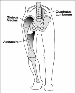
We have discussed our opinions of flexor dominance here many times and suffice it to say that increased corticospinal activity seems to have the double whammy effect of increased firing of the distal flexors due to a lack of input to the axial extensors in the rostral and caudal reticular formations respectively(possibly from decreased spindle and /or GTO input and/or mechanoreceptor dysafferentation?) and lack of reciprocal inhibition of the extensors from the increased firing of the flexors segmentally. Is it the cortical abnormailities and missed developmental windows seen in so many of these folks that drives this? These are the sorts of things that keep us up at night....

Physical treatment modalities (2) seem to help, we think most likely to plastic changes in the connective tissue. Orthotics may prove useful due to similar mechanisms, especially if there is an equinus deformity or forefoot to rearfoot abnormaility (9). More agressive (and invasive) measures like Botox, seem to not. An interesting study using whole body vibration (10) produced some immediate but short lived positive results. This really gets you thinking about joint and muscle mechanoreceptors and the cerebellum, and makes us think that perhaps we also should be looking (and treating) north of the foot. We could not find any studies looking at the effects of proprioceptive or vestibular exercises effects on this, but think it could be promising area of therapy and we will continue to employ them until our clinical results tell us otherwise. 


1. Barrow WJ, Jaworski M, Accardo PJ. Persistent toe walking in autism. J Child Neurol 2011;26(5):619-621

2. Harris NM. Multidisciplinary approach led to positive results for pediatric patient with idiopathic toe walking. Presented at the Association of Children’s Prosthetic-Orthotic Clinics Annual Meeting, Broomfield, CO, April 15, 2016.

3. Engelbert R, Gorter JW, Uiterwaal C, et al. Idiopathic toe-walking in children, adolescents and young adults: a matter of local or generalised stiffness? BMC Musculoskelet Disord. 2011;12:61.

4. Engström P, Tedroff K. The prevalence and course of idiopathic toe-walking in 5-year-old children. Pediatrics 2012;130(2):279-284.

5. https://tmblr.co/ZrRYjx1VV59rl

6. Williams, C. , Curtin, Wakefield and Nielsen Is idiopathic toe walking really idiopathic ? The motor skills and sensory processing abilities associated with idiopathic toe walking gait.  J Child Neurol 2014, 29:71

7. https://tmblr.co/ZrRYjx1WTNcdK

3. Is idiopathic toe walking really idiopathic ? The motor skills and sensory processing abilities associated with idiopathic toe walking gait.  J Child Neurol 2014, 29:71 Williams, C. , Curtin, Wakefield and Nielsen

8. Pomarino D, Ramirez Llamas J, Pomarino A. Idiopathic toe walking: tests and family predisposition. Foot Ankle Spec 2016;9(4):301-306

9. Herrin K, Geil M. A comparison of orthoses in the treatment of idiopathic toe walking: a randomized controlled trial. Prosthet Orthot Int 2016;40(2):262-269.

10. Williams CM, Michalitsis J, Murphy AT, et al. Whole-body vibration results in short-term improvement in the gait of children with idiopathic toe walking. J Child Neurol 2016;31(9):1143-1149.


Medieval "Turn Shoes": How we used to walk.

In the 1500's in Western Europe, shoes were different. People wore “turn shoes”, leather shoes that were made inside out then reversed for wear. This was likely the beginning of the use of molds to make shoes, carpenters up until the twentieth century would carve a wooden foot model of various sizes to model the process and standardize it.

These "Turn Shoes" were replaced by shoes with a frame construction as shoes changed to adapt to different environments, as streets changed.

The Turn shoes were basically a slip on or lace up thick leather sock. Thus, they were zero drop, soft, and provided much "feel" for the ground. Proprioception was obviously well appreciated.

We have spoken about the difference between heel strike and heel contact in walking. One can safely heel strike if barefoot on soft grass, but one cannot on the hard concrete or asphalt that we have covered much of our world with. Thus, if one were to wear "turn shoes" in our modern era, one would be forced to adapt to a heel contact or "heel kiss" on the ground, meaning, a more predominant forefoot loading style as described in this video.

What he describes, is largely not a choice, it was because they were in soft thin leather sock all day long, and even on wood or hard dirt packed floors and cart paths all day long, the heels would want some reprieve from heel "strike".

Screen Shot 2017-10-08 at 10.45.45 AM.png

Another way around this, to reduce heel strike, is to do it more naturally, by shortening the step and stride lengths a little, by keeping the body mass over the foot strike. "Chi Running" and "Chi walking" are based off of this principle. By moving the body mass forward with the foot, one has to naturally reduce heel strike. If one lags the body mass behind the foot however, the foot moves out in front, and heel strike begins to naturally (or shall we say, unnaturally) out in front, more heavily. This is not exactly desirable, for many reason.  Yet, since most of our shoes have some form of heel lift (a heel to forefoot drop), particular dress shoes (yes, even men's dress shoes, see photo), and even many modern day running shoes, the heel is essentially made more prominent (the heel rise essentially makes the brain think our heel (calcaneus) that much longer. This makes it easier, yet undesirable, to heel "strike" first. Oh what we have done for fashion !

He gets a few things wrong in the video, in terms of "ease" of walking, but largely it is decently done. One has to be careful if they perch the foot out in front like he does in the slower demonstration, in a plantarflexed ankle and foot, one can easily begin to lock up the knees. We often see this in teenagers in flip-flops.

-Shawn and Ivo, the gait guys

Video Block
Double-click here to add a video by URL or embed code. Learn more

More on Fatigue... When are you examining your patients?

You have probably read our posts from a day or so ago about fatigue and running. If not, see here and here.   In addition to the articles cited, it was based on this article here.

86546852.jpg

So how many times do we se someone in the clinic who have a problem, but it is not apparent at the time of exam? You know the scenario "I get this low back pain at mile 10" or "My knee hurts on the bike at mile 50". Our questions are

"So, when are you examining your patient?".

  • Are they fresh out of the box 1st thing in the morning, before their work out because that  is when you had an opening?
  • Is it after a long day with a different workout under different circumstances?
  • Is it right about at the time they usually have the issue?

The correct answer is "C". We like to say "if we can reproduce the pain, we can most likely figure out what the problem is and can usually come up with a solution or a different compensation". 

See your people around the time of the injury. If they get pain at mile 19, then have them run 18 prior to their visit and have them finish up in the office. If the knee pain is at mile 50 on the bike, have them do the last 10 on the trainer under your supervision. People will often have great mechanics until they begin to crumple. Your job is to see them at their worst, or watch their function deteriorate real time and try and come up with a solution. 

This concept is used all the time in exercise and stress testing. Why don't we use it with other than cardiovascular evaluations? The question eludes us. We often call this "pre fatigue" and use its all the time. You should too. The factors that separate a good clinician from a great one is outcomes. Be all you can be...


The Gait Guys


Dores H, Mendes L, Ferreira A, Santos JF. Symptomatic Exercise-induced Intraventricular Gradient in Competitive Athlete. Arquivos Brasileiros de Cardiologia. 2017;109(1):87-89. doi:10.5935/abc.20170075. FREE FULL TEXT

Biffi AAmmirati FCaselli GFernando FCardinale MFaletra EMazzuca VVerdile LSantini M.Usefulness of transesophageal pacing during exercise for evaluating palpitations in top-level athletes. Am J Cardiol. 1993 Oct 15;72(12):922-6. FREE FULL TEXT: http://www.ajconline.org/article/0002-9149(93)91108-T/pdf


Fatiguing your way to your injury? Endurance Injuries, Part 2

Screen Shot 2017-10-05 at 5.33.31 AM.png

Yesterday we wrote about the importance of endurance acquisition in preventing injuries. It is not a coincidence that many injuries sneak up on athletes in the later part of a game or event. Fatigue can predispose us to the variables that sent up compensation and injury, not always of course, but often.  

We felt it would be worthy work to look into a few other journal articles to make our case, not that it truly needed to be hammered out further, but we like to hammer.

We discovered that novice runner's (1) trunk inclination increased and ankle eversion increased with fatigue. Furthermore, as fatigue increased, it was noted to be prominent in the hip external rotators and hip abductors (2). We have discussed this ad nauseam over the years. Failure in these areas impact one's ability to hold sufficient limb rotation to ensure clean sagittal knee mechanics.  Challenges in these motions also lead to faults in foot targeting.  When these abductors and external rotators fatigue or weaken, hip adduction can often occur leading to undesirable medial foot targeting, hence narrow step width and our favorite soap box topic, the cross over gait. These issues become pronounced at the end of the run according to the Dierks study. However, in the 2nd Dierks (3) study these findings were challenged, "uninjured runners normally experience small alterations in kinematics when running with typical levels of exertion". Similarly, in the García-Pinillos study, (5) no major form failures were noted in endurance athletes that pushed their limits in another type of failure test, the HIIT (high intensity interval) workout. Dierks (3) remarked that "It remains unknown how higher levels of exertion influence kinematics with joint timing and the association with running injuries, or how populations with running injuries respond to typical levels of exertion.". 

None the less, these are just two studies, and there are others to refute it. We do however, challenge this. But, this is easy to do, because all day long in our clinics we see and hear the cases where there is correlation, because the people seeking us out are in fact "symptomatic" patients and not uninjured runners, so it is easy to lean in one biased direction from our end. Though, it bodes the bigger question off of this following statement, "uninjured runners normally experience small alterations in kinematics when running with typical levels of exertion", as to whether in time, these small alterations might lead to a symptomatic state. One can easily theorize that it is in fact this time variable that eventually leads these small alterations towards bigger ones that might become symptomatic. After all, every avalanche starts with a single snowflake, no offense to the snowflakes out there.

Shawn Allen, the gait guys

References:

1.  J Sci Med Sport. 2014 Jul;17(4):419-24. doi: 10.1016/j.jsams.2013.05.013. Epub 2013 Jun 19.
Kinematic changes during running-induced fatigue and relations with core endurance in novice runners. Koblbauer IF1, van Schooten KS2, Verhagen EA3, van Dieën JH2.

2. J Orthop Sports Phys Ther. 2008 Aug;38(8):448-56. doi: 10.2519/jospt.2008.2490. Epub 2008 Aug 1.
Proximal and distal influences on hip and knee kinematics in runners with patellofemoral pain during a prolonged run. Dierks TA1, Manal KT, Hamill J, Davis IS.

3. J Biomech. 2010 Nov 16;43(15):2993-8. doi: 10.1016/j.jbiomech.2010.07.001. 
The effects of running in an exerted state on lower extremity kinematics and joint timing. Dierks TA1, Davis IS, Hamill J.

4. Gait Posture. 2014;40(1):82-6. doi: 10.1016/j.gaitpost.2014.02.014. Epub 2014 Mar 4. 
Do novice runners have weak hips and bad running form? Schmitz A1, Russo K1, Edwards L1, Noehren B2.

5. J Strength Cond Res. 2016 Oct;30(10):2907-17. Do Running Kinematic Characteristics Change over a Typical HIIT for Endurance Runners?
García-Pinillos F1, Soto-Hermoso VM, Latorre-Román PÁ.

Gait is "all encompassing"

Screen Shot 2017-09-27 at 1.12.58 PM.png

Last week we did a presentation on some very classic, yet challenging, gait video case presentations. This slide was a big piece of our presentation. 
We discussed that there are volitional and non-volitional movements that accompany the adequate and appropriate postural system control.
If you want to hurt your brain, read this paper. 
But in a nutshell what this paper says is that we have a constant switching between steady state cortical neuron discharge and and non-steady state discharge. For example, when we are on a flat road, no obstacles ahead of us, nothing but boring open road, the system sort of runs on an automated program, making limb movements calculated off of a normal unchallenged baseline. But, if there are roots, rocks, curbs, bikes to dodge, puddles to hurdle etc, the volitional and postural systems must change their operation, and alter limb movements based off of those postural systems as we pay attention, and negotiate the obstacles. There is this delicate symphony occurring between automated posture, calculated posture, rhythmic limb movements. In other words, there are volitional, reactionary and anticipatory plans and adjustments occurring in the background at all times.
But, make no mistake, bad, faulty, inefficient motor patterns can become automated if injuries are left, if they are left partially rehabed, if we teach our clients faulty patterns by overloading them and forcing adaptive patterns to inappropriate load or fatigue. These modifications occur deep in the CNS, much in the premotor cortices, and take into account body schema (their correct or distorted perception of where they are, or their limbs are, in space). Build strength or endurance on an altered schema, one that might be present from an old injury, and one will build strength and endurance where one does not want them to go. Properly training clients, offering corrective exercise and the like is far deeper that just asking your client to load and get stronger, unless you wish to assume that their limitations and compensations are unimportant. This takes us right back to the asymmetry debate, which we know so many love to dive into. Asymmetry is the norm of course, just don't be the person creating more of it for your client.

"Adaptive gait control requires constant recalibration of walking pattern to navigate different terrains and environments. For example, motor cortical neurons do not exhibit altered discharge during steady-state locomotion, but altered discharge occurs when the experimental animal has to overcome obstacles. Loops from the motor cortical areas to the basal ganglia and the cerebellum may contribute to this purpose (ie, contribute to accurate and adaptive movement control that requires volition, cognition, attention, and prediction). In contrast, cortical processing seems unnecessary during the automatic execution of locomotion. Rather, high-level processing may occur in the systems between the basal ganglia, cerebellum, and brainstem in the absence of conscious awareness. - TAKAKUSAKI , Neurophysiology of Gait: From the Spinal Cord to the Frontal Lobe. Movement Disorders, Vol. 28, No. 11, 2013


Endurance and Injuries

S.E.S. , in that order.
We have been preaching this mnemonic for a decade now here at TGG. Skill first, then endurance, then strength. In other words, first move correctly/well, then move often (build a robust amount of endurance on that skill that you can maintain it throughout your activity without losing the skilled movement without fatiguing), then add strength to this patterned movement. Then rinse and repeat; add a higher skill, add endurance, add strength. Rinse repeat.
We tell this one to our athletes, distance runners in particular, because it is no surprise that most injuries come in the later miles, when fatigue sets in, and compensations have to make up the difference if the run continues. This is necessary and protective, but the wise choice is to never exceed the fatigue, but always be inching the endurance forward.
The question is, do you know where your risk threshold lives ? When are you moving the safety meter past the safe zone and into the risk zone ? Your tightness or pain, if you are lucky, and paying attention, may be your "check engine light" moment, again, if you are paying attention. Never dismiss the benefit of a 2 minute walk in the later part of a long run when a symptom creeps in, it just might get you enough recovery to push out that last 3-4 miles with the symptoms shut down again. If you are lucky. Listen to your body, it is your job.

From the study below:
"In conclusion, NOVICE runners showed larger kinematic adjustments when exhausted than COMPETITIVE (distance) runners. This may affect their running performance and should be taken into account when assessing a runner's injury risk."

-Shawn Allen, one of the gait guys

Reference:     https://www.ncbi.nlm.nih.gov/pubmed/28730917

Sports Biomech. 2017 Jul 21:1-11. doi: 10.1080/14763141.2017.1347193. [Epub ahead of print] Novice runners show greater changes in kinematics with fatigue compared with competitive runners. Maas E1, De Bie J1, Vanfleteren R1, Hoogkamer W2, Vanwanseele B1.


Podcast 128: Usain Bolt, Plantaris Tears, Arm Swing

Podcast links:

http://traffic.libsyn.com/thegaitguys/pod_128final.mp3

http://thegaitguys.libsyn.com/podcast-128-usain-bolt-plantaris-tears-arm-swing

https://www.thegaitguys.com/podcasts/


Key Tagwords:

usain bolt, plantaris tear, plantaris, sole lifts, heel lift, leg length, short leg, heel drop, shoeque, symmetry, asymmetry, sprinters, scoliosis, tendinopathy, achilles, runners, marathons, running injuries, arm swing

Our Websites:
www.thegaitguys.com
summitchiroandrehab.com   doctorallen.co     shawnallen.net


Our website is all you need to remember. Everything you want, need and wish for is right there on the site.
Interested in our stuff ? Want to buy some of our lectures or our National Shoe Fit program? Click here (thegaitguys.com or thegaitguys.tumblr.com) and you will come to our websites. In the tabs, you will find tabs for STORE, SEMINARS, BOOK etc. We also lecture every 3rd Wednesday of the month on onlineCE.com. We have an extensive catalogued library of our courses there, you can take them any time for a nominal fee (~$20).
 
Our podcast is on iTunes, Soundcloud, and just about every other podcast harbor site, just google "the gait guys podcast", you will find us.
 
Show Notes:

Superficial plantar cutaneous sensation does not trigger barefoot running adaptations.

https://www.ncbi.nlm.nih.gov/pubmed/28728130

Arm swing
http://www.medicalnewstoday.com/articles/173680.php

Usain bolt
https://mobile.nytimes.com/2017/07/20/sports/olympics/usain-bolt-stride-speed.html?referer=

Plantaris tears
https://www.ncbi.nlm.nih.gov/pmc/articles/PMC1978447/

How can feet relate to golf swing?

This 52 year old right handed gentleman presented with pain at the thoracolumbar junction after playing golf. He noticed he had a limited amount of “back swing” and pain at the end of his “follow through”.

Take a look a these pix and think about why.

Full internal rotation

Full internal rotation

full external rotation

full external rotation

full internal rotation

full internal rotation

full external rotation

full external rotation

neutral

neutral

neutral

neutral

Hopefully, in addition to he having hairy and scarred legs (he is a contractor by trade), you noted the following

  • Top: note the normal internal rotation of the right hip; You need 4 degrees to walk normally and most folks have close to 40 degrees. He also has internal tibial torsion.
  • second picture: loss of external rotation of the right hip. Again, you need 4 degrees (from neutral) of external rotation of the hip to supinate and walk normally.
  • third picture: normal internal rotation of the left hip; internal tibial torsion
  • 4th picture: limited external rotation of the left hip, especially with respect ti the amount of internal rotation present; this is to a greater degree than the right
  • last 2 pictures: note the amount of tibial varum and tibial torsion. Yes, with this much varum, he has a forefoot varus.

The brain is wired so that it will (generally) not allow you to walk with your toes pointing in (pigeon toed), so you rotate them out to somewhat of a normal progression angle. If you have internal tibial torsion, this places the knees outside the saggital plane. (For more on tibial torsion, click here.) If you rotate your extremity outward, and already have a limited amount of range of motion available, you will take up some of that range of motion, making less available for normal physiological function. If the motion cannot occur at the knee or hip, it will usually occur at the next available joint cephalad, in this case the spine.

The lumbar spine has a limited amount of rotation available, ranging from 1.2-1.7 degrees per segment in a normal spine (1). This is generally less in degenerative conditions (2).

Place your feet on the ground with your feet pointing straight ahead. Now simulate a right handed golf swing, bending slightly at the waist androtating your body backward to the right. Now slowly swing and follow through from right to left. Note what happens to your hips: as you wind back to the right, the left hip is externally rotating and the right hip is internally rotating. As you follow through to the left, your right, your hip must externally rotate and your left hip must externally rotate. Can you see how his left hip is inhibiting his back swing and his right hip is limitinghis follow through? Can you see that because of his internal tibial torsion, he has already “used up” some of his external rotation range of motion?

If he does not have enough range of motion in the hip, where will it come from?

he will “borrow it” from a joint more north of the hip, in this case, his spine. More motion will occur at the thoracolumbar junction, since most likely (because of degenerative change) the most is available there; but you can only “borrow” so much before you need to “Pay it back”. In this case, he over rotated and injured the joint.

What did we do?

  • we treated the injured joint locally, with manipulation of the pathomechanical segments
  • we reduced inflammation and muscle spasm with acupuncture
  • we gave him some lumbar and throacolumbar stabilization exercises: founders exercise, extension holds, non tripod, cross crawl, pull ups
  • we gave him foot exercises to reduce his forefoot varus: tripod standing, EHB, lift-spread-reach
  • we had him externally rotate both feet (duck) when playing golf

The Gait Guys. Helping you to store up lots “in your bank” of foot and gait literacy, so you can help people when they need to “pay it back”, one case at a time.

(1) http://www.ncbi.nlm.nih.gov/pmc/articles/PMC2223353/

(2) http://www.ncbi.nlm.nih.gov/pmc/articles/PMC3705911/

Subtle clues. Helping someone around their anatomy

tumblr_n9jt5dtWlc1qhko2so1_1280.jpg
tumblr_n9jt5dtWlc1qhko2so3_1280.jpg

This patient comes in with low back pain of years duration, helped temporarily with manipulation and activity. Her exam is relatively benign, save for increased lumbar discomfort with axial compression in extension and extension combined with lateral bending. Believe it or not, her abdominal and gluteal muscles (yes, all of them) test strong (no, we couldn’t believe it either; she is extremely regular with her exercises). She has bilateral internal tibial torsion (ITT) and bilateral femoral retro torsion (FRT). She has a decreased progression angle of the feet during walking and the knees do not progress past midlilne. There is a loss of active ankle rocker with gait, but not on the exam table; same with hip extension. 

We know she has a sweater on which obscures things a bit, but this is what you have to work with. Look carefully at her posture from the side. The gravitational line should pass from the earlobe, through the shoulder, greater trochanter and through or just anterior to the lateral malleolus.

In the top picture, can you see how her pelvis is anterior to this line? Do you see how it gets worse when she lifts her hands over her head (yes, they are directly over head)? This can signify many things, but often indicates a lack of flexibility in the lumbar lordosis; in this case, she cannot extend her lumbar spine further so she translates her pelvis forward. Most folks should have enough range of motion from a neutral pelvis and enough stability to allow the movement to occur without a significant change. Go ahead, we know you are curious, go watch yourself do this in a mirror and see if YOU change.

Looking at the this picture, can you pick out that she has a genu valgus? Look at the hips and look at the tibial angle.

Did you note the progression angle (or lack of) in her feet? This is a common finding (but NOT pathognomonic) in patients with internal tibial torsion. Notice the forefoot adductus on the right foot?

So what do we think is going on?

  • ITT and FRT both limit the amount of internal rotation of the thigh and lower leg. Remember you NEED 4 degrees of each to walk normally. Most folks have significantly more
  • if you don’t have enough internal rotation of the lower extremity, you will need to “create” it. You can do this by extending the lumbar spine (bottom picture, right) or externally rotating the lower extremity
  • Since her ITT and FRT are bilateral, she flexes the pelvis and nutates the pelvis anteriorly.
  • the lumbar facet joints should only carry 20% of load
  • she is increasing the load and causing facet imbercation resulting in LBP.

What did we do?

  • taught her about neutral pelvic positioning, creating more ROM in the lumbar spine
  • had her consciously alter her progression angle of her foot on strike, to create more available ROM in internal rotation
  • encouraged her to wear neutral shoes
  • worked on helping her to create more ankle rocker and hip extension with active drills and exercise (ie gait rehabilitation); shuffle walks, Texas walk, toes up walking, etc

why didn’t we put her in an orthotic to externally rotate her lower extremity? Because with internal tibial torsion, this would move her knee outside the saggital plane and create a biomechanical conflict at the knee and possibly compromising her meniscus.

Cool case, eh? We thought so. Keep on learning so your brain keeps expanding. If you are not growing your brain, you are shrinking it!

The Gait Guys

Your Gait Changes when you text....

GettyImages-534572749web-57a3087d3df78c3276b9fc80-2.jpg

Does texting alter your gait? It sure seems to slow you down, and according to this study, alter firing patterns of muscles about your ankle. Perhaps you are trying to preserve ankle rocker and maintain stability? It is interesting that ankle dorsiflexion actually increased and plantar flexion decreased.

"Young adults showed, overall, small gait modifications that could be mainly ascribable to gait speed reduction and a modified body posture due to phone handling. We found no significant alterations of ankle and knee kinematics and a slightly delayed activation onset of the left gastrocnemius lateralis. However, we found an increased co-contraction of tibialis anterior and gastrocnemius lateralis, especially during mid-stance. Conversely, we found a reduced co-contraction during terminal stance."


link to FREE FULL TEXT: https://www.ncbi.nlm.nih.gov/pmc/articles/PMC4579642/

More Foot Rocker Pathology Clues.

Is ankle rocker normal and adequate or is it limited ?  Is it limited in early midstance or late midstance ? How about at Toe off?  Is it even possible to distinguish this ? Well, we are splitting hairs now but we do think that it is possible. It is important to understand the pathologies on either end of the foot that can impact premature ankle rocker. 

Look at the photo above. You can see the clinical hint in the toe wear that this runner may have a premature heel rise. However, this is not solid evidence that every time you see this you must assume pathologic ankle rocker. The question is obviously, what is the cause.

Considerations:

1- weak anterior compartment, which is quite often paired with the evil neuroprotective tight calf-achilles posterior complex to offer the necessary sagittal protection at the ankle mortise.  This will cause premature heel rise from a posterior foot aspect.

2- rigid acquired blocked ankle rocker from something like “Footballer’s ankle”. This will also cause premature heel rise from a relatively posterior foot aspect.

3- there are multiple reasons for late midstance ankle rocker pathology. The client could completely avoid the normal pronation/supination phase of gait because of pain anywhere in the foot. For example, they could have plantar fascial pain, sesamoiditis, a weak first ray complex from hallux vaglus, they could have a painful bunion, they could be avoiding the collapse of a forefoot varus. There are many reasons but any of them can impair the timely pronation-supination phase in attempting to gain a rigid lever foot to toe off the big toe-medial column in “high gear” fashion. And when this happens the preparatory late midstance phase of gait can be delayed or rushed causing them to move into premature heel rise for any one of several reasons.  Rolling off to the outside and off of the lesser toes creates premature heel rise.  

4- And now for one anterior aspect cause of premature heel rise. This is obviously past the midstance phase but it can also cause premature heel rise. Turf toe, Hallux rigidus/limitus or even the dreaded fake out, the often mysterious Functional Hallux limitus (FnHL) can cause the heel to come up just a little early if the client cannot get to the full big toe dorsiflexion range.  

We could go on and on and include other issues such as altered Hip Extension Patterning, loss of hip extension range of motion, weak glutes, or even loss of terminal knee extension (from things like an incompleted ACL rehab, Osteoarthritis etc) but these are things for another time. Lets stay in the foot today.

All of these causes, with their premature heel rise component, will rush the foot to the forefoot and likely create Metatarsal head plantar loading and could cause forces appropriate enough to create stress responses to the bone. This abrupt forefoot loading thrust will often cause a reactive hammer toe effect.  Quite often just looking at the resting nature of a clients toes while they are lying down will show the underlying increase in neuro-protective hammering pattern (increased long toe flexor and short toe extensor activity paired with shortness of the opposing pairs which we review here in this short video link).  The astute observer will also note the EVA foam compressing of the shoe’s foot bed, and will also note the distal displacement of the MET head fat pad rendering the MET head pressures even greater osseously. 

Premature ankle rocker and heel rise can occur for many reasons. It can occur from problems with the shoe, posterior foot, anterior foot, toe off, ankle mortise, knee, hip or even arm swing pathomechanics.  

When premature heel rise and impaired ankle rocker rushes us to the front of the foot we drive the front half of the shoe into the ground as the foot plantarflexion is imparted into the shoe.  The timing of the normal biomechanical events is off and the pressures are altered.  instead of rolling over the forefoot and front half of the shoe after our body has moved past the foot these forces are occurring more so as our body mass is still over the foot. And the shoe can show us clues as to the torture it has sustained, just like in this photo case.

You must know the normal biomechanical gait events if you are going to put together the clues of each runner’s clinical mystery.  If you do not know normal how will you know abnormal when you see it ? If all you know is what you know, how will you know when you see something you don’t know ?

Shawn and Ivo, The Gait Guys … .  stomping out the world’s pathologic gait mechanics one person at a time. 

When you text while walking, you may be a hazard to yourself and others

pedestrian-london.jpg

Watch where you step... We have trouble while dual tasking with visual, attentional deficits. In other words, you don't see stuff. That's probably one of the reasons they are trying to ban texting while crossing the street in Honolulu.

"The results revealed that the size of visual field and visual acuity demand were varied across the visual task conditions. Approximately half of the visual cues provided during texting while walking were not perceived as compared to the visual task only condition. The field of regard loss also increased with increased dual-task cost of mobile phone use. Dynamic walking stability, however, showed no significant differences between the conditions. Taken together, the results demonstrate that the loss of situational awareness is unavoidable and occurs simultaneously with decrements in concurrent task performance. The study indicates the importance of considering the nature of attentional resources for the studies in dual-task paradigm and may provide practical information to improve the safe use of mobile phones while walking."


Lim J, Chang SH, Lee J, Kim K. Effects of smartphone texting on the visual perception and dynamic walking stability. Exerc Rehabil. 2017 Feb 28;13(1):48-54. doi: 10.12965/jer.1732920.460. eCollection 2017 Feb.  link to full text: https://www.ncbi.nlm.nih.gov/pmc/articles/PMC5331999/

http://www.npr.org/sections/thetwo-way/2017/07/29/540140824/its-now-illegal-to-text-while-crossing-the-street-in-honolulu

Cannabis users walk differently.

Screen Shot 2017-09-07 at 9.11.05 AM.png

We all have experienced or viewed the alcohol impaired gait at some point in our lives, the sloppy malcoordinated limb and torso movements. There are some classic observable characteristics there that many of us are familiar with.  But what about cannabis gait ?

"The research from the University of South Australia, published in the journal Drug and Alcohol Dependence, found those who smoke cannabis tend to move their shoulders less and elbows more as they walk. The pilot study also found marijuana users swing their knees more quickly during walking. The differences in gait were small and found in people who smoked a light or moderate amount of cannabis. Some changes were so small it was impossible for a specialist to detect."

However, the thing we found interesting was the papers final question, as to whether the subtle gait changes over a longer period of time would increase or become more apparent.

Not insinuating that Mystic Mac is a user, but he sure does help us hit our "reduced antiphasic gait" home with a glorious demo !

*We have seen this variation in arm swing gait many times before. We have discussed numerous times that when there is a reduction in the normal shoulder and pelvic "girdle" counter rotations, the normal antiphasic gait that presents us with the clearly obvious opposite arm-leg swings, we lose the ability to tap into these oscillations that afford us this free arm and leg swing.  So, when these girdle rotations are reduced, the limb movement has to come from further down into the limb, from elbow movement, a sort of casting the lower arm forward from biceps and triceps activity and from a kicking forward of the lower leg from quadriceps activity instead of hip flexion-extension activity.

We have mentioned this reduction in the normal antiphasic gait many times previously in our arm swing articles. Particularly, the reduction in the amplitude of the separation in the shoulder-pelvic girdle oscillations in those with spine pain. The more the spine is "twisted and wrung out" by these opposite swings, the more spinal motor unit compression, which can increase spine pain. Just search our blog for "arm swing" (30+ articles on the topic there). Thus the question remains , why does cannabis cause this same reduction?

Gait affects everything, and everything seems to affect our gait.

http://www.9news.com.au/national/2017/09/01/15/25/marijuana-users-walk-differently-australian-study-claims

Calf strength, the medial foot tripod, and pain in the great toe

Screen Shot 2017-09-06 at 10.21.01 AM.png

It has become evident that this component, the proper function of the 1st ray complex, is overlooked in some of the clinical world. Hallux joint pain is a difficult one to diagnose and treat at times. The source of pain and dysfunction can seemingly come from anywhere, but the more one understands the complex mechanics of this joint and regionally associate joints, the better clinical results one will achieve.  

One thing that has become recurrently obvious upon the many outside professional referrals that come though my office is the imbalance and/or weakness or endurance impairments in the posterior mechanism in relation to a painful 1st metatarsophalangeal joint (MTP). When I say posterior mechanism I am referring to the gastrocnemius, soleus, peronei, long flexors, and tibialis posterior namely. 

And, let me be clear, putting a theraband under the 1st metatarsal, encouraging your client to drive greater downward purchase of the head of the 1st MET during simulated foot tripod loading, does not necessarily help your client if their 1st MET is slightly more dorsiflexed. Do not be fooled by the flashy rehab guru tricks out there, proper clean function is achieved, not forced. If you have not earned it, you do not own it. 

It is quite simple really. If one does not have balanced function, including skill (motor pattern), endurance or strength of plantarflexion of the ankle, one cannot properly posture the first metatarsal (1st MET) in plantarflexion to sufficiently alter the sesamoid posturing underneath the metatarsal head, to sufficiently engage the unique eccentric axis (and it's necessary shift) of the 1st MTP to enable ample clean hallux dorsiflexion. Furthermore, without all this,  one will not be able to anchor the medial foot tripod properly.  This can lead to pain, functional hallux limitus, hallux rigidus to name a few. And, let me be clear, putting a theraband under the 1st metatarsal, encouraging your client to drive greater purchase of the head of the 1st MET during foot loading, does not necessarily help your client if their 1st MET is slightly more dorsiflexed. Do not be fooled by the flashy rehab guru tricks out there gang, proper clean function is achieved, not forced. 

A simple example might be a runner who fatigues the posterior mechanism in a long run. As the calf fatigues, they lose ample heel rise, thus ample plantarflexion of the 1st MET, thus proper posturing and translation of the sesamoids, thus successful eccentric axis shift, and thus clean dorsiflexion of the 1st MTP joint.  A player in a jumping sport who has less than ample strength of the posterior mechanism can have much the same issue at the resultant toe.  These are just garden variety examples.  But, should be clear that ample skill, endurance and strength (S.E.S.), our favorite mnemonic, of the posterior mechanism is necessary for pain free, functional toe off in the gait cycle or in jumping mechanics. 

If you are not systematically testing for these S.E.S. issues in the posterior mechanism, you are likely missing a major component in the proper posturing of the ankle and foot and thus proper functioning of the first ray complex and thus enabling clean function at the 1st MTP joint.  

(Sidebar rant: My past personal problems at this great toe joint started when a fellow chiropractor pulled on my toe many moons ago, for some random reason. It was the proverbial,  axial distraction "adjustment". The cavitation was heard around the world (the saliva inducing "pop" that fools many into blissful success), and my problems began.  I had painful dysfunction for many years after that for some strange reason, something was damaged but I was too stubborn and stupid to fix my own foot. I eventually remedied the problem through diving deeper into the complex mechanics of this joint and regionally associated areas. For this very intimate reason, it is why I am not one to perform this maneuver or recommend it. If we can be smarter in our understanding, we can be wiser in our interventions. Besides, axial distraction of this joint is not normal function of this joint. If I had a soap box to stand on for this topic, I would tell people to stop doing HVLA manipulations to this joint, mobilizations are more than ample to elicit a joint range response or a neurologic mechanoreceptor response. The more you understand this profoundly complicated and interesting joint, the 1st MTP joint, the more you will understand how to help your client. But, what do I know, I am just a dumb chiropractor, right Joe Rogan :) 

- Shawn Allen, the gait guys

Caveat Emptor: Foot placement is a complex thing.

"Understanding why we place a foot where we do can be a choice, eventually a habit, of perceived stability, of compensation and we like to say "it is a sliding scale between liabilities and economy". If you want more running economy, go for a narrow step width, but realize you are wrestling with its underlying liabilities.  The key is, one must have enough durability on the loading response of a narrow step width (cross over gait) to fend off the liabilities to reap the rewards of the improved economy. Forgo this principle, and it is caveat emptor."- Dr. Shawn Allen

Today we wanted to revisit a few topics and start to tie them together so that readers can perhaps more deeply bring the study discussed here today, into a deeper thought process.

Screen Shot 2017-08-30 at 7.41.43 AM.png

We have discussed the topic of gluteal pain in chronic low back pain clients previously, when the 2015 Cooper article was published ahead of print. Well, it came out in print (Euro Spine J) in 2016 so we wanted to revisit it with some more global thoughts. Those links are below. 
Basically, the article said that people with low back pain often have “Gluteus medius weakness and gluteal muscle tenderness are common symptoms in people with chronic non-specific LBP.”  
 

As we mentioned in our blog post last year, commenting on the "ahead of print" article, "it is often more on the side of pelvic frontal plane drift. The abdominals and spinal stabilizers also often test weak on this same side. We often see compromise of hip rotation stability as well because , since the hip is relatively adducting (because the pelvis is undergoing repeated frontal plane drift, hence no hip abduction) there is often a component of cross over gait phenomenon which can threaten rotation stability of the lower limb (type “cross over gait” into the search box of our tumblr blog for a landslide of work we have written on that phenomenon)."

This brings to mind this brief (14minute excerpt) from an old podcast we did (#109b, link below) on foot targeting, pelvis frontal plane drift, glute weakness and cross over gait. We brought together several concepts in that 14 minute span and it was on the topic from the Rankin article (link below).

If one is treating clients one must put all these concepts together (one should also have a deep grasp of the principles in this video ). One cannot have tunnel vision, one must embrace the entire picture neuromechanically. Foot targeting, gluteus medius activity, frontal plane pelvis drift or sway, cross over gait parameters, limb torsional issues, foot types and many more must all come into play if you are to truly get to the bottom of your clients problems. The approach must look at the loading and movement patterns at the very least, from foot to pelvis.  We would argue one should not stop there however, take your evaluation all the way into arm swing, thoracopelvic canister stability and more.  
We have pounded sand on the cross over gait and arm swing and the like for almost a decade now. As far as we know, we introduced, and if not, at the very least were the ones that dove deep into the cross over gait and its issues, and all of the attributes and functional pathologic pieces that go with it. We feel that if you fully understand the 40+ articles we have written on the cross over gait and arm and leg swing you will take your client and athlete evaluation to another level.  Understanding unconscious foot targeting is key in our opinion. "Understanding why we place a foot where we do can be a choice, eventually a habit, of perceived stability, of compensation and we like to say "it is a sliding scale between liabilities and economy". If you want more running economy, go for a narrow step width, but realize you are wrestling with its underlying liabilities.  The key is, one must have enough durability on the loading response of a narrow step width (cross over gait) to fend off the liabilities to reap the rewards of the improved economy. Forgo this principle, and it is caveat emptor. "

Shawn & Ivo, the gait guys

https://thegaitguys.tumblr.com/post/149177564774/podcast-109b-shorts-the-gluteus-medius-during

A neuromechanical strategy for mediolateral foot placement in walking humans. Rankin BL
J Neurophysiol. 2014 Jul 15;112(2):374-83. doi: 10.1152/jn.00138.2014. Epub 2014 Apr 30. 

Eur Spine J. 2016 Apr;25(4):1258-65. doi: 10.1007/s00586-015-4027-6. Epub 2015 May 26.
Prevalence of gluteus medius weakness in people with chronic low back pain compared to healthy controls.
Cooper NA1,2, Scavo KM3, Strickland KJ3, Tipayamongkol N3, Nicholson JD4, Bewyer DC4, Sluka KA3.

http://www.ncbi.nlm.nih.gov/pubmed/26006705

QL and Patellofemoral Pain?

photo credit: https://www.t-nation.com/training/training-disasters

photo credit: https://www.t-nation.com/training/training-disasters

"Subjects with PFP(patello femoral pain) have a higher prevalence of MTrPs (Myofascial trigger points) in bilateral GMe (gluteus medius)) and QL (quadratus lumborum) muscles. They demonstrate less hip abduction strength compared with controls, but the TPPRT (trigger point pressure release therapy, AKA ischemic compression) did not result in an increase in hip abduction strength. "

It is not surprising that when the hip is involved, the knee will be involved. As Dr. Allen often likes to say "the knee is basically in joint between 2 ball and socket joints ".

The gluteus medius and quadratus lumborum, along with the adductors are coronal plane stabilizers of the pelvis. They both have rotational components to their function as well affecting the hip directly for the former and lumbar spine for the latter. You can see our other QL articles about this here and here.

It is not much of a stretch to imagine that dysfunction of these muscles could result in trigger points and/or dysfunction of the knee (or foot for that matter ) could cause trigger points in these muscles.

Here is an article (1) examining trigger points in the gluteus medius and quadratus lumborum which, if you are familiar with Porterfield and DeRosa's work (2), are intimately linked during gait. We found it interesting that skin nick compression did not increase hip abduction strength where we find dry needling and intramuscular therapy often do.

Don't overlook these muscles and this important relationship.


  1. Roach, Sean et al.Prevalence of Myofascial Trigger Points in the Hip in Patellofemoral Pain Archives of Physical Medicine and Rehabilitation , Volume 94 , Issue 3 , 522 - 526link to free full text article: http://www.archives-pmr.org/article/S0003-9993(12)01079-9/fulltexthttp://www.archives-pmr.org/article/S0003-9993(12)01079-9/fulltext

  2. J. Porterfield, C. DeRosa (Eds.) Mechanical low back pain. 2nd ed. WB Saunders, Philadelphia; 1991


Not quite the QL, but close....

Screen Shot 2017-08-28 at 10.13.40 AM.png

We all see folks with low back pain and gait abnormalities. It is active during single limb support during stance phase of gait on the contralateral side (along with the external oblique) to elevate the ilium. This is coupled with the ipsilateral anterior fibers of the gluteus medius and minimus pulling the iliac crest toward the stable femur.

We found this FREE FULL TEXT while doing some quadratus lumborum research. It reminds us about things like scleratogenous pain (pain arising from tissues of like embryological origin with a common nerve innervation, like tendon, bone, muscle, etc) and other triggers for low back pain. We have needled this ligament with good result. Remember that this is an individual ligament making up a portion of the middle layer of the thoracolumbar fascia, and is not an aponeurosis of the lumbocostal fibers of the quadratus lumborum.


https://www.ncbi.nlm.nih.gov/pmc/articles/PMC5226660/

photo from: https://musculoskeletalkey.com/treatment-of-the-patient-with-chronic-pain/

A return to "the Kickstand Effect". So your foot is turned out, externally rotated ?

Amputee War Veteran Sergeant Christopher Melendez Became a Pro WrestlerRead more at http://www.craveonline.com/mandatory/1053779-standing-tall-how-amputee-war-veteran-christopher-melendez-beca#XeD2LrZ2xmtXQ6um.99

Amputee War Veteran Sergeant Christopher Melendez Became a Pro Wrestler
Read more at http://www.craveonline.com/mandatory/1053779-standing-tall-how-amputee-war-veteran-christopher-melendez-beca#XeD2LrZ2xmtXQ6um.99

Why is my foot turned out ?  A 3rd return to the solitary externally rotated foot.

Below you will find our 2 prior articles on this topic, but this is a relatable concept to other thing which we have embedded in many of our blog posts and podcasts over the last decade of sharing what we know.

In the photo above the brave Army Veteran Sergeant Melendez one can see the concept brilliantly as he only has one limb.  One can see the concept in full play, he must balance his body mass over one point, not two like the rest of us lucky folk.  In trying to balance over one point, if the foot is straight forward (if one is blessed with close to neutral torsional bone alignment) one will have good stability in the sagittal plane (forward /back) but will be at risk to fall, drift or sway into the frontal plane. Here Sergeant Melendez displays the foot and limb turn out into the frontal plane so that he can use the quadriceps to help him protect into that frontal plane, plus, by situating his base posture in more of an externally rotated position (likely losing internal rotation capability over time, unless forcibly maintained through specific exercises) he can more fully and skillfully engage all 3 divisions of the gluteus maximus and medius, and perhaps hamstrings and adductors and who knows what else, to maintain a more stable and likely less fatiguable posture. Go ahead, try it for yourself, this is easier to balance and maintain that a straight sagittal foot posturing. The one trouble he might have, is not deviating too much, or too often, into a frontal plane drift hip-pelvis posture. This will put much aberrant compressive load onto the roof of the femoral head-acetabular interval, where most of us begin a degenerative hip arthritis journey, unfortunately. 

Side note:   So you might think your client has FAI ?  Maybe start here, our thinking might lead you done a helpful path to get started. Search our blog for FAI as well.

here are the 2 prior articles on the topic, with video.  Watch for this one, it is everywhere out in the world, walking amongst us.  
Thank you for your service Sergeant Melendez.  Here is the article written by K. Thor Jensen, on Crave Online.  

https://thegaitguys.tumblr.com/post/14262793786/gait-problem-the-solitary-externally-rotated

https://thegaitguys.tumblr.com/post/40617674450/a-return-to-the-solitary-externally-rotated-foot

Shawn & Ivo, The Gait Guys

What do we have here and what type of shoe would be appropriate?

You are looking at a person with a fore foot varus. This means that the fore foot (ie, plane of the metatarsal heads) is inverted with respect to the rear foot (ie, the calcaneus withe the subtalar joint in neutral). Functionally translated, this means that they will have difficulties stabilizing the medial tripod (1st MET head) to the ground making the forefoot and arch unstable and likely rendering the rate and degree of pronation increased.

The incidence of this condition is 8% of 116 female subjects (McPoil et al, 1988) and 86% of 120 male and female subjects (Garbalosa et al, 1994), so it seems to happen happen more in males. We think this second number is inflated and those folks actually had a forefoot supinatus, which is much more common.

Fore foot varus occurs in 3 flavors:

  • compensated
  • uncompensated
  • partially compensated

What is meant by compensated, is that the individual is able to get the head of the 1st ray to the ground completely (compensated), partially, or, when not at all, uncompensated.What this means from a gait perspective ( for partially and uncompensated conditions) is that the person will pronate through the fore foot to get the head of the 1st ray down and make the medial tripod of the foot (ie, they pronate through the subtalar joint to allow the 1st metatarsal to contact the ground). This causes the time from mid-stance to terminal stance to lengthen and will inhibit resupination of the foot. 

Today we are looking at a rigid, uncompensated forefoot varus, most likely from insufficient talar head derotation during fetal development and subsequent post natal development. They will not get to an effective foot tripod. They will collapse the whole foot medially. These people look like severely flat-footed hyperpronators.


So, what do you do and what type of shoe is appropriate? Here’s what we did:

  • try and get the 1st ray to descend as much as possible with exercises for the extensor hallucis brevis and short flexors of the toes (see our videos on youtube)
  • create more motion in the foot with manipulation, massage mobilization to optimize what is available
  • strengthen the intrinsic muscles of the feet (particularly the interossei)
  • increase strength of the gluteus maximus and posterior fibers of the gluteus medius to slow internal rotation of the leg during initial contact to midstance
  • put them in a flexible shoe for the 1st part of the day, to exercise the feet and a more supportive; medially posted (ideally fore foot posted) shoe for the latter part of the day as the foot fatigues
  • monitor his progress at 3-6 month intervals
  • a rigid orthotic will likely not help this client and they will find it terribly uncomfortable because this is a RIGID deformity for the most part (the foot will not accommodate well to a corrective orthotic. Besides, the correction really has to be made at the forefoot anyways. 

Lost? Having trouble with all these terms and nomenclature? Take our national shoe fit program, available by clicking here.

The Gait Guys. Uber foot geeks. Separating the wheat from the chaff, with each and every post.

So you prescribe and fit orthotics you say ?

"It all matters, and quite possibly, if you do not know it all, you cannot help your client."

How about this then, you have someone with a rearfoot valgus with internal tibial torsion.  How are they going to load now? What if you throw in a valgus knee and femoral torsion variant?  Are they going to pronate more or less ? What if that person had just internal tibial torsion on one leg and not the other, yet they had 2 rearfoot valgus feet presentations.  Now what?

Ouch, that is a strong statement. It likely needs softened, but, there is some truth within those words. 

Last night we did our monthly lecture on www.onlineCE.com.  We had a packed room, biggest audience to date.  It is likely because people are realizing that the small stuff matters.  We talked for an hour on foot types and  how they present, how they potentially load, and how other mechanical issues above can impact how a foot type loads. 

We have all seen the pedographs like in the photo. The unwise depend on a static pedograph mapping for diagnostic help and God forbid that is all you use for making orthotics (that may only help if your client is  a professional stander), the more wise use the dynamic pedograph mapping to see how their client moves across the ground, and the wise use it as a mere piece of the data, combine it with a clinical exam, look far up into the biomechanical chain for other locomotive challenges that could change the dynamic loading pattern across the foot and ground.  What do we mean exactly ?  Well, a client with a rearfoot valgus foot type will load the heel and rest of the foot one way if they are doing a good job stacking the hip over the knee, and knee over the foot. But, if they have weakness in the hip affording a frontal plane drift of the pelvis over the foot, that is going to magnify the rearfoot valgus loading pattern (addendum: they could also tip into rearfoot varus posturing as well). That is just one example, of many.  In otherwords, it is the same foot type, but both of these are going to show a dynamic change in the loading pattern response. So, said another way, you cannot diagnose a foot type by the pedograph mapping. Nor should one make an orthotic for someone based off of a pedograph mapping, nor without an examination of the entire kinetic change.  What is your client able, and unable, to do? That is a big question, and when you start by asking those 2 questions, you get closer to the prize.  The pedograph only shows the static or dynamic pressures from the superincumbent load, it does not tell you if it is good or bad, and it does not tell you what they are doing, or why they are loading that way. It only shows the loading. Your job is to find out why they are loading that way, and then determine if that is part of their problem they have sought you out for.

So, does  your head spin now ? Does this suddenly make you sweat ? Do you realize you are missing pieces of the pie in helping your client?  Not yet maybe ?  How about this then, you have someone with a rearfoot valgus with internal tibial torsion. How are they going to load now? What if you throw in a valgus knee and femoral torsion variant? Are they going to pronate more or less ? What if that person had just internal tibial torsion on one leg and not the other, yet they had two rearfoot valgus feet presentations. Now what? Suddenly the loading is different in both feet and up the chains. There is likely going to be different challenges to limb spin control from side to side. This aberrant and asymmetrical loading is going to come up to a pelvis, upon which a single spinal column is trying to find a sound base of support and mobility to work and transfer loads from. 

And, what if this client also has some tibial varum on that same side ? What if they had external tibial torsion or some femoral torsional presentation on one side ?  You can see now how complicated this gets. And that is just on the structural components. What about the dynamic components ?  We here at The Gait Guys feel that this is all critical stuff to take into consideration and it is sometimes the stuff that is the tipping point between a successful management of a clients complaints, and unsuccessful.  

In closing, think about this. If you are sending out your orthotics for fabrication, have you conveyed this all to your fabricator ?  All they know is what a pedograph might show, and what the foot mold looks like. You have to provide them with all this other information, because essentially they are blind (this of course assumes your fabricator can mind juggle all the torsions, valgus/varus, pelvis drift loads etc,  oy vey ! That is hard to do !) This is why we do all of our modifications in office, in the rare case we need a temporary orthotic modification. But, we will aim to just correct what mechanics are aberrant and avoid the whole orthotic crutch when we are able. But lets face it, sometimes, for a period of time, we all need a crutch to get through a problem, to find better mechanics where we can strengthen from or gain protecting from temporarily.  That is what splints do, taping, crutches, braces, one might even argue what corrective exercises do. It is a path on the journey for your client, and sometimes they need help through the muddy parts.

And, don't be "that guy" that says orthotics are useless. They are a crutch , a tool. A small tool, one might argue that it should only be pulled out when the other tools are not working to get the job done.  Do not make them your first line of defense, except when that is called for.  After all, not all people were blessed with sufficient anatomical  and mechanical parts to avoid needing a crutch, so don't be "that guy" that preaches from that extreme, because it is not honest. Or, maybe, you just do not see the biomechanical messes we see in our clinics, that is quite the realistic possibility. 

Want to learn more about this kind of stuff? Keep up with our blog here. OR take some of our lecture recorded classes on www.onlineCE.com . We have a library of classes there for you to take anytime. And meet us once a month over there, every 3rd Wednesday. And, stay tuned for some new teaching gigs we have coming your way.

-Shawn and Ivo,  the gait guys