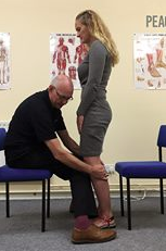
Proprioceptive Clues in Children’s Gait.

This goes along with Mondays post. We can learn a lot about gait from watching our children walk. An immature nervous system is very similar to one which is compensating meaning cheating around a more proper and desirable movement pattern; we often resort to a more primitive state when challenges beyond our ability are presented. This is very common when we lose some aspect of proprioception, particularly from some peripheral joint or muscle, which in turn, leads to a loss of cerebellar input (and thus cerebellar function). Remember, the cerebellum is a temporal pattern generating center so a loss of cerebellar sensory input leads to poor pattern generation output. Watch this clip several times and then try and note each of the following:

  • wide based gait; this is because proprioception is still developing (joint and muscle mechanoreceptors and of course, the spino cerebellar pathways and motor cortex)
  • increased progression angle of the feet: this again is to try and retain stability. External rotation allows them to access a greater portion of the glute max and the frontal plane (engaging an additional plane is always more stable).
  • shortened step length; this keeps the center of gravity close to the body and makes corrections for errors that much easier (remember our myelopathy case from last week ? LINK. This immature DEVELOPING system is very much like a mature system that is REGRESSING. This is a paramount learning point !)
  • decreased speed of movement; this allows more time to process proprioceptive clues, creating accuracy of motion


Remember that Crosby, Still, Nash and young song “Teach Your Children”? It is more like, “teach your parents”…


Proprioceptive clues are an important aspect of gait analysis, in both the young and old, especially since we tend to revert back to an earlier phase of development when we have an injury or dysfunction.


What did you notice? The Devil is in the details...

 Cavus foot? Loss of the transverse arch? Prominence of extensor tendons?

The question is: Why?

It’s about reciprocal inhibition. The concept, though observed in the 19th century, was not fully understood and accepted until it earned a Nobel prize for its creditor, Sir Charles Sherrington, in 1932. Simply put, when a muscle contracts, its antagonist is neurologically inhibited, So when your bicep contracts, your tricep is inhibited. This holds true whether you actively contract the muscle or if the muscle is irritated (causing contraction).

So how does this apply to this foot?

We see prominence of the extensor tendons (particularly the extensor digitorum brevis EDB; the longus would have caused extension at the distal interphalangeal joint). The belly of the muscle is visible, telling us that it is active. It is neurologically linked to the flexor digitorum brevis (FDB). This muscle, in turn, has slips which attach it to the abductor hallucis brevis (AHB) medially and the abductor digiti minimi (ADM) laterally. These muscles together form 2 triangles (to be discussed in another post) on the bottom of the foot, which lend to the stability of the foot and the arches, especially the transverse.

When the EDB fires, it inhibits the FDB, (which, in addition to flexing the MTP’s, assists in maintaining the arch). The EDB has an effect which drops the distal heads of the metatarsals as well (Hmm, think about all the people with met head pain) Now, look at the course of the tendons of the EDB. In a cavus foot, there is also a mild abductory moment, which flattens the arch. Conversely, the FDB in a cavus foot would serve to actually increase the arch, and would have a ,mild adductory moment. Net result? A flattened transverse arch.

Now look at the Flexor digitorum longus, overactive in tbis foot (as evidenced by the flexion of the distal interphalangeal joints, mild adduction of the toes (due to the change of direction of pull in a cavus foot) and lowering of the met heads due to hyperextesnion at the MTP joints ). This mm is reciprocally linked with the extensor digitorum longus. The prominence of the extensor tendons is do to increased activity of the EDB (go ahead, extend all your fingers and look at the tendons in your hand. Now flex the  DIP and IP joints and extend the MTP; see how they become more prominent?).

Reciprocal inhibition. It’s not just for dinner anymore…

We are and remain; The Gait Guys

Try THIS at home...

Screen Shot 2018-02-07 at 11.23.24 AM.png

Cool test, results you can see and some literature to back it up. If you are interested at all in proprioception, this is an interesting read.

So, the question for us is: "Does ankle dorsiflexion actually create more stability, like is purported?"

“The point is that if I make their ankle rigid this way, then they can more effectively use the balance mechanisms at their knees, hips, and proximally, because they’re on a stable base. My proposition is that their balance is actually normal, apart from that distal segment. When their ankle is stabilized, they use their knees more effectively, and they become less dependent on their eyesight to maintain their balance.”

http://lermagazine.com/article/afos-and-balance-issues-in-peripheral-neuropathy

More on Fatigue... When are you examining your patients?

You have probably read our posts from a day or so ago about fatigue and running. If not, see here and here.   In addition to the articles cited, it was based on this article here.

86546852.jpg

So how many times do we se someone in the clinic who have a problem, but it is not apparent at the time of exam? You know the scenario "I get this low back pain at mile 10" or "My knee hurts on the bike at mile 50". Our questions are

"So, when are you examining your patient?".

  • Are they fresh out of the box 1st thing in the morning, before their work out because that  is when you had an opening?
  • Is it after a long day with a different workout under different circumstances?
  • Is it right about at the time they usually have the issue?

The correct answer is "C". We like to say "if we can reproduce the pain, we can most likely figure out what the problem is and can usually come up with a solution or a different compensation". 

See your people around the time of the injury. If they get pain at mile 19, then have them run 18 prior to their visit and have them finish up in the office. If the knee pain is at mile 50 on the bike, have them do the last 10 on the trainer under your supervision. People will often have great mechanics until they begin to crumple. Your job is to see them at their worst, or watch their function deteriorate real time and try and come up with a solution. 

This concept is used all the time in exercise and stress testing. Why don't we use it with other than cardiovascular evaluations? The question eludes us. We often call this "pre fatigue" and use its all the time. You should too. The factors that separate a good clinician from a great one is outcomes. Be all you can be...


The Gait Guys


Dores H, Mendes L, Ferreira A, Santos JF. Symptomatic Exercise-induced Intraventricular Gradient in Competitive Athlete. Arquivos Brasileiros de Cardiologia. 2017;109(1):87-89. doi:10.5935/abc.20170075. FREE FULL TEXT

Biffi AAmmirati FCaselli GFernando FCardinale MFaletra EMazzuca VVerdile LSantini M.Usefulness of transesophageal pacing during exercise for evaluating palpitations in top-level athletes. Am J Cardiol. 1993 Oct 15;72(12):922-6. FREE FULL TEXT: http://www.ajconline.org/article/0002-9149(93)91108-T/pdf


tumblr_o586ok0qOo1qhko2so1_1280.jpg
tumblr_o586ok0qOo1qhko2so2_1280.jpg
tumblr_o586ok0qOo1qhko2so3_1280.jpg

Go ahead, take the shot.

This runner came in with ankle pain after running across the slope of the hill with the right foot uphill left foot down. She slipped on the ice and heard a pop. She presented to the office with minimal swelling, ankle pain on the right-hand side. Very little discoloration. She said that her ankle was “bent sideways” but reduced overtime as she crawled home to get help.

 She slipped on the ice and heard a pop. She presented to the office with minimal swelling, ankle pain on the right-hand side. Very little discoloration. She said that her ankle was “bent sideways” but reduced overtime as she crawled home to get help.

  The ankle was moderately swollen and tender at the medial and lateral malleoli with little gross deformity. She was not able to bear weight on that side without pain. We took the first picture (top) which didn’t look too bad. We could’ve stopped there thinking that it was just a bad sprain. But we didn't… We always take three views of an area so we don’t miss things. You can plainly see in the second and third views that she has involvement of the deltoid ligament as well as the more obvious distal fibula fracture.

We did some acupuncture to do reduce swelling at the patient’s request and contacted the orthopedists office for her, placed her in the mobilization brace and give her some crutches.

When in doubt, take the shot. It can make a huge difference clinically. 

Limitations: The powers of observation will help you.
Physical examination, FMS, DNS, gait analysis … . . these are all very important tools for the coach, trainer, therapist, clinician.  They will all offer information and lead the “th…

Limitations: The powers of observation will help you.

Physical examination, FMS, DNS, gait analysis … . . these are all very important tools for the coach, trainer, therapist, clinician.  They will all offer information and lead the “therapy giver” in a direction for intervention.  But when something doesn’t match up with the basic standard protocols, you have to go outside the standard box.  We have all been there and today is just a little reminder not to get caught up in the “proceedures” and merely running through protocol without an engaged brain putting the pieces together.  

Here we see 2 classic examples of deviations from the mean, the client on the left has drifted further outside the frontal plane because of tibial varum and a little genu varus.  The client on the right has imploded deep into the frontal plane via rigid pes planus foot collapse and genu valgum.  These will both affect your physical screenings for these clients. And keep in mind, and this is probably the most important point of today’s blog post, either client may have good or bad strategies around their anatomy.  In other words, some clients will have great compensations to limit further functional pathology, and some will have poor compensation strategies, and thus, both will have different physical exam findings, different screenings and different neuromotor patterns embedded deep into their CPGs (central pattern generators).   Put yet another way, all of the scenarios discussed may/will have varying screening assessment outcomes but for different reasons.  If you know the cause of these faults and the impaired neuro-recruitment patterns that are likely, your assessments will make more sense, and so will your exercise/therapy/rehab prescriptions.  If you do not understand the fundamental differences (ie long bone torsions or various femoral-neck shaft angles, foot types such as an uncompensated forefoot valgus etc) , one could prescribe therapies that will not address the underlying problems, rather they might address the compensations and strategies found with these client’s challenges.

It can get sloppy messy.  Wear a bib.

Dig for the roots, don’t mow the grass…… Shawn and Ivo, The Gait Guys

tumblr_mee0ffpHp51qhko2so1_1280.jpg
tumblr_mee0ffpHp51qhko2so2_r1_1280.jpg
tumblr_mee0ffpHp51qhko2so3_1280.jpg
tumblr_mee0ffpHp51qhko2so4_1280.jpg

“I’ll plead the 1st … ."   More foot geek stuff from The Gait Guys.

The 1st Ray that is!

The "1st ray” consists of the 1st metatarsal and the medial cunieform, essentially the long bones associated with the big toe. It is a functional unit we often refer to when discussing foot biomechanics.

You have heard us speak of the 1st ray needing to descend to form the medial tripod of the foot (tripod review: head of 1st metatarsal, head of 5th metetarsal, center of calcaneus). This action depends to some degree on the competency of the peroneus longus, which attaches from the upper lateral fibula and the associates interosseous membrane; curves around the lateral malleolus, crosses under the foot and attaches to the base of the 1st metatarsal and medial cunieform. The tibialis posterior is supportive to this action. This action is opposed (or modulated, for every Yin there is a Yang; it’s all about balance) is the tibialis anterior, which attaches to the top of the base of the 1st metatarsal and 1st cunieform.

As a result, 1st rays can be elevated or depressed. (here is a latin term to impress your friends with: Metatarsus Primus Elevatus, or elevation/dorsiflexion of the 1st ray/metatarsal). Clinically, we see more that are elevated, resulting in a faulty (collapsing) medial tripod of the foot. The important thing is isn’t necessarily its position, but rather its flexibility. The inflexible ones (isn’t it always?) are the problem children, because they result in altered (notice I didn’t say bad) biomechanics. The further we move from ideal, the closer we seem to move to some compensation pattern. The flexible ones are still a problem but we can control and dampen their rate of flexible collapse.

Generally speaking, a plantar flexed 1st ray that is rigid, has a tendency to throw your center of gravity (an often your knee) to the outside of the foot tripod (think of a rigid cavus foot) and a dorsiflexed to the inside of the foot tripod. Sure, there are LOTS of other factors, but we are talking in generalities here.

Look carefully at the images above and note the position of the 1st metatarsal heads. In the top set, the 1st is depressed (or plantarflexed). In the bottom set they are elevated (or dorsiflexed). Cool, eh? 

NOTE: please refrain from using the term “dropped metatarsal”. Nothing gets dropped, it is correctly stated as plantarflexed (rigid or flexible).

Be on the look out for these on your clinical exam.

Ivo and Shawn. Bringing you one step closer to foot geekdom each day!

copyright 2012 The Homunculus Group/The Gait Guys. All rights reserved. If you rip off our stuff, you will be plagued with the curse of Toelio…..

More on Leg Length Discrepancies

Hi Guys,

I hope you guys are well?

I have a question I hope you can help me with?

Last week I assessed an entire football team, and over 90% have some sort of Leg Length Discrepancy (LLD). I am working with the physiotherapist to improve their weaknesses, including using sole lifts.

My question is if it’s a tibial short leg, then a lift with align the knee and hip. But a lift in a leg with a short femur will align the pelvis but raise the knee higher than the other side. Would you still insert a sole raise, and if not, what would you do?

Kind Regards

Luke

____________

Hi Luke

Yes, you are correct in your assumption of the change in mechanics, and yes, most often, we prescribe a sole lift, if a lift is indicated. Keep in mind that if they are asymptomatic and test out well, a lift may not be indicated. Hope that helps. You can also search LLD on the blog; we have written extensively on it: http://thegaitguys.tumblr.com

Remember sole lifts will correct the LLD but it could shift the pelvis off further…….many LLDs are from pelvic asymmetry and core weakness, this encompasses hip rotation differences which is a typical response to the core and pelvis that is distorted. 
merely forcing a change at the Sole does not mean you are making the positive change at the top……however it may in some cases……you have to determine that with your evaluations.

Most folks legs are of symmetrical length……..the changes at the top (core / pelvis/ hip) is what throws the apparent length off.

i wish i had a good answer for your great insight……..but it is about
1- making the right changes……..so that all parts are in cooperation for the restoration change
2- that you are directing change and not a further body compensattion to the compensation you have forced…….(if it is in fact a forced compensation and not the correction you are hoping for)….. time and re-evals will determine this
3- after restoration and strengthenging you must quickly wean off the lifts from them
4- you are speaking of tibial and femoral short………those are structural short LLDs , make sure you know if you are dealing with functional or structural shortness

Hope that Helps

Ivo and Shawn

Part 2 of a case study from Northern Ireland. This video discusses the dynamic findings and how they correlate clinically with the history. Treatment recommendations are discussed as well.

Follow up question from a doctor…..

Thanks for the post. Interesting case study. Are most hernias at this point a result of overactive hip flexors? What would be your exercise dosage/prescription of the exercises mentioned in part 2?

The Gait Guys In our experience, most inguinal hernias are due to weakness of the lower abdominal wall, in this case, the external obliques, not being able to fire appropriately to guard against the load. Exercise would most likely progress along the lines of skill 1st (can he perform the exercise appropriately), endurance 2nd (increased reps to increase capillarization, myoglobin content, mitochondrial content; beginning with 8-12 reps and increasing to 5-10 sets daily) and strength last (low reps, high weight; dependent on progress)