Calf strength, the medial foot tripod, and pain in the great toe

Screen Shot 2017-09-06 at 10.21.01 AM.png

It has become evident that this component, the proper function of the 1st ray complex, is overlooked in some of the clinical world. Hallux joint pain is a difficult one to diagnose and treat at times. The source of pain and dysfunction can seemingly come from anywhere, but the more one understands the complex mechanics of this joint and regionally associate joints, the better clinical results one will achieve.  

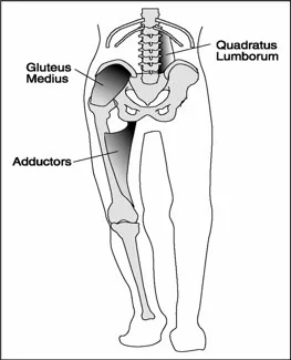
One thing that has become recurrently obvious upon the many outside professional referrals that come though my office is the imbalance and/or weakness or endurance impairments in the posterior mechanism in relation to a painful 1st metatarsophalangeal joint (MTP). When I say posterior mechanism I am referring to the gastrocnemius, soleus, peronei, long flexors, and tibialis posterior namely. 

And, let me be clear, putting a theraband under the 1st metatarsal, encouraging your client to drive greater downward purchase of the head of the 1st MET during simulated foot tripod loading, does not necessarily help your client if their 1st MET is slightly more dorsiflexed. Do not be fooled by the flashy rehab guru tricks out there, proper clean function is achieved, not forced. If you have not earned it, you do not own it. 

It is quite simple really. If one does not have balanced function, including skill (motor pattern), endurance or strength of plantarflexion of the ankle, one cannot properly posture the first metatarsal (1st MET) in plantarflexion to sufficiently alter the sesamoid posturing underneath the metatarsal head, to sufficiently engage the unique eccentric axis (and it's necessary shift) of the 1st MTP to enable ample clean hallux dorsiflexion. Furthermore, without all this,  one will not be able to anchor the medial foot tripod properly.  This can lead to pain, functional hallux limitus, hallux rigidus to name a few. And, let me be clear, putting a theraband under the 1st metatarsal, encouraging your client to drive greater purchase of the head of the 1st MET during foot loading, does not necessarily help your client if their 1st MET is slightly more dorsiflexed. Do not be fooled by the flashy rehab guru tricks out there gang, proper clean function is achieved, not forced. 

A simple example might be a runner who fatigues the posterior mechanism in a long run. As the calf fatigues, they lose ample heel rise, thus ample plantarflexion of the 1st MET, thus proper posturing and translation of the sesamoids, thus successful eccentric axis shift, and thus clean dorsiflexion of the 1st MTP joint.  A player in a jumping sport who has less than ample strength of the posterior mechanism can have much the same issue at the resultant toe.  These are just garden variety examples.  But, should be clear that ample skill, endurance and strength (S.E.S.), our favorite mnemonic, of the posterior mechanism is necessary for pain free, functional toe off in the gait cycle or in jumping mechanics. 

If you are not systematically testing for these S.E.S. issues in the posterior mechanism, you are likely missing a major component in the proper posturing of the ankle and foot and thus proper functioning of the first ray complex and thus enabling clean function at the 1st MTP joint.  

(Sidebar rant: My past personal problems at this great toe joint started when a fellow chiropractor pulled on my toe many moons ago, for some random reason. It was the proverbial,  axial distraction "adjustment". The cavitation was heard around the world (the saliva inducing "pop" that fools many into blissful success), and my problems began.  I had painful dysfunction for many years after that for some strange reason, something was damaged but I was too stubborn and stupid to fix my own foot. I eventually remedied the problem through diving deeper into the complex mechanics of this joint and regionally associated areas. For this very intimate reason, it is why I am not one to perform this maneuver or recommend it. If we can be smarter in our understanding, we can be wiser in our interventions. Besides, axial distraction of this joint is not normal function of this joint. If I had a soap box to stand on for this topic, I would tell people to stop doing HVLA manipulations to this joint, mobilizations are more than ample to elicit a joint range response or a neurologic mechanoreceptor response. The more you understand this profoundly complicated and interesting joint, the 1st MTP joint, the more you will understand how to help your client. But, what do I know, I am just a dumb chiropractor, right Joe Rogan :) 

- Shawn Allen, the gait guys

Caveat Emptor: Foot placement is a complex thing.

"Understanding why we place a foot where we do can be a choice, eventually a habit, of perceived stability, of compensation and we like to say "it is a sliding scale between liabilities and economy". If you want more running economy, go for a narrow step width, but realize you are wrestling with its underlying liabilities.  The key is, one must have enough durability on the loading response of a narrow step width (cross over gait) to fend off the liabilities to reap the rewards of the improved economy. Forgo this principle, and it is caveat emptor."- Dr. Shawn Allen

Today we wanted to revisit a few topics and start to tie them together so that readers can perhaps more deeply bring the study discussed here today, into a deeper thought process.

Screen Shot 2017-08-30 at 7.41.43 AM.png

We have discussed the topic of gluteal pain in chronic low back pain clients previously, when the 2015 Cooper article was published ahead of print. Well, it came out in print (Euro Spine J) in 2016 so we wanted to revisit it with some more global thoughts. Those links are below. 
Basically, the article said that people with low back pain often have “Gluteus medius weakness and gluteal muscle tenderness are common symptoms in people with chronic non-specific LBP.”  
 

As we mentioned in our blog post last year, commenting on the "ahead of print" article, "it is often more on the side of pelvic frontal plane drift. The abdominals and spinal stabilizers also often test weak on this same side. We often see compromise of hip rotation stability as well because , since the hip is relatively adducting (because the pelvis is undergoing repeated frontal plane drift, hence no hip abduction) there is often a component of cross over gait phenomenon which can threaten rotation stability of the lower limb (type “cross over gait” into the search box of our tumblr blog for a landslide of work we have written on that phenomenon)."

This brings to mind this brief (14minute excerpt) from an old podcast we did (#109b, link below) on foot targeting, pelvis frontal plane drift, glute weakness and cross over gait. We brought together several concepts in that 14 minute span and it was on the topic from the Rankin article (link below).

If one is treating clients one must put all these concepts together (one should also have a deep grasp of the principles in this video ). One cannot have tunnel vision, one must embrace the entire picture neuromechanically. Foot targeting, gluteus medius activity, frontal plane pelvis drift or sway, cross over gait parameters, limb torsional issues, foot types and many more must all come into play if you are to truly get to the bottom of your clients problems. The approach must look at the loading and movement patterns at the very least, from foot to pelvis.  We would argue one should not stop there however, take your evaluation all the way into arm swing, thoracopelvic canister stability and more.  
We have pounded sand on the cross over gait and arm swing and the like for almost a decade now. As far as we know, we introduced, and if not, at the very least were the ones that dove deep into the cross over gait and its issues, and all of the attributes and functional pathologic pieces that go with it. We feel that if you fully understand the 40+ articles we have written on the cross over gait and arm and leg swing you will take your client and athlete evaluation to another level.  Understanding unconscious foot targeting is key in our opinion. "Understanding why we place a foot where we do can be a choice, eventually a habit, of perceived stability, of compensation and we like to say "it is a sliding scale between liabilities and economy". If you want more running economy, go for a narrow step width, but realize you are wrestling with its underlying liabilities.  The key is, one must have enough durability on the loading response of a narrow step width (cross over gait) to fend off the liabilities to reap the rewards of the improved economy. Forgo this principle, and it is caveat emptor. "

Shawn & Ivo, the gait guys

https://thegaitguys.tumblr.com/post/149177564774/podcast-109b-shorts-the-gluteus-medius-during

A neuromechanical strategy for mediolateral foot placement in walking humans. Rankin BL
J Neurophysiol. 2014 Jul 15;112(2):374-83. doi: 10.1152/jn.00138.2014. Epub 2014 Apr 30. 

Eur Spine J. 2016 Apr;25(4):1258-65. doi: 10.1007/s00586-015-4027-6. Epub 2015 May 26.
Prevalence of gluteus medius weakness in people with chronic low back pain compared to healthy controls.
Cooper NA1,2, Scavo KM3, Strickland KJ3, Tipayamongkol N3, Nicholson JD4, Bewyer DC4, Sluka KA3.

http://www.ncbi.nlm.nih.gov/pubmed/26006705

QL and Patellofemoral Pain?

photo credit: https://www.t-nation.com/training/training-disasters

photo credit: https://www.t-nation.com/training/training-disasters

"Subjects with PFP(patello femoral pain) have a higher prevalence of MTrPs (Myofascial trigger points) in bilateral GMe (gluteus medius)) and QL (quadratus lumborum) muscles. They demonstrate less hip abduction strength compared with controls, but the TPPRT (trigger point pressure release therapy, AKA ischemic compression) did not result in an increase in hip abduction strength. "

It is not surprising that when the hip is involved, the knee will be involved. As Dr. Allen often likes to say "the knee is basically in joint between 2 ball and socket joints ".

The gluteus medius and quadratus lumborum, along with the adductors are coronal plane stabilizers of the pelvis. They both have rotational components to their function as well affecting the hip directly for the former and lumbar spine for the latter. You can see our other QL articles about this here and here.

It is not much of a stretch to imagine that dysfunction of these muscles could result in trigger points and/or dysfunction of the knee (or foot for that matter ) could cause trigger points in these muscles.

Here is an article (1) examining trigger points in the gluteus medius and quadratus lumborum which, if you are familiar with Porterfield and DeRosa's work (2), are intimately linked during gait. We found it interesting that skin nick compression did not increase hip abduction strength where we find dry needling and intramuscular therapy often do.

Don't overlook these muscles and this important relationship.


  1. Roach, Sean et al.Prevalence of Myofascial Trigger Points in the Hip in Patellofemoral Pain Archives of Physical Medicine and Rehabilitation , Volume 94 , Issue 3 , 522 - 526link to free full text article: http://www.archives-pmr.org/article/S0003-9993(12)01079-9/fulltexthttp://www.archives-pmr.org/article/S0003-9993(12)01079-9/fulltext

  2. J. Porterfield, C. DeRosa (Eds.) Mechanical low back pain. 2nd ed. WB Saunders, Philadelphia; 1991


Not quite the QL, but close....

Screen Shot 2017-08-28 at 10.13.40 AM.png

We all see folks with low back pain and gait abnormalities. It is active during single limb support during stance phase of gait on the contralateral side (along with the external oblique) to elevate the ilium. This is coupled with the ipsilateral anterior fibers of the gluteus medius and minimus pulling the iliac crest toward the stable femur.

We found this FREE FULL TEXT while doing some quadratus lumborum research. It reminds us about things like scleratogenous pain (pain arising from tissues of like embryological origin with a common nerve innervation, like tendon, bone, muscle, etc) and other triggers for low back pain. We have needled this ligament with good result. Remember that this is an individual ligament making up a portion of the middle layer of the thoracolumbar fascia, and is not an aponeurosis of the lumbocostal fibers of the quadratus lumborum.


https://www.ncbi.nlm.nih.gov/pmc/articles/PMC5226660/

photo from: https://musculoskeletalkey.com/treatment-of-the-patient-with-chronic-pain/

A return to "the Kickstand Effect". So your foot is turned out, externally rotated ?

Amputee War Veteran Sergeant Christopher Melendez Became a Pro WrestlerRead more at http://www.craveonline.com/mandatory/1053779-standing-tall-how-amputee-war-veteran-christopher-melendez-beca#XeD2LrZ2xmtXQ6um.99

Amputee War Veteran Sergeant Christopher Melendez Became a Pro Wrestler
Read more at http://www.craveonline.com/mandatory/1053779-standing-tall-how-amputee-war-veteran-christopher-melendez-beca#XeD2LrZ2xmtXQ6um.99

Why is my foot turned out ?  A 3rd return to the solitary externally rotated foot.

Below you will find our 2 prior articles on this topic, but this is a relatable concept to other thing which we have embedded in many of our blog posts and podcasts over the last decade of sharing what we know.

In the photo above the brave Army Veteran Sergeant Melendez one can see the concept brilliantly as he only has one limb.  One can see the concept in full play, he must balance his body mass over one point, not two like the rest of us lucky folk.  In trying to balance over one point, if the foot is straight forward (if one is blessed with close to neutral torsional bone alignment) one will have good stability in the sagittal plane (forward /back) but will be at risk to fall, drift or sway into the frontal plane. Here Sergeant Melendez displays the foot and limb turn out into the frontal plane so that he can use the quadriceps to help him protect into that frontal plane, plus, by situating his base posture in more of an externally rotated position (likely losing internal rotation capability over time, unless forcibly maintained through specific exercises) he can more fully and skillfully engage all 3 divisions of the gluteus maximus and medius, and perhaps hamstrings and adductors and who knows what else, to maintain a more stable and likely less fatiguable posture. Go ahead, try it for yourself, this is easier to balance and maintain that a straight sagittal foot posturing. The one trouble he might have, is not deviating too much, or too often, into a frontal plane drift hip-pelvis posture. This will put much aberrant compressive load onto the roof of the femoral head-acetabular interval, where most of us begin a degenerative hip arthritis journey, unfortunately. 

Side note:   So you might think your client has FAI ?  Maybe start here, our thinking might lead you done a helpful path to get started. Search our blog for FAI as well.

here are the 2 prior articles on the topic, with video.  Watch for this one, it is everywhere out in the world, walking amongst us.  
Thank you for your service Sergeant Melendez.  Here is the article written by K. Thor Jensen, on Crave Online.  

https://thegaitguys.tumblr.com/post/14262793786/gait-problem-the-solitary-externally-rotated

https://thegaitguys.tumblr.com/post/40617674450/a-return-to-the-solitary-externally-rotated-foot

Shawn & Ivo, The Gait Guys

What do we have here and what type of shoe would be appropriate?

You are looking at a person with a fore foot varus. This means that the fore foot (ie, plane of the metatarsal heads) is inverted with respect to the rear foot (ie, the calcaneus withe the subtalar joint in neutral). Functionally translated, this means that they will have difficulties stabilizing the medial tripod (1st MET head) to the ground making the forefoot and arch unstable and likely rendering the rate and degree of pronation increased.

The incidence of this condition is 8% of 116 female subjects (McPoil et al, 1988) and 86% of 120 male and female subjects (Garbalosa et al, 1994), so it seems to happen happen more in males. We think this second number is inflated and those folks actually had a forefoot supinatus, which is much more common.

Fore foot varus occurs in 3 flavors:

  • compensated
  • uncompensated
  • partially compensated

What is meant by compensated, is that the individual is able to get the head of the 1st ray to the ground completely (compensated), partially, or, when not at all, uncompensated.What this means from a gait perspective ( for partially and uncompensated conditions) is that the person will pronate through the fore foot to get the head of the 1st ray down and make the medial tripod of the foot (ie, they pronate through the subtalar joint to allow the 1st metatarsal to contact the ground). This causes the time from mid-stance to terminal stance to lengthen and will inhibit resupination of the foot. 

Today we are looking at a rigid, uncompensated forefoot varus, most likely from insufficient talar head derotation during fetal development and subsequent post natal development. They will not get to an effective foot tripod. They will collapse the whole foot medially. These people look like severely flat-footed hyperpronators.


So, what do you do and what type of shoe is appropriate? Here’s what we did:

  • try and get the 1st ray to descend as much as possible with exercises for the extensor hallucis brevis and short flexors of the toes (see our videos on youtube)
  • create more motion in the foot with manipulation, massage mobilization to optimize what is available
  • strengthen the intrinsic muscles of the feet (particularly the interossei)
  • increase strength of the gluteus maximus and posterior fibers of the gluteus medius to slow internal rotation of the leg during initial contact to midstance
  • put them in a flexible shoe for the 1st part of the day, to exercise the feet and a more supportive; medially posted (ideally fore foot posted) shoe for the latter part of the day as the foot fatigues
  • monitor his progress at 3-6 month intervals
  • a rigid orthotic will likely not help this client and they will find it terribly uncomfortable because this is a RIGID deformity for the most part (the foot will not accommodate well to a corrective orthotic. Besides, the correction really has to be made at the forefoot anyways. 

Lost? Having trouble with all these terms and nomenclature? Take our national shoe fit program, available by clicking here.

The Gait Guys. Uber foot geeks. Separating the wheat from the chaff, with each and every post.

So you prescribe and fit orthotics you say ?

"It all matters, and quite possibly, if you do not know it all, you cannot help your client."

How about this then, you have someone with a rearfoot valgus with internal tibial torsion.  How are they going to load now? What if you throw in a valgus knee and femoral torsion variant?  Are they going to pronate more or less ? What if that person had just internal tibial torsion on one leg and not the other, yet they had 2 rearfoot valgus feet presentations.  Now what?

Ouch, that is a strong statement. It likely needs softened, but, there is some truth within those words. 

Last night we did our monthly lecture on www.onlineCE.com.  We had a packed room, biggest audience to date.  It is likely because people are realizing that the small stuff matters.  We talked for an hour on foot types and  how they present, how they potentially load, and how other mechanical issues above can impact how a foot type loads. 

We have all seen the pedographs like in the photo. The unwise depend on a static pedograph mapping for diagnostic help and God forbid that is all you use for making orthotics (that may only help if your client is  a professional stander), the more wise use the dynamic pedograph mapping to see how their client moves across the ground, and the wise use it as a mere piece of the data, combine it with a clinical exam, look far up into the biomechanical chain for other locomotive challenges that could change the dynamic loading pattern across the foot and ground.  What do we mean exactly ?  Well, a client with a rearfoot valgus foot type will load the heel and rest of the foot one way if they are doing a good job stacking the hip over the knee, and knee over the foot. But, if they have weakness in the hip affording a frontal plane drift of the pelvis over the foot, that is going to magnify the rearfoot valgus loading pattern (addendum: they could also tip into rearfoot varus posturing as well). That is just one example, of many.  In otherwords, it is the same foot type, but both of these are going to show a dynamic change in the loading pattern response. So, said another way, you cannot diagnose a foot type by the pedograph mapping. Nor should one make an orthotic for someone based off of a pedograph mapping, nor without an examination of the entire kinetic change.  What is your client able, and unable, to do? That is a big question, and when you start by asking those 2 questions, you get closer to the prize.  The pedograph only shows the static or dynamic pressures from the superincumbent load, it does not tell you if it is good or bad, and it does not tell you what they are doing, or why they are loading that way. It only shows the loading. Your job is to find out why they are loading that way, and then determine if that is part of their problem they have sought you out for.

So, does  your head spin now ? Does this suddenly make you sweat ? Do you realize you are missing pieces of the pie in helping your client?  Not yet maybe ?  How about this then, you have someone with a rearfoot valgus with internal tibial torsion. How are they going to load now? What if you throw in a valgus knee and femoral torsion variant? Are they going to pronate more or less ? What if that person had just internal tibial torsion on one leg and not the other, yet they had two rearfoot valgus feet presentations. Now what? Suddenly the loading is different in both feet and up the chains. There is likely going to be different challenges to limb spin control from side to side. This aberrant and asymmetrical loading is going to come up to a pelvis, upon which a single spinal column is trying to find a sound base of support and mobility to work and transfer loads from. 

And, what if this client also has some tibial varum on that same side ? What if they had external tibial torsion or some femoral torsional presentation on one side ?  You can see now how complicated this gets. And that is just on the structural components. What about the dynamic components ?  We here at The Gait Guys feel that this is all critical stuff to take into consideration and it is sometimes the stuff that is the tipping point between a successful management of a clients complaints, and unsuccessful.  

In closing, think about this. If you are sending out your orthotics for fabrication, have you conveyed this all to your fabricator ?  All they know is what a pedograph might show, and what the foot mold looks like. You have to provide them with all this other information, because essentially they are blind (this of course assumes your fabricator can mind juggle all the torsions, valgus/varus, pelvis drift loads etc,  oy vey ! That is hard to do !) This is why we do all of our modifications in office, in the rare case we need a temporary orthotic modification. But, we will aim to just correct what mechanics are aberrant and avoid the whole orthotic crutch when we are able. But lets face it, sometimes, for a period of time, we all need a crutch to get through a problem, to find better mechanics where we can strengthen from or gain protecting from temporarily.  That is what splints do, taping, crutches, braces, one might even argue what corrective exercises do. It is a path on the journey for your client, and sometimes they need help through the muddy parts.

And, don't be "that guy" that says orthotics are useless. They are a crutch , a tool. A small tool, one might argue that it should only be pulled out when the other tools are not working to get the job done.  Do not make them your first line of defense, except when that is called for.  After all, not all people were blessed with sufficient anatomical  and mechanical parts to avoid needing a crutch, so don't be "that guy" that preaches from that extreme, because it is not honest. Or, maybe, you just do not see the biomechanical messes we see in our clinics, that is quite the realistic possibility. 

Want to learn more about this kind of stuff? Keep up with our blog here. OR take some of our lecture recorded classes on www.onlineCE.com . We have a library of classes there for you to take anytime. And meet us once a month over there, every 3rd Wednesday. And, stay tuned for some new teaching gigs we have coming your way.

-Shawn and Ivo,  the gait guys


Podcast 127: Tendinopathies, Tendon Pain & more.

Key Tagwords:

neuroscience, hip pain, tendonopathy, DNA, running, injuries, achilles, tendonitis, gait, shoecue

Show Links:

http://traffic.libsyn.com/thegaitguys/pod_127f.mp3

http://thegaitguys.libsyn.com/podcast-127-tendinopathies-tendon-pain-more


Our Websites:


www.thegaitguys.com

summitchiroandrehab.com   doctorallen.co     shawnallen.net

Our website is all you need to remember. Everything you want, need and wish for is right there on the site.
Interested in our stuff ? Want to buy some of our lectures or our National Shoe Fit program? Click here (thegaitguys.com or thegaitguys.tumblr.com) and you will come to our websites. In the tabs, you will find tabs for STORE, SEMINARS, BOOK etc. We also lecture every 3rd Wednesday of the month on onlineCE.com. We have an extensive catalogued library of our courses there, you can take them any time for a nominal fee (~$20).
 
Our podcast is on iTunes, Soundcloud, and just about every other podcast harbor site, just google "the gait guys podcast", you will find us.
 
Show Notes:

New neuron science
http://trendintech.com/2017/04/05/groundbreaking-study-reveals-neurons-communicate-in-a-way-we-never-imagined/

Exercise strengthens you DNA
http://www.mensfitness.com/training/build-muscle/exercise-strengthens-your-dna

Development of overuse tendinopathy: A new descriptive model for the initiation of tendon damage during cyclic loading
Tyler W. Herod, Samuel P. Veres
https://twitter.com/stijnbog/status/875270547562692608

The neuromechanical adaptations to Achilles tendinosis.
J Physiol. 2015 Aug 1;593(15):3373-87. doi: 10.1113/JP270220. Epub 2015 Jun 30.
Chang YJ1, Kulig K1.

Hip muscle strength is decreased in middle-aged recreational male athletes with midportion Achillestendinopathy: A cross-sectional study.
Phys Ther Sport. 2017 May;25:55-61. doi: 10.1016/j.ptsp.2016.09.008. Epub 2016 Sep 13.
Habets B1, Smits HW2, Backx FJG3, van Cingel REH4, Huisstede BMA5.

Changes of gait parameters and lower limb dynamics in recreational runners with achilles tendinopathy. Kim S1, Yu J2.
J Sports Sci Med. 2015 May 8;14(2):284-9. eCollection 2015 Jun.

Vibrations and strides
http://www.popsci.com/measuring-vibrations-runners-strides-could-prevent-muscle-injuries

ShoeCue product:
https://www.shoecue.com/?gclid=CKL1mI_e8tQCFZi4wAodgXMPJA

RULES of tendonopathies:
https://pbs.twimg.com/media/C3BbmWlXgAAg-ZA.png:large

Roger Enoka
http://www.humankinetics.com/products/all-products/neuromechanics-of-human-movement-5th-edition

http://onlinelibrary.wiley.com/doi/10.1002/jor.23629/abstract?campaign=wolacceptedarticle

Achilles Tendinitis?

You should read this study if you haven't already

We all treat different forms of achilles tendinitis and tendonosis. This landmark study uses loaded eccentrics and showed better tendon organization and decreased tendon thickness at follow up. 

Tendons do seem to respond better to tension and loaded eccentrics certainly seems to do the job. Though, this study is 2004 and much new research has leaned us all more toward looking at pain free isometrics , in other words, taking that tension in a pain free single range load and helping the tendon to reestablish appropriate stiffness. Tension and time under pain free load is the key, then expanding from that into more dynamic load challenges like eccentrics. But, as always, it is finding the load your client can pain free tolerate, get the organism to reload the tissue without threat and then build durability and tissue tolerance to load.

"Conclusions: Ultrasonographic follow up of patients with mid-portion painful chronic Achilles tendinosis treated with eccentric calf muscle training showed a localised decrease in tendon thickness and a normalised tendon structure in most patients. Remaining structural tendon abnormalities seemed to be associated with residual pain in the tendon."

Ohberg L, Lorentzon R, Alfredson H, Maffulli N. Eccentric training in patients with chronic Achilles tendinosis: normalised tendon structure and decreased thickness at follow up. British Journal of Sports Medicine. 2004;38(1):8-11. doi:10.1136/bjsm.2001.000284.

link to abstract: http://www.ncbi.nlm.nih.gov/pmc/articles/PMC1724744/

Does asymmetry matter ?

Does asymmetry matter ?

There has been some brilliant talk in the socialverse as of late that asymmetry doesn't matter. We believe these dialogues may be contextual for dialogue purposes (perhaps?) and we have no problem with that. I am sure we may approach our patients differently, though restoring pain free function is the goal. We have a problem layering more endurance, strength and power on asymmetry. Sure the client may feel better, but that is just because the threshold of the system is better, maybe. They have better armor, they are more durable, and thus further from the pain line, but the problem is undeniably still there, it is just protected. 
So, why not try to move closer to symmetry, if that gives pain relief, and then build strength, power, and endurance on those cleaner patterns ? Doesn't that make more sense ? One question we have, that science cannot prove (or disprove) is whether greater strength on asymmetry increases risk for injury ? Well, we think so, and we think that if for a given client, that strength and endurance built on a more symmetrical frame is likely to have less risk for injury. But, the verdict will always be out on that until we can clone folks.

We believe that driving toward symmetry much of the time does in fact matter. Is it going to happen 100%?, no, asymmetry is the rule in the human frame. We are talking about not driving deeper strength, power, endurance into an asymmetrical pattern that further puts strain into tissues not designed or apt to be favorable to the organism/joint/limb etc. We are putting together a written piece expressing some of our points of view further. We have found that when we drive our clients towards symmetry we often, not always, have to drive less strength and load into our clients to dampen the pain beast.

Stay tuned . . . .

https://youtu.be/0Jn2CESZ6jw

New shoe, old shoe. The rotation, it matters.

New shoe, old shoe. The rotation, it matters.

At this very moment i am responding to an email of a very sweet and extremely talented runner in Tasmania, I saw her months ago here in the USA as she travelled through. I find myself sharing a conversation with her at this very moment, one she likely knows, but one we all forget, or get lazy with. It is all about
"reducing one more risk factor, reducing one more sudden biomechanical change that can provoke changes in our loading response". 
This is nothing new for veteran Gait Guys brethren here, but we get 100's of newbies here each week, so it is good to remind all.
* Never underestimate the subtle changes in biomechanics that might come from a shoe change in a high mileage athlete. Sweat the small stuff, sometimes." Foam changes, foam loses its resilience with repeated compression cycles, foam deforms into your particular biomechanical loading habits. And sometimes your habitual loading cycles are subtle, but as the foam gives into them, the small thing mushrooms into a significant thing. IT can become a "tipping point" for your clients biomechanics. Something that was initially nothing, becomes something of significance. Help reduce your client's risk factors so you can stay focused on the things that matter, reduce those inner-mind rumbling thoughts of "i wonder if that is a factor". Take those off the table for all your clients, when possible.

We always want to get one more run in on a pair of shoes that is weak and limping its way into the finish line, on its final death throws. 
"Today's story: Bam, i got one more run in on these babies. 
Tomorrow's story: hey i wonder why i am having a little medial foot-arch-ankle pain today???" #facepalm
(not that this has anything to do with the client below, just slamming home my point)

"Dear _____:
Do you think switching to a newer pair of Zante's had any factor in this ? Did the shoe seems to guide the foot differently than the older pair ? Anything feel different ? Sometimes a fresh shoe today changes mechanics too much compared to the one you were just in yesterday (try in the future to have 2-3 pairs in rotation, switch up every run to a different one. Have one newer one in the rotation, another with 200 miles and one that is almost done. That way you are never burning down one shoe and then jumping in a new one. Always be finishing up on an older shoe and starting in on a new one, with one in the pocket in the middle wear milage.)
Now, onto your injury in question . . . . 
-Dr. Allen

Hearing and Gait Parameters

Here's an interesting study looking at the effects of her hearing on gait. Noticed that in children with hearing loss, walking speed was slower and this increased more with dual tasking. Muscle activities were greater as well, with respect to the medial gastroc, which is a strong he will adductor and supinator as well as vastus lateralis which internal swing is attenuating external rotation of the leg.

This implies that auditory cues and clues are important considerations during gait analysis and gait retraining.

"The findings indicated that gait speed in children with hearing loss was smaller than that in control group. Dual task resulted in a decreased walking speed of children with hearing loss. The activities of tibialis anterior muscle in terminal stance phase (p = 0.040), medial gastrocnemius muscle in loading response and initial swing phases (p < 0.05), and vastus lateralis muscle in the terminal stance and pre swing phases (p < 0.05) were greater in deaf group. In deaf children the gait speed was reduced and the muscle activity was increased with respect to those in control group. This altered gait speed and muscle activity is suggestive of a lower mechanical efficiency of gait in deaf children"

see our other posts on this here: https://thegaitguys.tumblr.com/search/sound


.https://www.ncbi.nlm.nih.gov/pubmed/28752319


The extra-articular hip impingements

There continues to be a plethora of research and dialogue on the femoral acetabular impingements (FAI), the intra-articular impingements.  But we must not forget about the extra-articular impingements about the hip. A common one we see is the Ischiofemoral variety whereby the quadratus femoris muscle gets pinched between the ischial tuberosity and the femur.  We wrote about it, see the link below.  This one gets mistake for proximal hamstring tendonopathies by some we suspect. We suspect, however this is pure speculation, that the two most common are #1 and #3.

This article outlines some of the common extra-articular impingement syndromes:

 1) Ischiofemoral impingement: as we discuss in our article

 "2) Subspine impingement: mechanical conflict occurs between an enlarged or malorientated anterior inferior iliac spine and the distal anterior femoral neck.

3) Iliopsoas impingement: mechanical conflict occurs between the iliopsoas muscle and the labrum, resulting in distinct anterior labral pathology.

4) Deep gluteal syndrome: pain occurs in the buttock due to the entrapment of the sciatic nerve in the deep gluteal space.

5) Pectineofoveal impingement: pain occurs when the medial synovial fold impinges against overlying soft tissue, primarily the zona orbicularis. "

Ischiofemoral Impingement
https://thegaitguys.tumblr.com/post/110564772099/ischial-femoral-impingement-you-have-to-know-what

Current concepts in the diagnosis and management of extra-articular hip impingement syndromes.  Nakano N1, Yip G1, Khanduja V2.
Int Orthop. 2017 Jul;41(7):1321-1328. doi: 10.1007/s00264-017-3431-4. Epub 2017 Apr 11.
https://www.ncbi.nlm.nih.gov/pubmed/28401279


Projecting, calculating, the next step.

Researchers have discovered that we most accurately hit targets when we see them 1 to 1.5 steps ahead of where we were. This is more difficult that it seems because we are making a plan, and at the same time we're making that plan, we're making a movement based on the stuff that we saw a second and half in the past according to the article by Erica Pandey.


Below this link, you will find our post on projecting and estimating steps. Much along the same lines but with a great video to set it up. Here is a lead in to that article we wrote long ago, on the strange steps at a Brooklyn subway station that everyone was tripping on.

 "At Brooklyn’s 36th Street subway stop, one of the steps is slightly higher than the others. Stairs have a standardized Rise and Run and when this is altered, specifically the Rise, funny things happen. Filmmaker Dean Peterson set up his camera to capture the stumbles and the video can be seen here http://vimeo.com/44807536 and above on our blog.

The dangerous step, it turns out (which has since this video been repaired), is apparently a half-inch higher than the others. Stairway design guidelines vary within a small range.  Guidelines call for risers to be a minimum of 6 inches and a maximum of 7 ¾ inches. The allowable variance between steps is 3/8 of an inch depending on the source you seek."   Read on  . . . . . 

https://thegaitguys.tumblr.com/post/44642195883/the-funny-problem-with-the-stairs-at-brooklyns

"When walking, the brain leads the body by one step" -Erica Pandey

https://www.axios.com/we-plan-one-step-ahead-when-we-walk-scientists-say-2465394429.html

Party over the Weekend?


So, the more you drink, the more you impair the CNS. The more you impair the CNS, the more dependent you become on peripheral mechanisms. A good reason to keep your vestibular system (alcohol changes the specific gravity of the endolymph), your visual system (long term use affects the option nerve directly and can cause involuntary saccades) and proprioceptive systems including the cutaneous mechanoreceptors (because you are relying on them more) intact. Watch what and how much you drink...

"Standing postural stability relies on input from visual, vestibular, proprioceptive and mechanoreceptive sensors. When the information from any of these sensors is unavailable or disrupted, the central nervous system maintains postural stability by relying more on the contribution from the reliable sensors, termed sensory re-weighting. Alcohol intoxication is known to affect the integrity of the vestibular and visual systems. The aim was to assess how mechanoreceptive sensory information contributed to postural stability at 0.00% (i.e. sober), 0.06% and 0.10% blood alcohol concentration (BAC) in 25 healthy subjects (mean age 25.1 years). The subjects were assessed with eyes closed and eyes open under quiet standing and while standing was perturbed by repeated, random-length, vibratory stimulation of the calf muscles. Plantar cutaneous mechanoreceptive sensation was assessed for both receptor types: slowly adapting (tactile sensitivity) and rapidly adapting (vibration perception). The correlation between recorded torque variance and the sensation from both mechanoreceptor types was calculated. The recorded stability during alcohol intoxication was significantly influenced by both the tactile sensation and vibration perception of the subjects. Moreover, the study revealed a fluctuating association between the subjects' vibration perception and torque variance during balance perturbations, which was significantly influenced by the level of alcohol intoxication, vision and adaptation. Hence, one's ability to handle balance perturbations under the influence of alcohol is strongly dependent on accurate mechanoreceptive sensation and efficient sensory re-weighting. 

Modig F, Patel M, Magnusson M, Fransson PA.Study II: mechanoreceptive sensation is of increased importance for human postural control under alcohol intoxication. Gait Posture. 2012 Mar;35(3):419-27. doi: 10.1016/j.gaitpost.2011.11.001. Epub 2011 Dec 27.
weighting.


We hope you are standing up while you read this….

A newborn’s brain is only about one-quarter the size of an adult’s. It grows to about 80 percent of adult size by three years of age and 90 percent by age five (see above). This growth is largely due to changes in individual neurons and their connections, or synapses.

The truth is, most of our brain cells are formed at birth, In fact, we actually have MORE neurons BEFORE we are born. It is the formation of synapses, or connections between neurons, that actually accounts for the size change (see 1st picture above). This is largely shaped by experience and interaction with the environment.

Do you think children’s brains are less active than adults? Think again, your 3 year old’s brain is twice as active as yours! It isn’t until later in life that you actually start dialing back on some of those connections and those pathways degenerate or fade away…a process scientists call “pruning”.

How does this apply to gait? Gait depends on proprioception, or body position awareness. Your brain needs to know where your foot is, what it is standing on and so on. Proprioception, as we have discussed in other posts, is subserved by muscle and joint receptors called mechanoreceptors (muscle spindles, golgi tendon organs and type 1-4 joint mechanoreceptors to be exact). This information is fed to 2 main areas of the brain: the cerebral cortex and the cerebellum. These 2 parts of the central nervous system are interconnected on many levels.

The cerebellum is intimately associated with learning. Try this experiment. you will need a tape recorder (guess we are showing our ages, digital recorder), a timer and a moderately difficult book.

Sit down and pick a section of the book to read. start the recorder and timer and read aloud for 2 minutes. Stop reading, stop the recorder and stop the timer.

Stand up, somewhere you won’t get hurt if you fall. Stand on 1 leg (or if available, stand on a BOSU or rocker board). Open the book to a different spot. Start the timer, the recorder and start reading again for 2 minutes.

Sit back down and grab a snack. Listen to the 2 recordings and pay attention to the way you sound when you were reading, the speed, fluency and flow of words. Now think about recall. Which passage do you remember better?

The brain works best at multitasking and balance and coordination activities intimately affect learning. Having children sit in a class room and remain stationary and listen to a lecture is not the best way to learn. We always tel our students to get up and move around…

This article looks at this relationship in a slightly different way.

We hope you are still standing : )


 Lopes VP, Rodrigues LP, Maia JA, Malina RM.Motor coordination as predictor of physical activity in childhood. Scand J Med Sci Sports. 2011 Oct;21(5):663-9. doi: 10.1111/j.1600-0838.2009.01027.x. Epub 2010 Mar 11

Abstract

This study considers relationships among motor coordination (MC), physical fitness (PF) and physical activity (PA) in children followed longitudinally from 6 to 10 years. It is hypothesized that MC is a significant and primary predictor of PA in children. Subjects were 142 girls and 143 boys. Height, weight and skinfolds; PA (Godin-Shephard questionnaire); MC (Körperkoordination Test für Kinder); and PF (five fitness items) were measured. Hierarchical linear modeling with MC and PF as predictors of PA was used. The retained model indicated that PA at baseline differed significantly between boys (48.3 MET/week) and girls (40.0 MET/week). The interaction of MC and 1 mile run/walk had a positive influence on level of PA. The general trend for a decrease in PA level across years was attenuated or amplified depending on initial level of MC. The estimated rate of decline in PA was negligible for children with higher levels of MC at 6 years, but was augmented by 2.58 and 2.47 units each year, respectively, for children with low and average levels of initial MC. In conclusion MC is an important predictor of PA in children 6-10 years of age.

Some Basic Tenets of Gait

Initial contact? Loading response? Mid stance? Terminal Stance? pre swing? Are these terms that are familiar to you? Hmmm. How about the wrong orthotic for someone with internal tibial torsion? Join us in this excerpt from a recent Gait and Needling seminar


https://vimeo.com/226733697