Leg length discrepancy

Badda Bing Badda Boom 

As you can see, this gal has an anatomical leg length discrepancy on the left side which is tibial. Can you see the other finding?

IMG_8565.jpg

Look carefully both of her feet. Can you see the bunion on the left but not on the right? This is a common finding on the shorter leg side of a true, anatomical leg length discrepancy it. Because of the leg length discrepancy, that foot is often in supination and the head of the first ray does not make it to the ground thus the pull of the adductor hallucis muscle pulls the great toe west.

IMG_8563.jpg

Look for bunions on the short leg side of true, anatomical leg length discrepancies.

IMG_8564.jpg

#lld #leglengthdifference #leglengthdiscrepancy #shortleg #bunions #bunion #gaitevaluation 

What happens when a ganglionectomy goes south

What happens when a ganglionectomy goes south?

- This patient had a ganglionectomy. Unfortunately, they tagged the joint capsule of the first MTP. By affecting the integrity of the capsule, as well as the surrounding musculature, she’s developed the beginnings of a hallux valgus (bunion) as well as hallux limitus (limited dorsiflexion of the first metatarsalphalangeal articulation). 

22625D64-2E36-43DB-AA5E-27E3220B7DDB.JPG

- she has degeneration of the first MTP as well as an osteophytic  crown at the distal aspect of the first metatarsal and tenderness over the capsule as well as the extensor hallucis tendon and proximal phalanx. 

-dorsiflexion is 30° on this side, 50 on the opposite side. We need about 50° to have adequate for foot rocker

– she also has moderate external tibial torsion, right greater than left and a left anatomically short leg secondary to a femur fracture.

– Since the mobility of the first ray was limited, we worked on first Ray mobility as well as exercises to descend the 1st ray, with acupuncture for pain control. 

-we are considering an orthotic to assist in raising the base and dropping the head of the first metatarsal to create more hallux dorsiflexion. When performed manually, she had a few extra degrees we would like to take advantage of. 

-We will keep you posted :-)

#ganglionectomy #footproblem #footproblems #halluxlimitus #bigtoewoes #bunion #bunions #bunionsurgery 

"You do not have a shoe problem, you have a "thing in the shoe problem", meaning, it is you."

We say this so often in our offices.
"You do not have a shoe problem, you have a "thing in the shoe problem", meaning, it is you."
Translation: compromised mechanics leading to tissue overloading.
But we all have to strongly consider that injury is a result of the loading you have not trained gradually into, failure to adapt and accommodate, excessive mileage without adequate tissue recovery,

From the article:
"So Napier and co-author Richard Willy from the University of Montana reviewed the highest-quality research featuring randomized controlled trials and systematic reviews.
"What we see is that there's really no high-level evidence that any running shoe design can prevent injuries," Napier said."

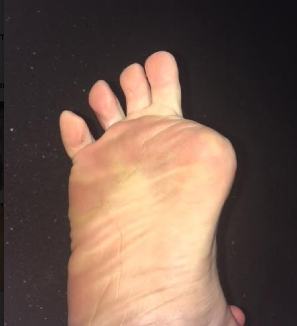
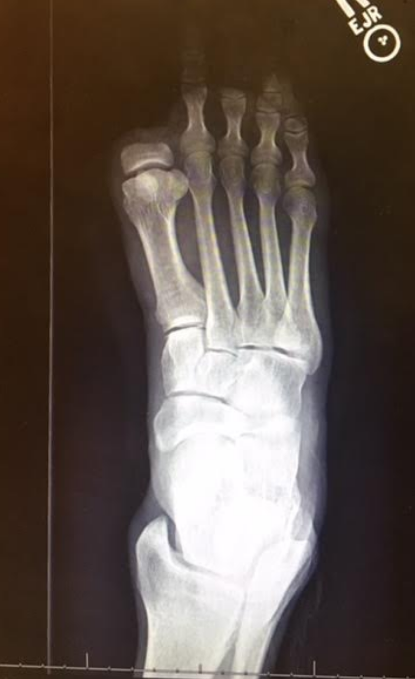
Now, to be honest, in our (the gait guys) opinion, there are times we do recommend a change in the foot wear for a client, and it is often because it appears to be working against someone mechanics and is a contributory factor in their injury or complaint. And sometimes that shoe recommendation is a temporary one, and sometimes a permanent one. We can use a shoe to help us get to a better/faster end point. After all, when we sprain an ankle sometime a brace or crutches are helpful and protective, of temporary value. A wisely chosen shoe can act the same if we are dealing with an acute achilles tendinopathy or a painful bunion for example. And in those cases we might recommend a shoe that can give us an assist. Sometime, when appropriate perhaps it is a shoe with a stronger medial post, perhaps one with a higher or lower heel drop/delta, or more or less stack height, or perhaps a mid/forefoot rocker built into the shoe. The truth is, people come in with functional or "fixed" pathology and sometimes pairing up a shoe to help us around some conflicting biomechanics can be temporarily, and sometimes permanently, helpful. But, the shoe is never the only answer, a wise clinician has many things they can utilize, all the way up the kinetic chain sometimes.
The more you know, the better you can assist someone.

Shawn Allen, one of the gait guys

#Nigg, #barefoot, #shoes, #stackheight, #heeldrop, #achillestendinitis, #bunion, #pronation, #supination, #running, #gait, #thegaitguys, #gaitanalysis, #gaitproblems, #gaitcompensation

Can the design of a running shoe help prevent injury? A B.C. researcher says he has the answer

Kelly Crowe · CBC News · Posted: Dec 15, 2018 9:00 AM ET

https://www.cbc.ca/news/health/running-shoe-injury-prevention-second-opinion-1.4947408?fbclid=IwAR3XaGPdgfQ68wj2N0tHqIamDdpYuxTIIL2LeudUd-doYN8YqQrIZI9-s9E

How does hallux valgus and bunion formation cause toe hammering?

Screen Shot 2018-11-13 at 4.36.12 PM.png

Photo: you need to recognize this predictable pattern.

When the hallux begins to become incompetent, from perhaps pain, hallux rigidus, hallux limitus, bunion and in this case hallux valgus with bunion (in this case rotational instability) when this incompetence kicks in, we must find stability elsewhere. One will often, unconsciously, begin to increase the flexor tone and pressure to try and find stability since one cannot get it sufficiently from the hallux anymore.

Screen Shot 2018-11-16 at 6.28.27 PM.png


Here you see the tenting up of the 2nd toe, from increased long flexor activity (FDL) over time.
And here is the interesting thing you will notice, over time, the 3rd toe will start the same strategy, then the 4th. We see this often. It is not set in stone, but we see it a lot.
Notice it in your clients feet. Teach them why they are getting hammer toes, flexible ones at first, and possibly rigid ones over time. Hammer toes can have many causes, this is just one.
Solution: find a way to help your client re-find better hallux and medial forefoot stability to halt the progression.

Part 2: The amputated hallux & the complex biomechanical fall-out from it.

Screen Shot 2018-08-10 at 8.10.05 AM.png

Last week we promised Part 2 to this case, the amputated big toe.
Here is part 2. These are the complicated biomechanical fall-outs, so grab a big mug o' coffee and have at it !

In review, this person (all photos and case premissioned in swap for insight) had the distal hallux removed because of a progressive melanoma on the big toe. Can you believe that ! This is one more reminder that the sun and regular dermatologist screenings are wise.
This person had a complaint of progressing right gluteal and QL pain, spasm, tone and some persistent pain now in the 2nd metatarsal as well as some shoe challenges. We discuss this case briefly in and upcoming podcast, #139 or #140 we believe.

Screen Shot 2018-08-10 at 8.10.19 AM.png

Before we add our final thoughts to this case, lets cap our post from last week.

-Without the hallux, we cannot wind up the windlass and shorten the distance between the first metatarsal and heel, thus the arch will splay (more permanently over time we suspect) and we cannot optimize the arch height.
This will promote more internal spin on that limb because of more midfoot pronation and poor medial foot tripod stabilization.
- More internal limb spin means more internal hip spin, and more demand (which might not be met at the glute level) and thus loads that are supposed to be buffered with hip stabilization, will likely be transferred into the low back, and or into the medial knee. Look for more quad protective tone if they cannot get it from the glutes. Troubles arise when we try to control the hip from quadriceps strategies, it is poorly postured to do so, but people do it everyday, *hint: most cyclists and distance runners to a large degree).
- anterior pelvis posturing on the right, perhaps challenging durability of the lower abdominals, hence suspect QL increased protective tone, possible low back tightness or pain depending on duration of activities
- These factors are likely related to his complaints in the right gluteal and low back/QL area.

Now, onto our next thoughts.

- when the hallux is incompetent, in this case absent, there are few other choices to gain forefoot purchase on the ground other than more flexion gripping of the 2nd toe (then the 3rd, then 4th). This is a progressing "searching" phenomenon for forefoot stability and without the function of the big fella, the 2nd toe will begin a hammering phenomenon, often, but not always. We would not be surprised to see hammer toe development in this case, but this person is now very aware of it, and can at least now fight that battle with increased awareness. There is some mild evidence of this on the side lateral photo.

- We are happy to see that the proximal phalange was spared. The adductor hallucis is inserted medially there, and this will help to reduce bunion generation risk (medial metatarsal drift). Comparing the photo and the radiograph is a great example of how far back/proximal the 1st MTP joint is. One could easily assume that the entire hallux was resected from the photo, but the radiograph shows otherwise.

Screen Shot 2018-08-10 at 8.22.36 AM.png

- Toe off is obviously going to be compromised. The patient cannot adequately stabilize the 1st metatarsal (MET) and this will mean a compromised foot tripod, medial foot/tripod splay, arch pronation control challenges but toe off stabilization is going to have to be met by the 2nd and 3rd digits, as discussed above. They are not suited to be the major players here, they are synergistic to this end. Do not be surprised to see one of 2 strategies at toe off here:

1. heavy medial foot tripod toe off, dropping into the void and this maximize the internal spin challenges and minimizing the requisite foot supination stiffness generation phase that should be normal at toe off

2. avoidance of the above, with a forced conscious forefoot lateral toe off, a supinatory strategy, to avoid internal limb spin, more toe hammering, and the lurch heavily and abruptly off of the right foot and onto the left limb.

Screen Shot 2018-08-10 at 8.10.27 AM.png

3. taking #2 further, any time there is perceived challenges or deficits in strength, endurance, proprioception, balance, power and the like, the brain often will create a premature departure off of said limb, creating a requisite premature loading onto the opposite limb. This can cause a phenomenon well loosely refer to "catching" in the contralateral quadriceps mechanism. These clients, with their abrupt loading pattern onto the opposite limb will most often have troubles getting into initial gluteal hip stabilization strategies, and thus default into a quadriceps strategy, that in time can lead to quad shortness and increased tone, which can cause more compression across the patellofemoral joint and cause knee pain. This is more of a compression/loading response issue rather than tracking phenomenon, which we see at the typical diagnosis. We often look for causes in the opposite limb for contralateral knee pain. IT is quite often there if you are looking hard enough for it. Fix the problem, not the symptom.
There is a long host of other things than can arise from here, including heavy contralateral (in this case left sided) foot loading challenges, often more forefoot initial loading, and all of the problems than can arise when this pattern is cyclical, but that would take this post far too deep and long. So, . . . . another time.

Screen Shot 2018-08-10 at 8.09.47 AM.png

4. Shoe fit, we could make the case that a shoe that nicely hugs the forefoot, as opposed to a wide toe box'ed shoe, could help fight off the risk of 1st metatarsal abduction and thus bunion formation risk. However, one cannot dismiss the wider toe box giving the remaining toes a better environment to engage without hammering with over use of long flexors. We might suggest a trial of an elastic sleeve, one often used for plantar fascitis symptom management, placing a snug one around the forefoot when ambulating. This could help keep that metatarsal snug and stop the bunion-like drift we would be watching for.

have at it gang, cases like this are far and deep and require deep understanding of normal and abnormal biomechanics, and the rabbit hole deep myriad of compensations that can be engaged.

have a great weekend !

Shawn and Ivo

One of life's great mysteries....Some folks will do what they want anyway....

The origins of the species, gravity and women...Just a few of life mysteries. Reading this article (1) made us sad in many ways. It's like smoking. You know it's bad for you but you keep doing it. Why? The mystery remains to us.

Vanity seems to often trump biomechanics, as we see in pencil skirts (see our post here), droopy pants (see here)  and high heels (here).

Yet, here is yet another study about women, heels and bunion surgery. 

"Almost two thirds (31) of the 50 patients who said they wanted to go back to wearing heels after surgery did so, and 24 of these women said their postoperative use equaled or exceeded the frequency of their preoperative wear. There were no differences between pre- and postoperative heel heights.

In the study, women older than 65 years were more likely than younger women to report high-heel use prior to hallux valgus surgery.

However, 58.5% of study participants reported difficulty with heel wear, and 13.9% said they had significant restriction, and couldn’t wear anything without pain but custom orthopedic shoes or slippers. Most women (86%) were able to return to comfortable shoes after surgery with minimal or no discomfort; 27.7% said their footwear choice was unrestricted, meaning they could wear both comfortable shoes and heels with minimal discomfort. The 23 women older than 65 years were twice as likely to report significant restriction as those in the younger cohort; compared by operative type, patients who had the most extensive procedures had the highest rates of restriction. The findings were published in June by the World Journal of Methodology. (2)"

Bunions are believed to be caused by an inability to anchor the 1st ray and the untoward action of the adductor hallucis, acting from the transverse and oblique insertions more proximally on the foot, make the hallux head west. This is under the purview of the peroneus longus, extensor hallucis brevis as well as the short flexors of the lesser toes (see here).

The components of supination are plantar flexion, inversion and adduction. Why would you continue to wear a shoe with a narrow toe box that forces the big toe medially and that puts you in plantar flexion? We won't even begin to talk about the loss of ankle rocker.....

We guess folks will continue to do what they will do....


1. Robinson C, Bhosale A, Pillai A. Footwear modification following hallux valgus surgery: The all-or-none phenomenon. World J Methodol 2016;6(2):171-180.

2. http://lerfoothealth.com/archives/2016/most-women-who-want-to-wear-heels-after-bunion-surgery-do-so/

Hallux valgus can affect the entire kinetic chain(s)


Here at The Gait Guys we have been mentioning hallux valgus in many different clincial and biomechanical scenarios over the years.  Inability to stabilize this all critical joint is a severe handicap for the recipient. Not only is there a lateral drift of the hallux (big toe) which has its own challenges, but clients have a rotational stability challenge that makes anchoring the distal 1st metatarsal extremely difficult.  Often clients have few other options other than to begin strategies into lesser toe hammering and even flexion hammering of the hallux itself which does little than to further create the rotational vectors about the metatarsal head.  This is one of the most difficult problems to address let alone a remote changes of correction.  Surgery, when absolutely the last resort, has its own set of challenges to say the least.  
Impairing of the hallux-metatarsal interval makes toe off inefficient and can often lead to instability and pain that begins to impair the medial foot tripod, splay of the forefoot-rearfoot relationship, challenges the tibialis posterior and contributes to hip extension motor pattern impairment and thus gluteal function. These are all realms we have beaten into our readers heads over and over for years. 
The background of this study was "The aim of our study was to compare spatiotemporal parameters and lower limb and pelvis kinematics during the walking in patients with hallux valgus before and after surgery and in relation to a control group."
Here were their summary highlights from the study, things we have been saying for years and and could not agree with more:

Hallux valgus deformity is not only a problem of the foot's structure and function.
•Hallux valgus affects the entire lower limb and the pelvis motion during walking.
•Hallux valgus surgery itself solves only problems related with skeletal alignment.
•Hallux valgus surgery does not solve dynamic related problems that occur during walking.
•Hallux valgus surgery solves only consequences and not causes.


Hallux valgus surgery affects kinematic parameters during gait

Jitka Klugarova
http://www.clinbiomech.com/article/S0268-0033(16)30154-1/abstract?platform=hootsuite

tumblr_nka9cydN3k1qhko2so3_400.jpg
tumblr_nka9cydN3k1qhko2so1_400.jpg
tumblr_nka9cydN3k1qhko2so4_500.jpg
tumblr_nka9cydN3k1qhko2so2_1280.jpg

The partial truth about the Foot Tripod. The HEXApod.

The gait guys have talked about the foot tripod for a very long time. But the truth of the matter is that it is really a HEXApod. HEXA means 6. And when the foot is properly orientated and engaged on the ground, the 5 metatarsal heads and the heel should all be engaged with the ground, truly making it an asymmetrical hexapod. In an ideal scenario, the foot would be most stable if it looked like the strange symmetrical hexapod above with the contact points equally distributed around a center point. But that is not the human foot and this version of a hexapod is far simpler and likely inferior to the foot hexapod when human locomotion is to be attempted. The human foot is engineering marvel when it works properly.  

Perhaps the best example of what I mean by the foot being a HEXApod is in the pressure diagram above. In that first picture, on the right of that picture, we see multiple pressure points under the metatarsal heads of the right foot.  Minus the missing 1st metatarsal head pressure point (taken over by increased flexor hallucis longus activity represented by increased pressure at the big toe),  this pretty much confirms that the foot is not a tripod, rather a hexapod. The theory of the tripod, the 1st and 5th metatarsal heads and the heel, is only crudely accurate and honest. In this picture case, this person has increased lateral foot weight bearing (possibly why the 1st MET head pressure is absent) and possibly represented by pressure under the base of the 5 metatarsal. This is not normal for most people and if this person could get the 1st MET head down, they might even have a HEPTApod foot structure because of the 5th metatarsal base presentation (which sometimes represents peroneal muscle weakness). 

Where did we lead you astray after all these years of “tripod” talk ? We have always discussed the foot tripod. We have always discussed the imperative need to keep the limb’s plumb line forces within the area represented by the tripod.  If your forces fall more laterally within the tripod, as in this first discussed picture, one is at increased risk of inversion events and the myriad of compensations within the entire body that will occur to prevent that inversion. So again, why the tripod?  Well, it is easier to understand and it serves our clients well when it comes to finding active foot arch restoration as seen in this video of ours here.  But, the truth of the matter is that all of the metatarsal heads should be on the ground. The 2nd METatarsal is longer, the 3rd a little shorter, and the 4th and 5th even a little short than those. With the 1st MET shorter, these 5 form a kind of parabolic arc if you will. Each metatarsal head still should contact the ground and then each of those metatarsals should be further supported/anchored by their digits (toes) distally.  So the foot is actually more truly a HEXAPOD. Take the old TRIPOD theory we have always spoken about and extend a curved line beyond the forefoot bipod points (1st and 5th metatarsals) to incorporate contact points on the 2, 3 and 4th metatarsal heads. These metatarsals help to form the TRANSVERSE arch of the foot. It is this transverse arch that has given us the easily explainable foot TRIPOD because if a line is drawn between just the shorter 1st and 5th metatarsals, we do not see contact of the 2-4 metatarsal heads when we only look for pressure between these two bipod landmarks, but the obvious truth is that the 2-4 metatarsals are just longer and extend to the ground further out beyond this theoretical line drawn between the 1st and 5th MET heads.   

So, the foot is a HEXAPOD. Make no mistake about it. It is more stable than a tripod because there are more contact points inside the traditionally discussed foot tripod model. And frankly, the tripod theory is just a lie and just too fundamentally simple, unless you are an American 3 toed woodpecker.

Dr. Shawn Allen,     www.doctorallen.co

one of the gait guys

tumblr_nhdevsLYyN1qhko2so3_400.jpg
tumblr_nhdevsLYyN1qhko2so4_r1_250.jpg
tumblr_nhdevsLYyN1qhko2so1_1280.jpg
tumblr_nhdevsLYyN1qhko2so2_250.jpg

Keeping it Objective.

For clinicians and some die hard foot geeks, we often like to keep things objective. What could be more objective than an angular measurement? A few important measurements when examining or radiographing feet can give us information about clinical decision making (not that we suggest radiographs for mensuration purposes unless you are a surgeon, but when they are already available, why not put them to good use ?). When things fall outside the accepted range, or appear to be heading that way, these numbers can help guide us when to intervene. 

Hallux valgus refers to the big toe headed west (or east, depending on the foot and your GPS). In other words, the proximal and distal phalanyx of the great toe (hallux) have an angle with the 1st metatarsal shaft of typically > 15 degrees. This angle, called the Hallux Valgus Angle (HVA above) is used to judge severity, often for surgical intervention purposes but can guide conservative management as well. 

Metatarsus Primus Varus (literally, varus deformity of the 1st metatarsal) often accompanies Hallux Valgus. It describes medial deviation of the 1st metatarsal shaft, greater than 9 degrees. This angle is called the intermetatarsal angle and is measured by the angle formed by lines drawn parallel along the long axis of the 1st and 2nd metatarsal shafts. 

One other measurement is the Distal Metatarsal Articular Angle, which measures the angle between the metatarsal shaft and the base of the distal articular cap (ie, where the cartilage is) of the 1st metatarsal. This typically should be less than 10 degrees, preferably less than 6 degrees. Remember, these are static angles, things can change with movement, engagement, weight bearing strategies and shoes. What you see statically does not always predict dynamic angles and joint relationship.s

Are you doing surgery? Perhaps, as a last resort. Hallux valgus and metatarsus primus varus can be treated conservatively.

How do you do that?

The answer is both simple and complex.

The simple answer is: anchor the head of the 1st ray and normalize foot function. This could be accomplished by:

  • EHB exercises to descend the head of the 1st metatarsal
  • exercise the peroneus longus, to assist in descending the head of the 1st metatarsal
  • short flexor exercises, such as toe waving, to raise the heads of the lesser metatarsals relative to the 1st
  • work the long extensors, particularly of the lesser metatarsals to create balance between the flexors and extensors
  • consider using a product like “Correct Toes” to normalize the pull of the muscles and physically move the proximal and distal phalanyx of the hallux
  • wear shoes with wide toe boxes, to allow the foot to physically splay
  • consider using an orthotic with a 1st ray cut out, to help descend the head of the 1st metatarsal

This is by no means an exhaustive list and you probably have some ideas of your own. 

The complex answer is that in the above example, we have only included conservative interventions for the foot and have not moved further up the kinetic (or neurological chain). Could improving ankle rocker help create more normal mechanics? Would you accomplish this by working the anterior leg muscles, the hip extensors, or both? Could a weak abdominal external oblique be contributing? How about a faulty activation pattern of the gluteus medius? Could a congenital defect or genetic be playing a role? We have not asked “What caused this to occur in the 1st place?”

Examine your patients and clients. Understand the biomechanics of what is happening. Design a rehab program based on your findings. Try new ideas and therapies. it is only through our failures that we can truly learn.

The Gait Guys

references used:

http://www.bjjprocs.boneandjoint.org.uk/content/90-B/SUPP_II/228.3

http://www.slideshare.net/ANALISIS/hallux-valgus-2008-pp-tshare

http://www.orthobullets.com/foot-and-ankle/7008/hallux-valgus

http://www.slideshare.net/bahetisidharth/hallux-valgus-31768699?related=1

tumblr_nhbuqbFrTD1qhko2so1_250.png
tumblr_nhbuqbFrTD1qhko2so2_500.png
tumblr_nhbuqbFrTD1qhko2so3_r1_1280.jpg

When the big guy heads medially….Game Changer

Lately we have been seeing a lot of bunions (hallux valgus). While doing some research on intermetatarsal angles (that’s for another post) we came across the nifty diagram you see above. 

Regardless of the cause, as the 1st metatarsal moves medially, there are biomechanical consequences. Lets look at each in turn. 

  • the EHB (extensor hallucis brevis) axis shifts medially. this muscle, normally an extensor of the proximal phalanyx, now becomes more of an abductor of the hallux. It’s secondary action of assisting the descent of the head of the 1st metatarsal no longer happens and it actually moves the base of the proximal phalanyx posteriorly, altering the axis of centration of the joint, contributing to a lack of dorsiflexion of the joint and a hallux limitus
  • Abductor hallucis becomes more of a flexor, as it moves to the plantar surface of the foot. Remember, a large percentage of people already have this muscle inserting more on the plantar surface of the foot (along with the medial aspect of the flexor hallucis brevis), so in these folks, it moves even more laterally, distorting the proximal phalanx along its long axis (ie medially) see this post here for more info
  • Flexor hallucis brevis moves more laterally. Remember this muscle houses the sesamoid bones before inserting onto the base of the proximal phalannx; the medial blending with the abductor hallucis and the lateral with the adductor hallucis. Because the sesamoid bones have moved laterally, they no longer afford this muscle the mechanical advantage they did previously and the axis of motion of the 1st metatarsal phalangeal joint moves dorsally and posterior, contributing to limited dorsiflexion of that joint and a resultant hallux limitis. The lateral movement of the sesamoids also tips the long axis of the 1st metatarsal and proximal phalanyx into eversion. In addition, the metatarsal head is exposed and is subject to the ground reactive forces normally tranmittted through the sesamoids; often leading to metatarsalgia. 
  • Adductor hallucis: this muscle now has a greater mechanical advantage  and because the head of the 1st ray is not anchored, acts to abduct the hallux to a greater degree. The now everted position of the hallux contributes to this as well

As you can see, there is more to the whole than the sum of the parts. Bunions have many biomechanical consequences, and these are only a small part of the big picture. Take you time, learn your anatomy and examine everything that has a foot!

See you in the shoe isle…

Ivo and Shawn

pictures from: http://www.orthobullets.com/foot-and-ankle/7008/hallux-valgus and http://www.stepbystepfootcare.com/faqs/nakedfeet/

tumblr_n9t3r07m8O1qhko2so5_r1_1280.png
tumblr_n9t3r07m8O1qhko2so1_500.png
tumblr_n9t3r07m8O1qhko2so2_1280.jpg
tumblr_n9t3r07m8O1qhko2so3_250.jpg
tumblr_n9t3r07m8O1qhko2so4_250.jpg

Did you see this in our recent blog post here ? a reader made us look closer. Did you catch it ?
The clients right foot appears to have a dropped 1st met head. (we hate this term, because it is not accurate and is a sloppy clinical description). In this still photo it appears plantarflexed.  But in this video, consider the descended 1st met head as due to the disuse or weakness of the EHL muscle (extensor hallucis longus) of the 1st toe. Or, is this in fact a compensated forefoot varus ? Sure looks like it. But with all that anterior compartment weakness (as we discussed in the previous blog post link above) it could just be a mirage. In the photo above, in a normal foot the rearfoot plane (greenline) should parallel the forefoot line (orange line). In this case, in this actively postured foot (thus some inaccuracy here, we are merely making a teaching point from the photo) the upslope of the orange line suggests a forefoot varus. This would be true if the first Metatarsal head also was on this line, but you can see that it has its own idea. This represents, in theory (regarding this photo), a compensated forefoot varus. But remember, this client is  holding the foot actively in this posture. A true hands on assessment is needed to truly define a Forefoot varus, and whether it is anatomic, flexible, rigid or in many cases, just a learned functional posturing from weakness of the flexor/extensor pairing of the 1st metatarsal complex or from other weaknesses of the other forefoot evertors.  It gets complicated as you can see.

As always, knowledge of the anatomy and functional anatomy allows for observation, and observation leads to understanding, which leads to answers and then remedy implementation. Our thoughts, knowing the case, is that this is a functional appearance illusion of a compensated forefoot varus due to the EHL, EDL and tibialis anterior weakness (anterior compartment) and how they play together with the flexors. One must be sure to assess the EHL when examining the foot. Test all of the muscles one by one.  We have been talking about toe extensors for a long time, they can be a paramount steering wheel for the forefoot and arch posture. Podcast 71 talks about this Forefoot varus, and you should care.
In a 2009 study by Reynard et al they concluded: 

  • “The activity of extensor digitorum longus muscle during the swing phase of gait is important to balance the foot in the frontal plane. The activation of that muscle should be included in rehabilitation programs.” (1)

here is the video again.

Have a burning desire to learn more about forefoot varus, here are 25 blog post links from our last few years. And/or you can take our National Shoe Fit program (downloadable links below).

Knowing what you are seeing during your exam and gait analysis can only truly come from coupling your observations with a clinical exam.  Anything less is speculation and guess work.  It is gambling, and this is not Vegas baby, this is someone’s health.

Shawn and Ivo, The Gait Guys

________________

National Shoe Fit Certification Program:

Gait Guys online /download store (National Shoe Fit Certification and more !) :

http://store.payloadz.com/results/results.aspx?m=80204

1. Foot (Edinb). 2009 Jun;19(2):69-74. Epub 2008 Dec 31. Foot varus in stroke patients: muscular activity of extensor digitorum longus during the swing phase of gait.  Reynard F, Dériaz O, Bergeau J.

Other web based Gait Guys lectures:

www.onlinece.com   type in Dr. Waerlop or Dr. Allen,  ”Biomechanics”Reference

tumblr_n4swdbdmHS1qhko2so2_1280.jpg
tumblr_n4swdbdmHS1qhko2so1_1280.jpg

The case of the missing toes.

OK, a bit dramatic but as you can see in the plantar view above, all you can see is the toe pads, the rest of the digit shafts are hidden.  

This is a classic example of a foot imbalance. We have talked about this many times before but the attached video link here  ( http://youtu.be/IIyg7ejYNOg ) shows it very well.  Read on.

There is shortness and increased resting tone in the short toe extensors (EDB, extensor digitorum brevis) and long toe flexors (FDL=flexor dig. longus) with insufficiency in the short flexors and long extensors. This pairing creates a hammer toe effect.  In the video, you can see that these toes are showing early hammering characteristics, but not yet rigid ones. The key word there is, “yet” so this is still a correctable phenomenon at this point.  You can also clearly see the distal migration of the metatarsal fat pad. The fat pad has migrated forward of the MET heads and is being pulled forward by the excess tension in the long toe flexors. As this imbalance in the toe flexors and extensors develops, the forefoot mechanics get impaired and the lumbricals (which anchor off off the FDL) become challenged. Their contributory biomechanics, amongst other things, help to keep the fat pad in place under the metatarsal heads. You can see in this video link above that by proximally migrating (towards the heel) just the fat pad back under the MET heads the resting mechanics of the toes changes, for the better.  

Remember the other functions of the lumbricals ?  their other major functions, namely: thinking from a distal to proximal orientation (a closed chain mode of thinking), they actually plantarflex the metatarsal on the fixed phalynx, assist in dorsiflexion of the ankle, and help to keep the toes from clawing from over recruitment of the flexor digitorum longus.

Here is another blog post we did on a similar presentation.http://thegaitguys.tumblr.com/post/14766494068/a-case-of-plantar-foot-pain-during-gait-this

Proper balance of the toe flexors and extensors, and their harmony with lumbricals and fat pad amongst other things will give healthy long flat toes that can help the proximal biomechanics of the foot.  If you have neuromas, metatarsalgia, hammer toes, claw toes, migrating toes, bunions or hallux valgus amongst many other things, this might be a good place to start.   

There are exercises that can help this presentation, but understanding “the why” is the first step.

Shawn and Ivo

The Gait Guys

Hallux Varus: The anti-bunion. Thinking of bunion surgery ? This could be a complication if things go sour.
Hallux varus, when the big toe drifts medially, is a real problem. It is typically an acquired problem from a hallux valgus/bunion surgery go…

Hallux Varus: The anti-bunion. Thinking of bunion surgery ? This could be a complication if things go sour.

Hallux varus, when the big toe drifts medially, is a real problem. It is typically an acquired problem from a hallux valgus/bunion surgery gone awry.  (This post will not delve into some of the suspected culprits of this problem including Mc Bride, Scarf, Chevron or Akin osteotomy etc but that would be some of the reader’s next steps into diving deeper into this problem. Surgical procedures to the 1st ray was one of the gait guys senior orthopedic residency thesis topics, hence we now hate this topic !). 
This deformity can be rigid or flexible.  This case seen in the photo walked into our office recently.  These are not all that common and you won’t see many of them, but you do need to know they exist and where they can come from, how to cope with them and what issues you will need to understand (ie. footwear, talked about below) to assist your client. 
Hallux varus can be painful, uncomfortable and even debilitating in some cases.  Sometimes they necessitate fixation to realign the hallux bone along a more reasonable alignment with the shaft of the 1st metatarsal. 
 
Early correction seems critical because the linear and rotational forces at work generating the deformity can eventually lead to a further progressing deformity that can be even more problematic. When left unaddressed more drastic and radical corrective interventions seem necessary, including but not limited to, resection of the base of the proximal phalanx, fusions and tendon transfers. However, newer surgical procedures are coming along proposing things like reconstruction of the lateral stabilising components of the first metatarsophalangeal (MTP) joint. 
 
So here at The Gait Guys we like to ask the big, and sometimes obvious, questions.  What is toe off in walking and running gait going to look like in this hallux varus case ?  Well, one has to consider that the normal linear and rotational forces are now changed.  This means that the normal eccentric axis of the 1st MPT joint involved is going to very likely be changed. This means that the clearance of the base of the phalanx could be impaired and lead to painful binding, grinding or locking of the toe prior to reaching the adequate range of dorsiflexion for normal toe off. Additionally, the toe may act functionally unstable as the rotational forces remain unchecked leading to joint instability. Naturally, the medial foot tripod will be impaired and since the big toe acts in part like a kickstand to help support and fixate the 1st metatarsal (medial tripod), pronation forces can remain unchecked and beyond normal.  Naturally the foot will attempt to shift the tripod stability elsewhere and often this goes to the 2nd metatarsal commonly found with hammering of the digit in an attempt to help with stability through increased long flexor tone (FDL). Pain with a hallux varus can be a bigger complaint than the unsightly surgical outcome.
 
There is so much more to this topic. We could go on for at least another 50 pages on this topic (as our thesis reminds us) but volume is not the point of today’s task. It was to bring something new to light for our brethren here at The Gait Guys.  In the photo above, you see drift of the lesser toes, seemingly to follow the big toe. What you need to know is that this is not typical, however not impossible one could propose. This client had some other forefoot procedures done that were largely, although not exclusively, related to that lesser digit drift. Regardless, this is a client that is in some amount of foot trouble. They had good mobility of the 1st MTP joint, so full toe off was possible but because of the instability and uncontrollable rotational forces the joint was painful. A simple intervention made her life infinitely more comfortable, moving her into rigid rocker bottomed shoes.  Dansko clogs for work, and ROCS shoes for walking.  This left us with a very happy client. Not bad, all things considered.  In the mean time we will watch for deformity progression even though the patient could not be urged to have another surgery probably even if their life depended upon it. 
 
In summary, being a patient can be difficult. These days, more than ever it seems, one needs to do their homework and be their own advocate.  Prior to surgery several consults should have taken place, risk and rewards should have been discussed, realistic outcomes dialogued and perhaps most of all questioning whether surgery needed to be on the table in the first place. Remember, surgery is most wisely selected in cases of neurologic decline and excessively painful and further detrimental biomechanics (ie. unaddressed ACL deficiency eventually promoting secondary instability with time). If there are ways around either, they should be explored. Cosmetic correction should never be on the table, and in the case of the foot, nor should poor shoe choices that promote problems.
Does this foot look like your foot ? 
There are a few subtle issues here. At first glance this foot looks half-way decent but upon further observation you should note the subtle drift of all of the toes.  In the foot, the toe that delineates abducti…

Does this foot look like your foot ? 

There are a few subtle issues here. At first glance this foot looks half-way decent but upon further observation you should note the subtle drift of all of the toes.  In the foot, the toe that delineates abduction and adduction of the toes is the 2nd toe. The 2nd toe is considered the anatomic middle of the digits and forefoot. Any toe or movement that moves away from the 2nd toe is abduction and any movement towards the 2nd toe is adduction. This is obviously different than in the hand where the 3rd digit is the reference digit.  

In this foot, look at the shape of the 2nd and even the 3rd digit, they have a curve to them. Remember, form follows function and the dead give away here is that the hallux (the big toe) is drifting into adduction towards the 2nd digit. This is referred to as early hallux valgus and it is accompanied by early evidence of a bunion at the medial foot at the metatarsophalangeal joint.  When the shaft of the hallux is not in line with the shaft of the metatarsal long bone we get the angulation between the two causing the hallux valgus.  This is often from excessive pronation (either rearfoot, midfoot and/or forefoot) that collapses the tripod, splays the distal MET head via its dorsiflexion, and the development of complicated long and short hallux flexor muscle dysfunction as well as abductor hallucis (transverse and oblique head) disfunction further driving the hallux pull medially.  When the distal toes are engaged on the ground and there is still forefoot pronation occurring through the medial tripod support, the toes will be forced into a twist or spin, and in time you will get toes that appear drifted or windswept like these toes appear.  A similar phenomenon occurs at the lateral foot and a Tailor’s bunion begins to occur there as the forefoot begins to widen as the MET heads separate and the toes funnel medially (often provoked to do so by pointed footwear).  

We can also see the 4th and 5th toes curl under from the probably weak lateral head of the quadratus plantae thus encouraging unopposed oblique pull of the long flexors of the digits (FDL). See this post here for an explanation of this phenomenon.  

This is a fairly typical foot that we see in our practices.  This is not a far-gone foot but one has to catch this foot at this stage or it is rather difficult to resuscitate back to a healthy foot. Like a spinal scoliosis, once a bunion and  hallux valgus gets too far, it becomes an issue of symptom management rather than repair.  Hallux abduction must be retaught, tripod skills must be retaught, intrinsic foot muscle strength must be regained as well as strength and endurance of the tibialis anterior and toe extensors to help raise the arch again and control pronation. Sometimes a temporary orthotic can help the person to passively regain some degree of competent tripod while homework earns the changes. In some cases, an orthotic needs to be a permanent intervention if tripod stability cannot be adequately achieved.  But, we never give up and neither should you or your client, amazing things can happen over long periods of time when correction is forced.

There is plenty of life left in this foot, but you have to get to it quickly and get them in lower heeled shoes if tolerable and ones with a wider toe box.  Support the midfoot with an orthotic or built up foot bed, if necessary, but don’t leave it there. It is a crutch, and even crutches are intended to be put aside at some point. 

Shawn and Ivo, The gait guys

Pod 43. Achilles problems, Neurology of watching sports, PEDS, hip joint centration.

Pod 43. Achilles problems, Neurology of watching sports, PEDS, hip joint centration, risks of swaddling babies and so much more. Join us today for this great podcast !

A. Link to our server:

http://thegaitguys.libsyn.com/podcast-43-achilles-problems-neurology-of-watching-sports-peds-hip-joint-centration-risks-of-swaddling-babies-and-so-much-more-join-us-today-for-this-great-podcastB.

iTunes link:

https://itunes.apple.com/us/podcast/the-gait-guys-podcast/id559864138

C. Gait Guys online /download store (National Shoe Fit Certification and more !) :

http://store.payloadz.com/results/results.aspx?m=80204

D. other web based Gait Guys lectures:

www.onlinece.com   type in Dr. Waerlop or Dr. Allen,  ”Biomechanics”

________________________________________

* Today’s show notes:

Neuroscience pieces:
1. Brief Exposure to Performance-Enhancing Drugs May Be Permanently ‘Remembered’ by Muscles
 
 Brief exposure to anabolic steroids may have long lasting, possibly permanent, performance-enhancing effects, shows  … .

2- Watching is like doing http://www.theglobeandmail.com/technology/science/go-neurons-go-science-explains-why-it-hurts-to-be-a-leafs-fan-sports-diehards-are-wired-that-way/article15214848/

This summer, Australian researchers at the University of Western Sydney published a study in which volunteers lounged comfortably in reclining chairs and watched a bland video of someone walking and running. The faster the person on the screen ran, the higher the pulse and breathing rates of the spectators rose, along with  … .

3. Economy and rate of carbohydrate oxidation during running with rearfoot and forefoot strike patterns.
4. Radiolab.org    
5. Neuromuscular strategies for lumbopelvic control during frontal and sagittal plane movement challenges differ between people with and without low back pain.
6. Achilles: How Much Energy Does Your Achilles Tendon Store? Stiffer tendons help you run more efficiently, but it’s not clear how.
7. Ivo: blog post on toe extensors, the neuromechanics behind it
8. From onlineCE.com, last weeks course
9. Hip centration principles……. principles of accessory motions
from a blog reader
11 Q: can metatarsalgia be caused by ITband tighness ?
 
12. Disclaimer
13. National Shoe Fit program and our Payloadz store
14. Take a monthly course from us at www.OnlineCe.com
 
15. Blog reader:
Guys I feel a little ridiculous asking this ? considering the amount of time I’ve spent reading your info but here goes: I understand the concept of the foot tripod and it’s importance for stabilization and balance when static or during single leg with eg squats, but when should the tripod be utilized during the normal gait cycle?
16 .From a blog reader:
Hello Gentlemen, I was wondering if you could point me in the right direction in terms of addressing a Tailor’s bunion on the 5th met. Thank you!!

Podcast 39: Ankle mobilizations, Plyos & Bunions

Risks and Understanding Band assisted Ankle mobilizations, bunion correction, Plyo jumps on inclines and more !

A. Link to our server:

http://thegaitguys.libsyn.com/podcast-39-ankle-mobilizations-plyos-bunions

B. iTunes link:

https://itunes.apple.com/us/podcast/the-gait-guys-podcast/id559864138

C. Gait Guys online /download store (National Shoe Fit Certification and more !) :

http://store.payloadz.com/results/results.aspx?m=80204

D. other web based Gait Guys lectures:

www.onlinece.com   type in Dr. Waerlop or Dr. Allen,  ”Biomechanics”

* Today’s show notes:

Neuroscience:
1.Emma Adam, a Northwestern professor and an expert on sleep in adolescents and young adults, said , “Sleep has effects on cognition, your attention, your memory, your mood, your metabolism, your appetite — it affects so many different things.”
 
2.  Eye tracking technology:
3. From Mashable: 3 Days With a Posture-Correcting Wearable Gadget
4. Band assisted ankle mobilizations. Do you know what you are doing ?
5.  Why we prefer a low ramp delta shoe, when tolerable
6. From a Blog reader:
I have a patient who is suffering MS… . 
DISCLAIMER !
7.  Bunions
 I am a fitness instructor, and teach mostly barefoot classes…Pilates, yoga, willPower & grace. One of my students came to me originally because she has had bunion surgery, and wanted to regain alignment and strength in her feet. She is doing well with her big toe, but due to compensations made for the bunion, she has this pronounced  protrusion of the lateral edge of the foot by her 5th toe and metatarsal.  It looks larger in person than it does in the photo and is painful for her.  What is the cause and are there specific exercises for her?
Thanks for any insight,
Suzy 
Bloomington, IN
8. From Men’s health magazine: Doing plyo jumps on an incline !?
9. From the field doc: Dr. Rothbart himself !
Dear Dr Allen and Dr Waerlop, … . 
I thought you might be interested in my definition of normal vs abnormal pronation (and supination):
11. 
J Manipulative Physiol Ther. 2013 Jul-Aug;36(6):359-63. doi: 10.1016/j.jmpt.2013.06.002. Epub 2013 Jul 3.

Effect of customized foot orthotics in addition to usual care for the management of chronic low back pain following work-related low back injury.

The findings showed that patients in this study with chronic, nonspecific low back pain following work-related low back injury had greater improvement in short-term outcomes with orthotics and UC than with UC alone.

Lebron James and his funky toes. We have the scoop as to what is going on.

http://bleacherreport.com/articles/1757693-everybody-look-at-lebron-james-toesimage

This is what happens when you get too much short extensor tone and/or strength in the digits of the foot.  Now this is his trailing foot and he has moved into toe off so he should be activating his toe extensors and the tibialis anterior (ie. the anterior compartment) to create clearance for that foot so that he doesn’t catch the toes on the swing through phase of gait.  In this case we do not see alot of ankle dorsiflexion (which we should see at this point) so we are  seeing a compensation of perhaps increased short extensor (of the toes) activity.  

We also see what appears to be a drifting of the big toe (the hallux) underneath the 2nd toe. This often happens when a bunion or hallux valgus is present.  Now we do not see a bunion present here but the viewing angle is not optimal however it does appear that there is a slight drift of the hallux big toe towards the lesser toes . We are not sure if we would qualify this as hallux valgus, and if so it is mild, but none the less we see a slight lateral drift. What is interesting is that despite the obvious activity of the lesser toes short extensor muscle (EDB) we do not see a simultaneous activity of the short extensor of the hallux (EHB, extensor hallucis brevis). Does he need to do our exercise ? See video link here ! 

And so, when the lesser toes are in extension as we see here and the big toe is not moving into extension, and when that is simultaneously combined with even a little hallux valgus tendency, the big toe will drift underneath the lesser toes as we see here, even appearing to push the 2nd toe further into extension.  

As for his little toe, well, Dr. Allen  has one just like it so perhaps he missed his calling in the NBA. Some folks just do not have as plantarward orientation of the 5th toe and so it migrates upward (dorsally) a little. This can be from birth but it can also come from trauma. But in time because the toe is not more plantar oriented, the dorsal muscles (the extensors) become more dominant and the toe just starts to take on this kind of appearance and orientation. It will reduce significantly when the foot is on the ground and the extensors are turned off, but it looks more shocking during the swing phase because of the extensor dominance in that phase.

This kind of presentation if left unchecked can lead to hammer toes, plantar fat pad migration distally exposing the metatarsal heads to more plantar forces without protection and a host of other problems.  Lebron needs to do our Shuffle Walk Exercise to get more ankle rocker (dorsiflexion) and also work to increase his long toe extensors (EDL) and lumbricals.  This will flatten his toes and improve mechanical leverage.  Remember, if you gait better foot function with increased ankle dorsiflexion you will get more hip extension and more glute function.  But does the big fella really need to jump any higher? We are sure he would accept being faster though … .  who wouldn’t ?

Fee for today’s long distance consult: …  Lebron, lets say 10,000$ and we will call it even.  Sound good ?  But a lifetime of prettier, stronger and more functional toes……priceless. Have  your people contact our people.  (Ok, we don’t have people, but we do have an email address here on our blog !).

Shawn and Ivo, The Gait Guys.  Even helping the elite, little by little.

More Foot Rocker pathology Clues.
Is ankle rocker normal and adequate or is it limited ?  Is it limited in early midstance or late midstance ? How about at Toe off?  Is it even possible to distinguish this ? Well, we are splitting hairs now but we d…

More Foot Rocker pathology Clues.

Is ankle rocker normal and adequate or is it limited ?  Is it limited in early midstance or late midstance ? How about at Toe off?  Is it even possible to distinguish this ? Well, we are splitting hairs now but we do think that it is possible. It is important to understand the pathologies on either end of the foot that can impact premature ankle rocker. 

Look at the photo above. You can see the clinical hint in the toe wear that this runner may have a premature heel rise. However, this is not solid evidence that every time you see this you must assume pathologic ankle rocker. The question is obviously, what is the cause.

Considerations:

1- weak anterior compartment, which is quite often paired with the evil neuroprotective tight calf-achilles posterior complex to offer the necessary sagittal protection at the ankle mortise.  This will cause premature heel rise from a posterior foot aspect.

2- rigid acquired blocked ankle rocker from something like “Footballer’s ankle”. This will also cause premature heel rise from a relatively posterior foot aspect.

3- there are multiple reasons for late midstance ankle rocker pathology. The client could completely avoid the normal pronation/supination phase of gait because of pain anywhere in the foot. For example, they could have plantar fascial pain, sesamoiditis, a weak first ray complex from hallux vaglus, they could have a painful bunion, they could be avoiding the collapse of a forefoot varus. There are many reasons but any of them can impair the timely pronation-supination phase in attempting to gain a rigid lever foot to toe off the big toe-medial column in “high gear” fashion. And when this happens the preparatory late midstance phase of gait can be delayed or rushed causing them to move into premature heel rise for any one of several reasons.  Rolling off to the outside and off of the lesser toes creates premature heel rise.  

4- And now for one anterior aspect cause of premature heel rise. This is obviously past the midstance phase but it can also cause premature heel rise. Turf toe, Hallux rigidus/limitus or even the dreaded fake out, the often mysterious Functional Hallux limitus (FnHL) can cause the heel to come up just a little early if the client cannot get to the full big toe dorsiflexion range.  

We could go on and on and include other issues such as altered Hip Extension Patterning, loss of hip extension range of motion, weak glutes, or even loss of terminal knee extension (from things like an incompleted ACL rehab, Osteoarthritis etc) but these are things for another time. Lets stay in the foot today.

All of these causes, with their premature heel rise component, will rush the foot to the forefoot and likely create Metatarsal head plantar loading and could cause forces appropriate enough to create stress responses to the bone. This abrupt forefoot loading thrust will often cause a reactive hammer toe effect.  Quite often just looking at the resting nature of a clients toes while they are lying down will show the underlying increase in neuro-protective hammering pattern (increased long toe flexor and short toe extensor activity paired with shortness of the opposing pairs which we review here in this short video link).  The astute observer will also note the EVA foam compressing of the shoe’s foot bed, and will also note the distal displacement of the MET head fat pad rendering the MET head pressures even greater osseously. 

Premature ankle rocker and heel rise can occur for many reasons. It can occur from problems with the shoe, posterior foot, anterior foot, toe off, ankle mortise, knee, hip or even arm swing pathomechanics.  

When premature heel rise and impaired ankle rocker rushes us to the front of the foot we drive the front half of the shoe into the ground as the foot plantarflexion is imparted into the shoe.  The timing of the normal biomechanical events is off and the pressures are altered.  instead of rolling over the forefoot and front half of the shoe after our body has moved past the foot these forces are occurring more so as our body mass is still over the foot. And the shoe can show us clues as to the torture it has sustained, just like in this photo case.

You must know the normal biomechanical gait events if you are going to put together the clues of each runner’s clinical mystery.  If you do not know normal how will you know abnormal when you see it ? If all you know is what you know, how will you know when you see something you don’t know ?

Shawn and Ivo, The Gait Guys … .  stomping out the world’s pathologic gait mechanics one person at a time. 


Why alignment of the big toe is so critical to gait, posture, stabilization motor patterns and running.

There are two ways of thinking about the arch of the foot when it comes to competent height.  One perspective is to passively jack up the arch with a device such as an orthotic, a choice that we propose should always be your last option, or better yet to access the extrinsic and intrinsic muscles of the foot (as shown in this video) to compress the legs of the foot tripod and lift the arch dynamically.  Here today we DO NOT discuss the absolute critical second strategy of lifting the arch via the extensors as you have seen in our “tripod exercise video” (link here) but we assure you that regaining extensor skill is an absolute critical skill for normal arch integrity and function.  We like to say that there are two scenarios going on to regain a normal competent arch (and that does not necessarily mean a high arch, a low arch can also be competent….. it is about function and less about form): one scenario is to hydraulically lift the arch from below and the other scenario is to utilize a crane-like effect to lift the arch from above. When you combine the two, you restore the arch function.  In those with a flat flexible incompetent foot you can often regain normal alignment and function.  But remember, you have to get to the client before the deforming forces are significant enough and have been present long enough that the normal anatomical alignments are no longer possible. For example, a hallux valgus with a large bunion (this person will never get to the abductor hallucis sufficiently) or a progressively collapsed arch that is progressively becoming rigid or semi-rigid.

Think about these concepts today as you watch your clients walk, run or exercise.  And then consider this study below on the critical importance of the abductor hallucis muscle after watching our old video of Dr. Allen’s competent foot.  

CONCLUSIONS AND CLINICAL RELEVANCE:

The abductor hallucis muscle acts as a dynamic elevator of the arch. This muscle is often overlooked, poorly understood, and most certainly rarely addressed. Understanding this muscle and its mechanics may change the way we understand and treat pes planus, posterior tibial tendon dysfunction, hallux valgus, and many other issues that lead to a challenge of the arch, effective and efficient gait. Furthermore, its dysfunction and lead to many aberrant movement and stabilization strategies more proximally into the kinetic chains.

*From the article referenced below,  “Most studies of degenerative flatfoot have focused on the posterior tibial muscle, an extrinsic muscle of the foot. However, there is evidence that the intrinsic muscles, in particular the abductor hallucis (ABH), are active during late stance and toe-off phases of gait.“

We hope that this article, and the video above, will bring your focus back to the foot and to gait for when the foot and gait are aberrant most proximal dynamic stabilization patterns of the body are merely strategic compensations.

Study RESULTS:

All eight specimens showed an origin from the posteromedial calcaneus and an insertion at the tibial sesamoid. All specimens also demonstrated a fascial sling in the hindfoot, lifting the abductor hallucis muscle to give it an inverted ‘V’ shaped configuration. Simulated contraction of the abductor hallucis muscle caused flexion and supination of the first metatarsal, inversion of the calcaneus, and external rotation of the tibia, consistent with elevation of the arch.

http://www.ncbi.nlm.nih.gov/pubmed/17559771

Foot Ankle Int. 2007 May;28(5):617-20.

Influence of the abductor hallucis muscle on the medial arch of the foot: a kinematic and anatomical cadaver study.

Wong YS. Island Sports Medicine & Surgery, Island Orthopaedic Group, #02-16 Gleneagles Medical Centre, 6 Napier Road, Singapore, 258499, Singapore.