Part 2: The amputated hallux & the complex biomechanical fall-out from it.

Screen Shot 2018-08-10 at 8.10.05 AM.png

Last week we promised Part 2 to this case, the amputated big toe.
Here is part 2. These are the complicated biomechanical fall-outs, so grab a big mug o' coffee and have at it !

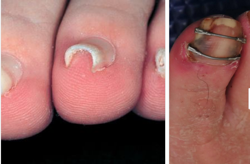
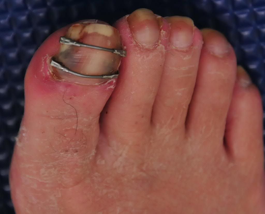
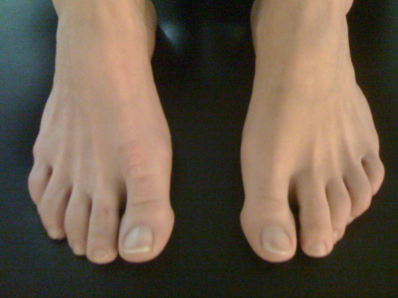
In review, this person (all photos and case premissioned in swap for insight) had the distal hallux removed because of a progressive melanoma on the big toe. Can you believe that ! This is one more reminder that the sun and regular dermatologist screenings are wise.
This person had a complaint of progressing right gluteal and QL pain, spasm, tone and some persistent pain now in the 2nd metatarsal as well as some shoe challenges. We discuss this case briefly in and upcoming podcast, #139 or #140 we believe.

Screen Shot 2018-08-10 at 8.10.19 AM.png

Before we add our final thoughts to this case, lets cap our post from last week.

-Without the hallux, we cannot wind up the windlass and shorten the distance between the first metatarsal and heel, thus the arch will splay (more permanently over time we suspect) and we cannot optimize the arch height.
This will promote more internal spin on that limb because of more midfoot pronation and poor medial foot tripod stabilization.
- More internal limb spin means more internal hip spin, and more demand (which might not be met at the glute level) and thus loads that are supposed to be buffered with hip stabilization, will likely be transferred into the low back, and or into the medial knee. Look for more quad protective tone if they cannot get it from the glutes. Troubles arise when we try to control the hip from quadriceps strategies, it is poorly postured to do so, but people do it everyday, *hint: most cyclists and distance runners to a large degree).
- anterior pelvis posturing on the right, perhaps challenging durability of the lower abdominals, hence suspect QL increased protective tone, possible low back tightness or pain depending on duration of activities
- These factors are likely related to his complaints in the right gluteal and low back/QL area.

Now, onto our next thoughts.

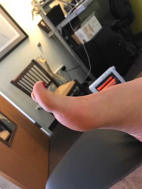
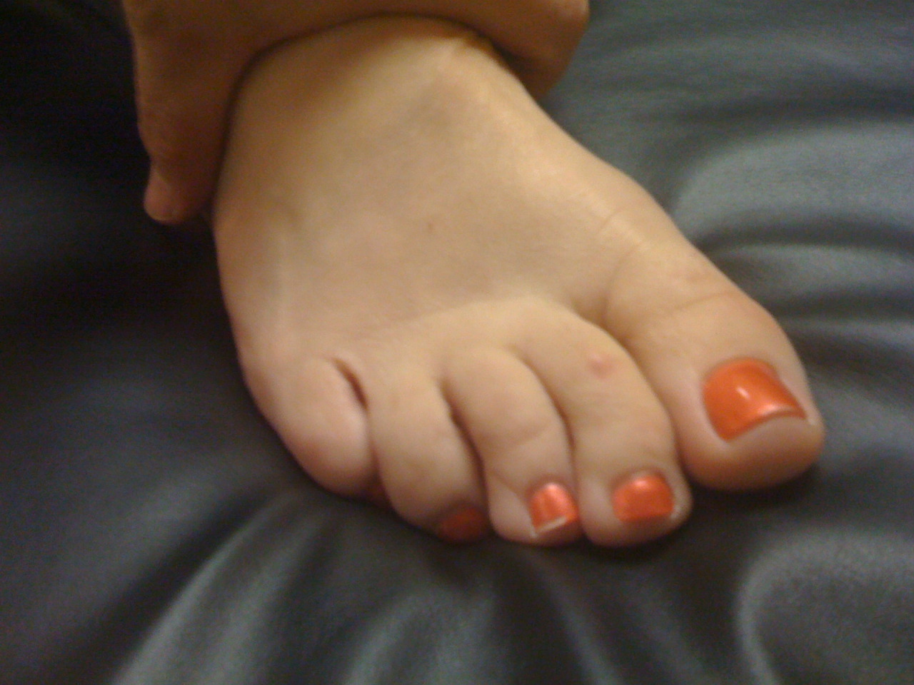
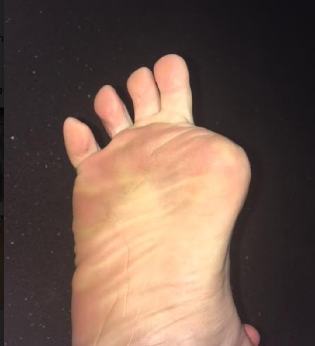
- when the hallux is incompetent, in this case absent, there are few other choices to gain forefoot purchase on the ground other than more flexion gripping of the 2nd toe (then the 3rd, then 4th). This is a progressing "searching" phenomenon for forefoot stability and without the function of the big fella, the 2nd toe will begin a hammering phenomenon, often, but not always. We would not be surprised to see hammer toe development in this case, but this person is now very aware of it, and can at least now fight that battle with increased awareness. There is some mild evidence of this on the side lateral photo.

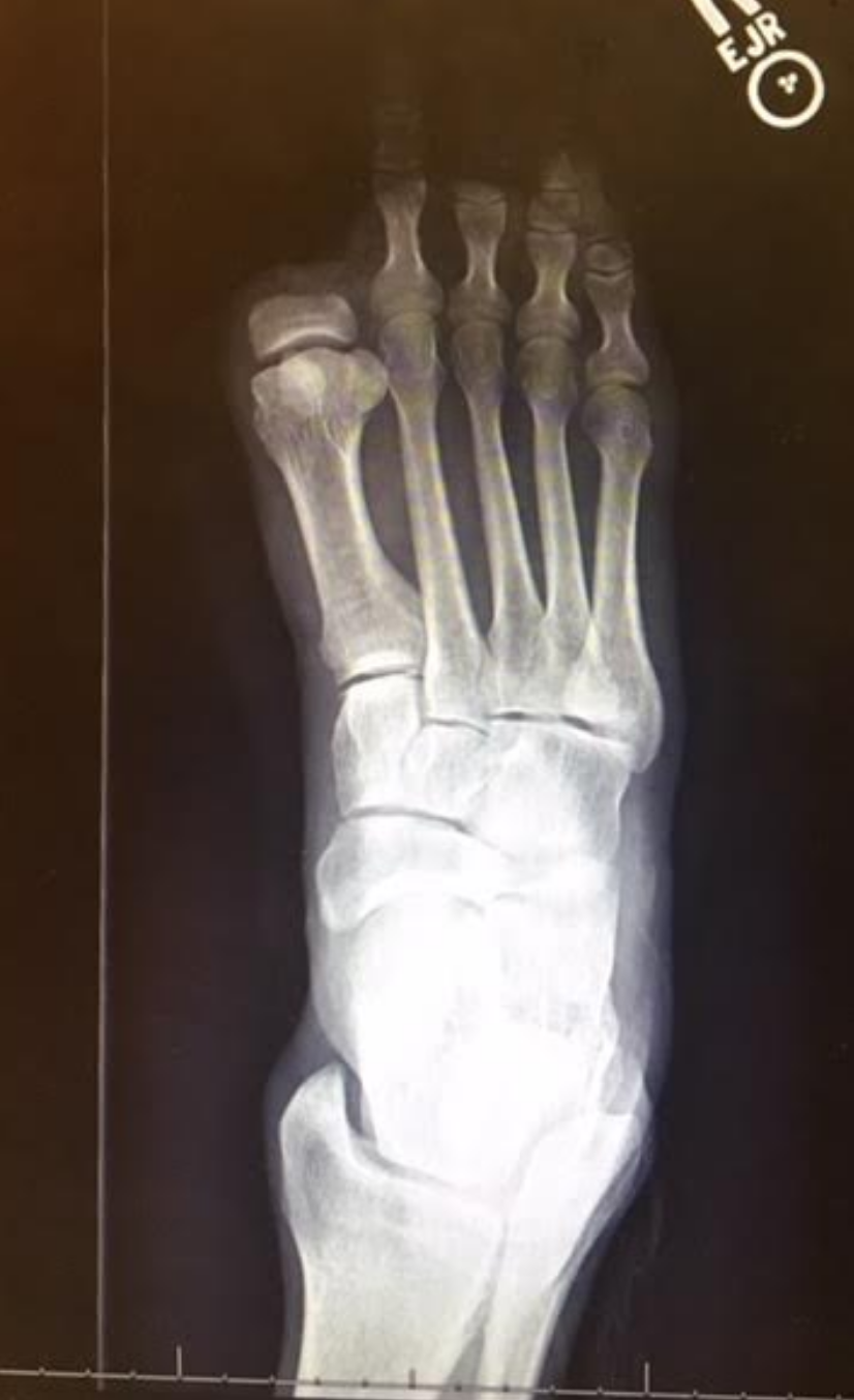
- We are happy to see that the proximal phalange was spared. The adductor hallucis is inserted medially there, and this will help to reduce bunion generation risk (medial metatarsal drift). Comparing the photo and the radiograph is a great example of how far back/proximal the 1st MTP joint is. One could easily assume that the entire hallux was resected from the photo, but the radiograph shows otherwise.

Screen Shot 2018-08-10 at 8.22.36 AM.png

- Toe off is obviously going to be compromised. The patient cannot adequately stabilize the 1st metatarsal (MET) and this will mean a compromised foot tripod, medial foot/tripod splay, arch pronation control challenges but toe off stabilization is going to have to be met by the 2nd and 3rd digits, as discussed above. They are not suited to be the major players here, they are synergistic to this end. Do not be surprised to see one of 2 strategies at toe off here:

1. heavy medial foot tripod toe off, dropping into the void and this maximize the internal spin challenges and minimizing the requisite foot supination stiffness generation phase that should be normal at toe off

2. avoidance of the above, with a forced conscious forefoot lateral toe off, a supinatory strategy, to avoid internal limb spin, more toe hammering, and the lurch heavily and abruptly off of the right foot and onto the left limb.

Screen Shot 2018-08-10 at 8.10.27 AM.png

3. taking #2 further, any time there is perceived challenges or deficits in strength, endurance, proprioception, balance, power and the like, the brain often will create a premature departure off of said limb, creating a requisite premature loading onto the opposite limb. This can cause a phenomenon well loosely refer to "catching" in the contralateral quadriceps mechanism. These clients, with their abrupt loading pattern onto the opposite limb will most often have troubles getting into initial gluteal hip stabilization strategies, and thus default into a quadriceps strategy, that in time can lead to quad shortness and increased tone, which can cause more compression across the patellofemoral joint and cause knee pain. This is more of a compression/loading response issue rather than tracking phenomenon, which we see at the typical diagnosis. We often look for causes in the opposite limb for contralateral knee pain. IT is quite often there if you are looking hard enough for it. Fix the problem, not the symptom.
There is a long host of other things than can arise from here, including heavy contralateral (in this case left sided) foot loading challenges, often more forefoot initial loading, and all of the problems than can arise when this pattern is cyclical, but that would take this post far too deep and long. So, . . . . another time.

Screen Shot 2018-08-10 at 8.09.47 AM.png

4. Shoe fit, we could make the case that a shoe that nicely hugs the forefoot, as opposed to a wide toe box'ed shoe, could help fight off the risk of 1st metatarsal abduction and thus bunion formation risk. However, one cannot dismiss the wider toe box giving the remaining toes a better environment to engage without hammering with over use of long flexors. We might suggest a trial of an elastic sleeve, one often used for plantar fascitis symptom management, placing a snug one around the forefoot when ambulating. This could help keep that metatarsal snug and stop the bunion-like drift we would be watching for.

have at it gang, cases like this are far and deep and require deep understanding of normal and abnormal biomechanics, and the rabbit hole deep myriad of compensations that can be engaged.

have a great weekend !

Shawn and Ivo

Pincer Toe nails: You've seen them; did you know what they were and how they got that way? Or, did you dismiss them?

Screen Shot 2018-04-06 at 8.13.23 AM.png

We think Hitomi’s hypothesis is correct. Here is why (this is paraphrased from our blog post on subungal hematomas and our revolutionary thinking on why they occur and it seems to fit well with pincer nail formation as well).

… when the skin is pulled at a differential rate over the distal phalange (from gripping of the toes rather than downward pressing through the toe pad) there will be a net lifting response of the nail from its bed as the skin is drawn forward of the backward drawn phalange (there is a NET movement of skin forward thus lifting the nail from its bedding). For an at-home example of this, put your hand AND fingers flat on a table top. Now activate JUST your distal long finger flexors so that only the tip of the fingers are in contact with the table top (there will be a small lifting of the fingers). There should be minimal flexion of the distal fingers at this point. Note the spreading and flattening of the nail. Now, without letting the finger tip-skin contact point move at all from the table, go ahead and increase your long flexor tone/pull fairly aggressively. You are in essence trying to pull the finger backward into flexion while leaving the skin pad in the same place on the table. Feel the pressure building under the distal tip of the finger nail as the skin is RELATIVELY drawn forward.] This is fat pad and skin being drawn forward (relative to the phalange bone being drawn backward) into the apex of the nail. Could this be magnifying the curvature of the nail and not offsetting the “automatic curving and shrinkage” function of the nail ? We think it is quite possible.

We have more to say on this topic, the above is just an excerpt of our blog post. More here, in the link below

Too much extensor tone: The banana toe.

IMG_7618.JPG

Too much extensor tone.
We are often talking about the subtle balanced relationship of the long and short toe flexors and extensors. We often discuss that hammer toes are too much long flexor and short extensor tone (with too little in the short flexor and long extensor).
Here we see the opposite. We see too much long extensor tone (note the upward banana-shaped orientation of the big toe). When this foot is on the ground, the pad and distal 1/2 of the big toe does not even touch the ground, standing or in gait. IF you look closely at the blown up pic, you can sort of see (sorry, should have taken more pics) the increased callus development in the contact area of the short flexor attachment (FHB, flexor hallucis brevis).

IMG_7620.PNG

This relationship is the opposite of the above with hammer toes. Too much long extensor, too much short flexor, and not enough long flexor and short extensor. These clients need more homework for long flexor and short extensor. This is one of the reasons why we developed the exercise below in the youtube link.

Unilateral heightened toe extensor tone.

Look at this foot. What do you see ? See the asymmetry ? This is a perfect case to prove our point, for those out there that love the short foot exercise, that insist on towel scrunches, marble pick-ups, or just mere foot rolling on the ball. These things are useless in some cases, arguably to us, much of the time actually. This is about having sufficient foot integrity, normal heel rocker, ankle rocker, forefoot rocker mechanics, and especially in this case, a NORMAL balance between the long and short flexors and extensors. These 4 must work together in harmony, and this is clearly not happening on the left foot. Head on over to this Archived blog post from 2014, and learn what is wrong here. One has to understand it, to fix it. And throwing a short foot regimen, or pilates foot work at it or even more flexor tone into this foot will not fix this. Exercise prescription is supposed to be specific, not a shot gun approach of "try this exercise", lets see if it helps. A 5th grader can give that advice, sadly it is more the industry norm at times. Yes, every exercise is a test, but do not be mistaken that every test is the exercise.

https://thegaitguys.tumblr.com/post/85726861424/unilateral-heightened-toe-extensor-tone-what-do

Unilateral heightened toe extensor tone.

What do we have here ? Well, it is obvious. The left foot is showing increased short extensor tone (EDB: extensor digitorum brevis) and heightened long flexor tone (FDL: flexor digitorum longus). This is the classic pairing for hammer toe development.  We also know from this post (link) and from this post (link) that this presentation is closely related with lumbrical weakness and distal fat pad migration.

So, at an assessment took we like to play games. Mental games to be precise. When we see something like this we immediately begin the mental gyrations of “what could have caused this, and what could this in turn be causing”. Remember, what you see is often not the problem, rather your clients compensation around the problem.  In this case, what goes through your mind ?  Without deep thought, our knee jerk thoughts are:

  • possible loss of ankle rocker dorsiflexion (the increased EDB tone can be recruited to help drive more ankle dorsiflexion indirectly)
  • plantar intrinsic weakness ?
  • flip flops or slip on shoes where the heel is riding up and down inside the shoe/sloppy fit ?  (initiating a gripping response from the FDL)
  • weak tib anterior (recruiting EDB to help)
  • weak peroneus tertius (recruiting EDB again)
  • Ankle /foot instability (more FDL gripping will help gain ground purchase)
  • lateral ankle instablity (same thing, more gripping)
  • Weak gastrosoleus (since the FDL is a posterior compartment neighbor it can kick into high gear and help with posterior comparment function, we have a whole video case based around this issue, check this out ! )
  • premature departure off of the good side leg, and thus an abrupt loading response onto this affected side can challenge the frontal plane of the body and thus require more grip response at the foot level.
  • how about simple weakness of the lumbricals or FDB , the short flexors. The long flexors will have to make up for it and present like this.  
  • the list goes on and on … .

These are just some quick cursory thoughts, and by NO means a complete exhaustive list.  Just some quick thoughts.

But what about hip function ?  if ankle rocker is blocked in terminal stance and the FDL fire like this what will that do to hip extension ? Well, heel rise will be premature because of the limitation and thus hip extension will be abbreviated. Thus glute function will be impaired to a degree.  This can become a viscous cycle, each feeding off of each other.

This diagnostic stuff is a tricky and difficult game. If you think you can diagnose or fix a problem from just changing what you see you are mistaken, unless you like driving compensation patterns and future injuries into your clients.   There must be a hands on examination and assessment with an intact educated brain attached to the process.

Just some mental gymnastics for you today.  

Dr Shawn Allen, one of the gait guys

Short leg and Pronation

Dr Allen was ON FIRE on tonites onlinece.com lecture Biomechanics 322). Hope you will join us again (or next time if you missed us). We talked about many of the aspects of a static exam and how it effects weight bearing in the foot. The word "short leg" came up more than once, and yes, from Dr Allen : )

Remember, as the foot pronates more on one side, the center of gravity will move medially. You will often see more toe clenching (and resultant quadratus plantae weakness) on the more pronatory side and more toe elongation on the more supinatory side. You will often also see more splay and elongation on the pronatory side, and less elongation and less splay on the supinatory side. Remember, these are guidelines and not rules, and there are ALWAYS exceptions.

Screen Shot 2018-01-27 at 8.00.13 AM.png

Flexible hammer toes

IF you've been with us all along you know this one by heart. All you have to do is test it to confirm (never assume, ever !). 
And remember, sometimes people present with their problem, and sometimes they present with a layered compensation to strategize and cope with the underlying problem. It is your job to determine that.
You must recognize here:
flexible hammer toe early development. It is a compensation for weakness somewhere in the chain. It is a gripping strategy to make up for something somewhere. It is not normal, it is a clue.

1- over activity of the short extensors & and long flexors
2- under activity of the long extensors & and short flexors
3- thusly, distal displacement of the Metatarsal fat pad complex
4- and lotsa other things (but we will not detract from the major cursory observation point here today)

Loading this forefoot is troubled, in many ways...... so many that we will not go it here. We have written about these things in long form so many times, just head over to the blog and search. This client did not have any foot or ankle issues, they had impaired hip rotation and extension and were complaining of low back pain. They have obviously been coping through the entire chain for awhile. Be Sherlock Holmes in your practice today, look for the clues.

-Dr. Allen

Podcast 112: Strengthening the foot's arch


Interested in our stuff ? Want to buy some of our lectures or our National Shoe Fit program? Click here (thegaitguys.com or thegaitguys.tumblr.com) and you will come to our websites. In the tabs, you will find tabs for STORE, SEMINARS, BOOK etc. We also lecture every 3rd Wednesday of the month on onlineCE.com. We have an extensive catalogued library of our courses there, you can take them any time for a nominal fee (~$20).
 

Show links:
http://traffic.libsyn.com/thegaitguys/pod_112f.mp3
* and on iTunes, Soundcloud, and just about every other podcast harbor site, just google "the gait guys podcast", you will find us.
 

Show notes:

Job security, become so good and so unique that Ai cant replace your skills as a doctor
http://www.techinsider.io/age-of-ems-machines-will-take-over-all-jobs-2016-8

How prosthetics are working now, and will in the future
and why you should be scared
http://thenextweb.com/insider/2016/08/04/researches-think-we-may-have-to-protect-our-brains-from-hackers-in-a-few-years/

Open talk about how coordination is the first strength changes someone notes. It comes before true strength is achieved. It is neurologic, and its can feel decievingly safe, but it is a lie.

Foot Strengthening ?
https://drjohnrusin.com/advanced-strength-training-for-feet/

http://www.jospt.org/doi/abs/10.2519/jospt.2016.6482?platform=hootsuite&

Impaired Foot Plantar Flexor Muscle Performance in Individuals With Plantar Heel Pain and Association With Foot Orthosis Use

Tags:
foot arch, foot intrinsics, short foot, yoga toes, gastrocnemius, soleus, heel pain, hammer toes, correct toes, foot exercises, thegaitguys, squatting, gait, gait analysis, gait assessment,  orthotics, prosthetics
 

Toe sardines. What have we done to our feet ?

Note that form follows function. If you are observant, you will see the deformation of the 5 digit, just like in this case as the quadratus weakens and the long flexors dominate. The toe begins to spin laterally, and thus the plantar toe pad begins to deform medially, look closely, you can see that here in the video.

Does this look like your foot ? There are a few subtle issues here. 

In the foot, the toe that delineates abduction and adduction of the toes is the 2nd toe. The 2nd toe is considered the anatomic middle of the digits and forefoot. Any toe or movement that moves away from the 2nd toe is abduction and any movement towards the 2nd toe is adduction. This is obviously different than in the hand where the 3rd digit, the one you use during road rage, is the reference digit. Next time you are questioned, tell them you threw them your reference finger, not “the bird”, it is a more accurate descriptor.

In this foot, note how neatly and tightly packed the cute little toes are, all snuggled up to their brothers and sisters. Remember, form follows function. Obviously function has been low on these fellas, at least in abduction.  This often comes from snug toe box footwear and lack of abduction (toe spread) use.  But make no mistake, this is a weak foot.

Today we wish to really focus your attention to an old topic, just a revisit. We can see the 4th and 5th toes curl under from the probably weak lateral head of the quadratus plantae thus encouraging unopposed oblique pull of the long flexors of the digits (FDL). See this post here for an explanation of this phenomenon.  There is also obvious imbalance between the long and short flexors and extensors in these toes, the long flexors are expressing more tone, and that means the long extensors are deprived. 

Note that form follows function. If you are observant, you will see the deformation of the 5 digit, just like in this case as the quadratus weakens and the long flexors dominate. The toe begins to spin laterally, and thus the plantar toe pad begins to deform medially, look closely, you can see that here in the video. This spin can carry the toe nail so far laterally sometimes that the nail can begin to touch the ground during gait and cause painful nail lifting with even some losing the nail. 

There is plenty of life left in this foot, but you have to get to it quickly and get them in lower heeled shoes if tolerable and ones with a wider toe box.  The client needs to be retaught how to access the toe extensors and abductors. Lumbrical retraining, which is a recurrent topic here on our blog, should also be instituted. 

Shawn Allen, one of the gait guys

How quickly does your brain start working when you start your client evaluation ?  Answer: as quickly as possible.Stepping up to the plate today for the first pitch I see this.  Do you see the ever so slight evidence of “possible” right toe extensor…

How quickly does your brain start working when you start your client evaluation ?  

Answer: as quickly as possible.

Stepping up to the plate today for the first pitch I see this.  

Do you see the ever so slight evidence of “possible” right toe extensor tone ? It is subtle but it is staring you right in the face if you care to embrace the subtle clue.  From this angle you can see more of the fat pad and plantar aspect of the RIGHT forefoot. The very next thing we did was look at the dorsum of the foot, and yup, more prominent extensor tone, short extensors to be specific.  The next question was why was I seeing this ? Short flexors usually pair with long toe flexors, and that means gripping the ground and distal displacement of the fat pad and even sometimes lumbrical inhibition or frank weakness. Possible attempts to gain more foot purchase on the ground ? Hmmmmm, perhaps.  The client had right hip pain and right lateral sesamoid pain.  Time for an examination to find out the “whys” and then fix things.

The powers of observation should always get your clinical juices flowing. Even the smallest of things should help guide you or at least clue you in to things.

tumblr_ntnuoif8Tj1qhko2so1_r1_500.jpg
tumblr_ntnuoif8Tj1qhko2so2_r1_1280.jpg

Pincher nails.  Who knew !?

Written by Dr. Shawn Allen

*note: there are two photos here in today’s blog post, look for the side scroll arrows and click on the small box in the upper left corner if you cannot see the photo

 We have seen this one for years in our clinics but we never got around to researching it and pondering the condition more deeply.  Here is our mantra for today, Form follows Function.

Studies seem to be undecided on the cause of this entity. Some suggest that pincer nails are caused by lack of upward mechanical forces on the toe pad where others remark that they can be observed amongst the healthy mechanical walkers. Hitomi’s study suggests that the affected toes fail to receive adequate physical stimulation from proper toe and forefoot loading. Please read on, this gets interesting.

According to Hitomi’s study, in both the barefoot and shod state,

“the pincer nail group had significantly lower pressure on the first toe than the control group. In both the barefoot and shod state, the peak pressure area was mostly the metatarsal head area in the pincer nail group, whereas it was mostly the first toe area in the control group. Binomial logistic regression analysis revealed that peak pressure area was a significant risk factor for pincer nail development.”

This seems to suggest that there is insufficient or aberrant use of downward pressure on the toes and into the toe pads. Hitomi speaks of the locale of the peak pressure, seemingly proposing from this study that it should not be under the metatarsal heads. This, in our experience and thinking, could suggest that more long flexor dominance is present. This long flexor activity seems to create some disfunction not only in the activity of the lumbrical muscles but also altered pressures in the metatarsal (MET) heads.  It certainly alters distal toe pressures which can alter skin and nail responses (see our blog post on subungal hematomas for more on this topic where we discuss principles of counter pressure and shear forces). We try to teach a “spread and reach with long flat toes” approach to our clients in correcting bad habits such as toe hammering and gripping (which are often a result of flawed biomechanics elsewhere).

The nail bed is very rich in vasculature (hence the cause of the dreaded hematoma, the black toenail) and nerve endings.  The nail bed is a derivative of the epidermis containing keratin which gives it its hard nature. The nail consists of the nail plate, the nail fold, the nail matrix, the sterile matrix and the hyponychium. There are many factors that go into the formation of a normal nail, including blood flow, nutrition, local neurogenic factors and not to forget, mechanical loading issues. Failure of any of these issues can lead to softening, brittle, thinning, diseased or malformed nails. The nail grows from a nail root in front of the cuticle and grows distally at a slow but (usually) steady rate.  It is interesting to note that the long extensor tendon (EDL) attachment is close to the proximal nail bed root area thus it brings forward thinking of possible imbalances between long and short flexors and extensor tendons/muscles and their patterns of imbalance in toe gripping and hammering that could cause a change in function which could drive a change in form.  We have all heard it, form follows function, why should this area be any different ?

Hitomi also mentioned something interesting in his study, the observation that bed ridden clients seem to have a predilection to pincer toes.  This at least seems to fit the aberrant loading patterns, in this case an absence of. The study also started some interesting thinking in us when it mentioned a hypothesis,

“that human nails are constitutively equipped with an automatic shrinkage function that allows them to adapt to daily upward mechanical forces.”

This was a fascinating hypothesis to us. It seems to make sense. If constant downward pressure on the toe pads were present, the toe nails would always be undergoing a flattening and spreading response so it could make sense that the nails have a built in curve and shrinkage function offsetting and adapting to the constant distorting pressures (the flattening and spreading forces).  Hence, some possible clarity in Hitomi’s hypothesis that pincer nails are caused by lack of (and in our thinking, distorted) upward mechanical forces on the toe pad.  And, when those distorting pressures are placed elsewhere (ie. the MET heads or tips of the toes as in our subungal hematoma hypothesis) or faulting gripping or hammering loading the automatic shrinkage function is left to dominate.

We think Hitomi’s hypothesis is correct. Here is why (this is paraphrased from our blog post on subungal hematomas and our revolutionary thinking on why they occur and it seems to fit well with pincer nail formation as well).

…  when the skin is pulled at a differential rate over the distal phalange (from gripping of the toes rather than downward pressing through the toe pad) there will be a net lifting response of the nail from its bed as the skin is drawn forward of the backward drawn phalange  (there is a NET movement of skin forward thus lifting the nail from its bedding).  For an at-home example of this, put your hand AND fingers flat on a table top. Now activate JUST your distal long finger flexors so that only the tip of the fingers are in contact with the table top (there will be a small lifting of the fingers). There should be minimal flexion of the distal fingers at this point. Note the spreading and flattening of the nail.  Now, without letting the finger tip-skin contact point move at all from the table, go ahead and increase your long flexor tone/pull fairly aggressively. You are in essence trying to pull the finger backward into flexion while leaving the skin pad in the same place on the table. Feel the pressure building under the distal tip of the finger nail as the skin is RELATIVELY drawn forward.]   This is fat pad and skin being drawn forward (relative to the phalange bone being drawn backward) into the apex of the nail. Could this be magnifying the curvature of the nail and not offsetting the “automatic curving and shrinkage” function of the nail ? We think it is quite possible.

So, there you have it. We will dive deeper on this topic another time, but after reading Hitomi’s study our brain’s started buzzing because we had discussed this process similarly a few years back in our Subungal Hematoma blog post.

And, if you are thinking about chronic repeated ingrown toe nails with this clinical entity, your thoughts are clearly on a logical path.  There is a correlation it seems.

And, as for the horrific metal bar correction you see in the other photo above, this too is new to our eyes.  It seems rather medieval, something one might see in the gallows of yesteryear.  And if that doesn’t curl your hair and make you nauseated, try looking at what this one guy did, a DIY remedy (caution, not for the feint of heart). https://www.mja.com.au/journal/2005/182/4/diy-pincer-nail-repair-brace-yourself

ShawnAllen, one of the gait guys

References:

Foot loading is different in people with and without pincer nails: a case control study  Hitomi Sano1*, Kaori Shionoya2 and Rei Ogawa1  Journal of Foot and Ankle Research 2015, 8:43

The Bouncy Gait: Premature heel rise gait. Taking another look.

This is a great video example of a premature heel rise during gait. You should be able to clearly see it on the left foot (and this was toned down after we brought it to his awareness!).  The heel rise occurs early in the stance phase of gait, instead of the late stance phase.

We have talked about this bouncy type vertically oriented gait many times in blog posts and in our podcasts.  This is a pretty prevalent problem in the world, mostly because so many people have impaired ankle rocker/dorsiflexion from weak anterior compartments and short/tight posterior compartments.  None the less, for the majority, this is a pathologic gait pattern and it will impart undue stress into the posterior mechanism (calf-achilles complex). Just think about it, this person is going vertical at or prior to the tibia achieving 90degrees (perpendicular to the ground) instead of continuing to progress the tibia to 110+ degrees to enable normal timely pronation and foot biomechanical events.  This is not a normal gait. Period. This will change the function of the entire posterior chain upward. 

If you want to see another great example  from the frontal plane, check out this cute video representation of a vertial/premature heel rise bouncy gait. 

This gait style is caused by a premature heel rise from joint range limitation and/or from premature engagement of the gastrosoleus (and sometimes even the long toe flexors, you will see them hammering and curled in many folks). It can be a learned habitual pattern and nothing more, we have even seen it even in child-parental gait modeling in our offices. These people will never get to NORMAL full late-midstance of gait (without biomechanical compromise) and thus never achieve full hip extension nor adequate ankle dorsiflexion / ankle rocker. The gait cycle is an orchestrated symphony of timely events and when one or several timely events are omitted or impaired the mechanics are passed into other areas for compensation. This vertical gait style is very inefficient in that the gluteals cannot adequately power into hip extension into a forward progression drive, because the calf is prematurely generating vertical movement through ankle plantarflexion.  This strategy is sometimes deployed because the person actually is significantly ankle dorsiflexion (ankle rocker) deficient.  Meaning, they hit the limitations of dorisflexion and in order to progress forward they first have to go vertical.  This vertical motion, because they are moving into ankle plantarflexion, re-buys more ankle dorsiflexion range which then can be used if they so choose. Obviously, the remedy is to find the functional deficit, remove it and retrain the pattern.  There are a whole host of other problems that go with this compensation pattern but we wanted our mission to stay focused today.  Remember, this is usually a subconscious motor pattern compensation. Is it like the toe walking issue we talked about last week (post link here) ? It is similar in some ways and can have primitive and postural motor pattern implications. We will follow up the “Idiopathy Toe Walking Gait: Part 2” shortly but we wanted to strategically put this blog post ahead of it, because there are similar characteristics and implications. Trust us, there is a method to our madness :)

Shawn and Ivo

The Gait Guys

tumblr_nd32fvOzjx1qhko2so3_r1_1280.png
tumblr_nd32fvOzjx1qhko2so2_1280.png
tumblr_nd32fvOzjx1qhko2so1_1280.png
tumblr_nd32fvOzjx1qhko2so4_r1_500.png

Part 2: “Standing on Glass” Static Foot/Pedograph Assessment

* note (see warning at bottom): This is a static assessment dialogue. One cannot, and must not, make clinical decisions from a static assessment. The right and left sides are indicated by the R and L circled in pink. There are 4 photos here today.

Blue lines: Last time we evaluated possible ideas on the ORANGE lines here, it would be to your advantage to start there. 

We can see a few noteworthy things here in these photos. We have contrast-adjusted the photo so the pressure areas (BLUE) are more clearly noted. There appears to be more forefoot pressure on the right foot (the right foot is on the readers left), and more rearfoot pressure on the left (not only compare the whiteness factor but look at the displacement of the calcaneal fat pad (pink brackets). There is also noticeably more lateral forefoot pressure on the left. There is also more 3-5 hammering/flexion dominance pressure on the left.  The metatarsal fat pad positioning (LIME DOTS represent the distal boundary) is intimately tied in with the proper lumbrical muscle function  (link) and migrates forward toward the toes when the flexors/extensors and lumbricals are imbalanced. We can see this fat pad shift here (LIME DOTS). The 3-5 toes are clearly hammering via flexor dominance (LIME ARROWS), this is easily noted by visual absence of the toe shafts, we only see the toe pads. Now if you remember your anatomy, the long flexors of the toes (FDL) come across the foot at an angle (see photo). It is a major function of the lateral head of the Quadratus plantae (LQP) to reorient the pull of those lesser toe flexors to pull more towards the heel rather than on an angle. One can see that in the pressure photos that this muscle may be suspicious of weakness because the toes are crammed together and moving towards the big toe because of the change in FDL pull vector (YELLOW LINES). They are especially crowding out the 2nd toe as one can see, but this can also be from weakness in the big toe, a topic for another time. One can easily see that these component weaknesses have allowed the metatarsal fat pad to migrate forward. All of this, plus the lateral shift weight bearing has widened the forefoot on the left, go ahead, measure it. So, is this person merely weight bearing laterally because they are supinating ? Well, if you read yesterday’s blog post we postulated thoughts on this foot possibly being the pronated one because of its increased heel-toe and heel-ball length. So which is it ? A pronated yet lateral weight bearing foot  or a normal foot with more lateral weight bearing because of the local foot weaknesses we just discussed ? Or is it something else ? Is the problem higher up, meaning, are they left lateral weight bearing shift because of a left drifted pelvis from weak glute medius/abdominal obliques ?  Only a competent clinical examination will enlighten us.

Is the compensation top-down or bottom up, or both in a feedback cycle trying to find sufficient stability and mobility ? These are all viable possibilities and you must have these things flowing freely through your head during the clinical examination as you rule in/rule out your hands-on findings.  Remember, just going by a screen to drive prescription exercises from what you see on the movement screen is not going to necessarily fix the problem, it could in fact lead one to drive a deeper compensation pattern. 

Remember this critical fact.  After an injury or a long standing problem, muscles and motor patterns jobs are to stabilize and manage loads (stability and mobility) for adequate and necessary movement. Injuries leave a mark on the system as a whole because adaptation was necessary during the initial healing phase. This usually spills over during the early movement re-introduction phase, particularly if movement is reintroduced too early or too aggressively.  Plasticity is the culprit. Just because the injury has come and gone does not mean that new patterns of skill, endurance, strength (S.E.S -our favorite mnemonic), stability and mobility were not subsequently built onto the apparently trivial remnants of the injury.  There is nothing trivial if it is abnormal. The forces must, and will, play out somewhere in the body and this is often where pain or injury occurs but it is rarely where the underlying problem lives.

Come back tomorrow.  We will try to bring this whole thing together, but remember, it will just be a theory for without an exam one cannot prove which issues are true culprits and which are compensations. Remember, what you see is often the compensatory illusion, it is the person moving with the parts that are working and compensating not the parts that are on vacation.  See you tomorrow friends !

Shawn and ivo, the gait guys

* note: This is a static assessment dialogue. One cannot, and must not, make clinical decisions from a static assessment. As in all assessments, information is taken in, digested and then MUST be confirmed, denied and/or at the very least, folded into a functional and clinically relevant assessment of the client before the findings are accepted, dismissed and acted upon. As we always say, a gait analysis or static pedograph-type assessment (standing force plate) is never enough to make decisions on treatment to resolve problems and injuries. What is seen and represented on either are the client’s strategies around clinical problems or compensations.  Today’s photo and blog post are an exercise in critical clinical thinking to get the juices flowing and to get the observer thinking about the client’s presentation and to help open up the field to questions the observer should be entertaining.  The big questions should be, “why do i see this, what could be causing these observances ?”right foot supinated ? or more rear and lateral foot……avoiding pronation ?

The “Standing on Glass” Static Foot/Pedograph Assessment: Part 1
* note: This is a static assessment dialogue. One cannot, and must not, make clinical decisions from a static assessment. As in all assessments, information is taken in, di…

The “Standing on Glass” Static Foot/Pedograph Assessment: Part 1

* note: This is a static assessment dialogue. One cannot, and must not, make clinical decisions from a static assessment. As in all assessments, information is taken in, digested and them MUST be confirmed, denied and/or at the very least, folded into a functional and clinically relevant assessment of the client before the findings are accepted, dismissed and acted upon. As we always say, a gait analysis or pedograph-type assessment is never enough to make decisions on treatment to resolve problems and injuries. What is seen and represented on either are the client’s strategies around clinical problems or compensations.  Today’s photo and blog post are an exercise in critical clinical thinking to get the juices flowing and to get the observer thinking about the client’s presentation and to help open up the field to questions the observer should be entertaining.  The big questions should be, “why do i see this, what could be causing these observances ?”

* note the right and left sides by the R and L circled in pink.

ORANGE lines: The right foot appears to be shorter, or is it that the left is longer (see the lines and arrows drawing your attention to these differences)? A shorter foot could be represented by a supinated foot (if you raise the arch via the windlass mechanism you will shorten the foot distance between the rear and forefoot). A longer foot could be represented by a more pronated foot.  Is that what we have here ? There is no way to know, this is a static presentation of a client standing on glass. What we should remember is that the goal is always to get the pelvis square and level.  If an anatomically or functionally short leg is present, the short leg side MAY supinate to raise the mortise and somewhat lengthen the leg.  In that same client, they may try to meet the process part way by pronating the other foot to functionally “shorten” that leg.  Is that what is happening here ? So, does this client have a shorter right leg ? Longer left ?  Do you see a plunking down heavily onto the right foot in gait ? Remember, what you see is their compensation.  Perhaps the right foot is supinating, and thus working harder at the bottom end of the limb (via more supination), to make up for a weak right glute failing to eccentrically control the internal spin of the leg during stance phase ? OR, perhaps the left foot is pronating more to drive more internal rotation on the left limb because there is a restricted left internal hip rotation from the top ? Is the compensation top-down or bottom up ? These are all viable possibilities and you must have these things flowing freely through your head during the clinical examination as you rule in/rule out your hands-on findings.  Remember, just going by a FMS-type screen to drive prescription exercises from what you see on a movement screen is not going to necessarily fix the problem, it could in fact lead one to drive a deeper compensation pattern. You can be sure that Gray Cook’s turbo charged brain is juggling all of these issues (and more !) when he sees a screen impairment, although we are not speaking for him here.

Remember this critical fact.  After an injury or a long standing problem, muscles and motor patterns jobs are to stabilize and manage loads (stability and mobility) for adequate and necessary movement. Injuries leave a mark on the system as a whole because adaptation was necessary during the initial healing phase. This usually spills over during the early movement re-introduction phase, particularly if movement is reintroduced too early or too aggressively.  Plasticity is the culprit. Just because the injury has come and gone does not mean that new patterns of skill, endurance, strength (S.E.S -our favorite mnemonic), stability and mobility were not subsequently built onto the apparently trivial remnants of the injury.  There is nothing trivial if it is abnormal. The forces must, and will, play out somewhere in the body and this is often where pain or injury occurs but it is rarely where the underlying problem lives.

Come back tomorrow, where we will open your mind into the yellow, pink, blue and lime markings on the photo. Are the hammering toes (lime) on the left a clue ? How about the width of the feet (yellow) ? The posturing differences of the 5th toe to the lateral foot border ?  What about the static plantar pressure differences from side to side (blue)? Maybe, just maybe, we can bring a logical clinical assumption together and then a few clinical exam methods to confirm or dis-confirm our working diagnostic assumption.  See you tomorrow friends !

Shawn and ivo, the gait guys

Unilateral heightened toe extensor tone.
What do we have here ? Well, it is obvious. The left foot is showing increased short extensor tone (EDB: extensor digitorum brevis) and heightened long flexor tone (FDL: flexor digitorum longus). This is the …

Unilateral heightened toe extensor tone.

What do we have here ? Well, it is obvious. The left foot is showing increased short extensor tone (EDB: extensor digitorum brevis) and heightened long flexor tone (FDL: flexor digitorum longus). This is the classic pairing for hammer toe development.  We also know from this post (link) and from this post (link) that this presentation is closely related with lumbrical weakness and distal fat pad migration.

So, at an assessment took we like to play games. Mental games to be precise. When we see something like this we immediately begin the mental gyrations of “what could have caused this, and what could this in turn be causing”. Remember, what you see is often not the problem, rather your clients compensation around the problem.  In this case, what goes through your mind ?  Without deep thought, our knee jerk thoughts are:

  • possible loss of ankle rocker dorsiflexion (the increased EDB tone can be recruited to help drive more ankle dorsiflexion indirectly)
  • plantar intrinsic weakness ?
  • flip flops or slip on shoes where the heel is riding up and down inside the shoe/sloppy fit ?  (initiating a gripping response from the FDL)
  • weak tib anterior (recruiting EDB to help)
  • weak peroneus tertius (recruiting EDB again)
  • Ankle /foot instability (more FDL gripping will help gain ground purchase)
  • lateral ankle instablity (same thing, more gripping)
  • Weak gastrosoleus (since the FDL is a posterior compartment neighbor it can kick into high gear and help with posterior comparment function, we have a whole video case based around this issue, check this out ! )
  • premature departure off of the good side leg, and thus an abrupt loading response onto this affected side can challenge the frontal plane of the body and thus require more grip response at the foot level.
  • how about simple weakness of the lumbricals or FDB , the short flexors. The long flexors will have to make up for it and present like this.  
  • the list goes on and on … .

These are just some quick cursory thoughts, and by NO means a complete exhaustive list.  Just some quick thoughts.

But what about hip function ?  if ankle rocker is blocked in terminal stance and the FDL fire like this what will that do to hip extension ? Well, heel rise will be premature because of the limitation and thus hip extension will be abbreviated. Thus glute function will be impaired to a degree.  This can become a viscous cycle, each feeding off of each other.

This diagnostic stuff is a tricky and difficult game. If you think you can diagnose or fix a problem from just changing what you see you are mistaken, unless you like driving compensation patterns and future injuries into your clients.   There must be a hands on examination and assessment with an intact educated brain attached to the process.

Just some mental gymnastics for you today.  

Shawn and Ivo

the gait guys

tumblr_mwzu9gTBzS1qhko2so1_1280.jpg
tumblr_mwzu9gTBzS1qhko2so2_400.gif
tumblr_mwzu9gTBzS1qhko2so3_r1_1280.jpg
tumblr_mwzu9gTBzS1qhko2so4_r1_1280.jpg

Is your 5th toe curled under ? What do you do when “this little piggy” can’t go wee wee wee all the way home.

Have a look at the 4 photos above.  You will see this curling of the lesser toes quite often in your practice, and when you know what it means it can help to guide your thinking, both from a diagnostic and treatment perspective.  

You should have noticed in the photos that the 4th and 5th toes curl under and are hyper-flexed, and this is at rest.  So, what does this mean ?

It means that the long flexors are overactive, the extensors are underactive, and the adduction pull of the long flexors is unopposed by the under appreciated quadratus plantae muscle.

Look at the clinical drawing. The quadratus plantae has 2 heads, a medial head and a lateral head.  Being able to clinically test these two heads will give you much insight into the function of the foot and when you see these outer two toes curling under, as you see in the photo, you will always see weakness of the lateral head of the quadratus plantae.  

The quadratus plantae arises from two heads separated from each other by the long plantar ligament. The medial head is larger and more muscular, attached to the medial calcaneus;  the lateral head is smaller and more tendinous, attaching to the lateral border of the inferior surface of the calcaneus and the long plantar ligament.  The two portions join and end in a flattened band which inserts into the lateral, upper and under surfaces of the tendons of the flexor digitorum longus, usually the second, third, and fourth toes.

But this time, if you have studied the drawing, you should notice the oblique line of pull of the long flexors.  This should in fact create this undesirable curling effect of the lateral two toes since they are so far out on the oblique line of pull. However, if you look at the insertion of the lateral head of the quadratus plantae you should be able to conclude that this head is designed to offset this oblique pull of the outer two long flexor tendons.  The quadratus creates a posterior pull on the outer long flexor tendons ensuring that the curling effect (as seen in the photo) is nullified. Thus, we have a clinical presentation of a weak lateral head of the quadratus plantae (and probably a few others which we will not discuss here so as to not dilute the purpose of today’s post). Now you just have to figure out why it is weak or if there is a biomechanical reason for its insufficiency

  • is there a foot type presenting itself that makes it difficult for this muscle to create sufficient posterior pull to offset the tremendous leverage of the long flexors? Maybe a forefoot varus, which gives the flexor tendons a mechanical advantage or a forefoot valgus which puts the quadratus plantae at a mechanical disadvantage? (Taking our National Shoe Fit Certification Program will help you get closer to understanding many of these issues.)
  • Are their other anatomical variants like an increased forefoot width or bunions (medial or tailor’s)
  • is there excessive rear or midfoot pronation?
  • Shoe choice problem ?

Some folks do have adequate function of the quadratus plantae. Note the lovely feet in the last picture … .  they must have strong lateral quadratus plantae and abductors of the lateral foot and toes ! And, they have great toe separation, thus great intrinsic interossei muscles, and nice flat toes (great balance between flexors and extensors).

So, what do you do?

  • you could do a surgery, amputate or fuse some of the joints to make them look better. Extreme for a problem like this
  • you could ignore the issue and hope it goes away. (in all likelihood it will worsen)
  • you could give them long flexor, toe scrunching Towel-curling, marble-grasping exercises , like you see all over the internet…and give the flexor digitorum longus even more of a mechanical advantage, and make the problem worse
  • you could give them exercises to increase the function of the long extensors, which would increase the mechanical advantage of the quadratus plantae. like the shuffle walk; lift, spread and reach and tripod standing exercises (hmm…sounding better)
  • be a real clinician and in addition to looking at the foot, look north of the foot to see what might be causing the problem (loss of ankle rocker, insufficient gluteal activity, loss of internal rotation of the hip, etc) Hmmm; sounding like a good idea too…

The Gait Guys. Hammering it home, day after day, about the importance of gait and giving you clues to be a better _________ (insert athlete, coach, trainer, clinician, shoe fitter, rehab specialist…).

Podcast 44: New knee ligaments and Ankle Rocker

The newly discovered knee ligament, ankle rocker, hammer toes, yoga, joint flexibility and more ! Download Podcast # 44 today !

A. Link to our server:

http://thegaitguys.libsyn.com/podcast-44-new-knee-ligaments-and-ankle-rocker

B. iTunes link:

https://itunes.apple.com/us/podcast/the-gait-guys-podcast/id559864138

C. Gait Guys online /download store (National Shoe Fit Certification and more !) :

http://store.payloadz.com/results/results.aspx?m=80204

D. other web based Gait Guys lectures:

www.onlinece.com   type in Dr. Waerlop or Dr. Allen,  ”Biomechanics”

________________________________________

* Today’s show notes:

Neuroscience:
New ligament discovered in the human knee
http://www.sciencedaily.com/releases/2013/11/131105081352.htm
3. Brain and Motion
‘Anklebot’ Helps Determine Ankle Stiffness
8. Blog reader:
richies77 asked a questionHi, Incredible source of information. I have severe arthritis in the 2nd toe of my left foot. I have very little dorsiflexion and this has caused my hip flexor to become chronically, extremely tight. This has twisted my entire spine and made me pretty much disabled. I’ve been offered orthotics and perhaps rocker shoes but do you think surgery is the only way to bring back correct balance to my spine? Does anything else actually work? Thank you!
9. In the News:
Yoga and the Brain:
11. another blog reader:
What should I start doing for early cerebellar atrophy symptoms? I’m 6'5 195 and an athlete
 
12. CADENCE and BAREFOOT


Why alignment of the big toe is so critical to gait, posture, stabilization motor patterns and running.

There are two ways of thinking about the arch of the foot when it comes to competent height.  One perspective is to passively jack up the arch with a device such as an orthotic, a choice that we propose should always be your last option, or better yet to access the extrinsic and intrinsic muscles of the foot (as shown in this video) to compress the legs of the foot tripod and lift the arch dynamically.  Here today we DO NOT discuss the absolute critical second strategy of lifting the arch via the extensors as you have seen in our “tripod exercise video” (link here) but we assure you that regaining extensor skill is an absolute critical skill for normal arch integrity and function.  We like to say that there are two scenarios going on to regain a normal competent arch (and that does not necessarily mean a high arch, a low arch can also be competent….. it is about function and less about form): one scenario is to hydraulically lift the arch from below and the other scenario is to utilize a crane-like effect to lift the arch from above. When you combine the two, you restore the arch function.  In those with a flat flexible incompetent foot you can often regain normal alignment and function.  But remember, you have to get to the client before the deforming forces are significant enough and have been present long enough that the normal anatomical alignments are no longer possible. For example, a hallux valgus with a large bunion (this person will never get to the abductor hallucis sufficiently) or a progressively collapsed arch that is progressively becoming rigid or semi-rigid.

Think about these concepts today as you watch your clients walk, run or exercise.  And then consider this study below on the critical importance of the abductor hallucis muscle after watching our old video of Dr. Allen’s competent foot.  

CONCLUSIONS AND CLINICAL RELEVANCE:

The abductor hallucis muscle acts as a dynamic elevator of the arch. This muscle is often overlooked, poorly understood, and most certainly rarely addressed. Understanding this muscle and its mechanics may change the way we understand and treat pes planus, posterior tibial tendon dysfunction, hallux valgus, and many other issues that lead to a challenge of the arch, effective and efficient gait. Furthermore, its dysfunction and lead to many aberrant movement and stabilization strategies more proximally into the kinetic chains.

*From the article referenced below,  “Most studies of degenerative flatfoot have focused on the posterior tibial muscle, an extrinsic muscle of the foot. However, there is evidence that the intrinsic muscles, in particular the abductor hallucis (ABH), are active during late stance and toe-off phases of gait.“

We hope that this article, and the video above, will bring your focus back to the foot and to gait for when the foot and gait are aberrant most proximal dynamic stabilization patterns of the body are merely strategic compensations.

Study RESULTS:

All eight specimens showed an origin from the posteromedial calcaneus and an insertion at the tibial sesamoid. All specimens also demonstrated a fascial sling in the hindfoot, lifting the abductor hallucis muscle to give it an inverted ‘V’ shaped configuration. Simulated contraction of the abductor hallucis muscle caused flexion and supination of the first metatarsal, inversion of the calcaneus, and external rotation of the tibia, consistent with elevation of the arch.

http://www.ncbi.nlm.nih.gov/pubmed/17559771

Foot Ankle Int. 2007 May;28(5):617-20.

Influence of the abductor hallucis muscle on the medial arch of the foot: a kinematic and anatomical cadaver study.

Wong YS. Island Sports Medicine & Surgery, Island Orthopaedic Group, #02-16 Gleneagles Medical Centre, 6 Napier Road, Singapore, 258499, Singapore. 

tumblr_mpq5inFe9Z1qhko2so2_1280.jpg
tumblr_mpq5inFe9Z1qhko2so3_1280.jpg
tumblr_mpq5inFe9Z1qhko2so4_1280.jpg
tumblr_mpq5inFe9Z1qhko2so5_1280.jpg
tumblr_mpq5inFe9Z1qhko2so6_1280.jpg

The Rigid Flat Foot: Do you know what you are actually dealing with ?

In these 5 photos of a client with a flat arch we see some great opportunity to discuss some of the clinical issues and clinical thinking that needs to occur.  As usual we write our blog posts on the fly with a principle at hand that we want to drive home, or in this case “into the ground”.  There are many more clinical issues with this type of foot and its problems, so today’s list and dialogue is not meant to be exhaustive.  But, if you take one thing away from this case, it should be that not all flat feet can take a stability shoe or an orthotic. So, if you are in the mind set that “when it is flat, jack it up (the arch)” and “when it is high (the arch), cushion it” hopefully you will open your eyes a bit to the reality that it just is not that simple.  IF you want to learn more about these issues we have purposefully put together the National Shoe Fit program for stores and doctors/therapists so they can learn more about the anatomy of the feet and shoes and how to pair them up to create the best recipe for a person.  

Now, onto this case.

In this case you should notice a few things. 
1- the rigidity of the flat foot as portrayed in the photo where we are pushing up with our thumb on what once was the peak of the arch (yes, there are 5 photos in this case, click on one to enlarge or scroll) . We are attempting to push up, but the midfoot is completely rigid. This is a classic Rigid Flat Foot Deformity, A Rigid Pes Planus if you will. 

2- There is a prominence at the navicular bone, both top (dorsal) and bottom (plantar) aspects of the foot (see photo of my hand with finger and thumb indicating these areas). The plantar prominence is the actual naviular bone (mostly) that has become weight bearing (termed “weight bearing  navicular” and crudely by some as a dropped navicular, a term we dislike). And the dorsal prominence is a dorsal crown of osteophytes. This means a dorsal ridge of bone has formed at the navicular-1st cuneiform bone/joint interval because of the constant and repetitive compression of the two against each other dorsally as midfoot arch collapse occurred repeatedly and then became a fixed permanent entity.

3- The hyper dorsiflexion range at the 1st MTP joint (the big toe). This range is excessive at actually was able to exceed 90 degrees (see photo) !  Even at rest the hallux (big toe) is extended suggesting the volume of dorsiflexion it gets all the time.  By the way, there was little to no hallux 1st MPJ joint plantarflexion (downward bend), not in a foot this flat. In fact most of that is from the contracture of the short extensors of the toes as noted by the photo showing the hammer toe formation (hammer toe = contractured short extensor myotendon, and to the long flexors as well). Hammer toes are almost always seen in a flat foot presentation, to a degree.

Now, lets put some things together (but a reminder, this is a single principle today, there are many more issues here).

Today’s Principle: Passing the Buck

Normally we need to have just slightly greater than 90 degrees of ankle mortise dorsiflexion to progress the body over the ankle.  Put in other words, we need to be able to get the tibia slightly past vertical (perpendicular to the ground, hence 90+ degrees). Depending on the reference, anywhere from 15-25 degrees past that 90 degree vertical, thus 105 to 120 degrees) is the goal.

If an ankle cannot get that range, the range must be achieved either proximal or distal to that joint, ie. Passing the Buck beyond the ankle mortise joint.  Proximally, one can hyperextend the knee to enable the body mass to pass sagittally over the ankle but a better strategy (arguably) is to compensate distally via collapsing the arch and pronate more than normally through the midfoot putting undue stress and strain into the plantar fascia and over time eventually collapsing the arch and creating the dorsal and plantar bony prominences we mentioned in #2. By dropping the arch, the subtalar joint exceeds its ranges and the talus and navicular collapse medially and plantarwards. 
When the arch drops to the planus stage the tibia is passively thrust forward achieving the necessary forward tibial progression to get body over and past the ankle to enable forward progression. 
Remember, this pes planus will dorsiflex the long metatarsal bone (meaning make it parallel to the ground). This will screw up the 1st Metatarsal-phalangeal joint function and  impair the Windlass Mechanism of Hicks at the big toe (translation, it will impair the sesamoids, possibly leading to sesamoiditis, and change the normal toe function and its tendons.  This is seen both in the pes planus foot and in hallux rigidus turf toe presentations where the big toe loses its  normal ranges as compared to this case here).

So, the normal range can as for the buck to be passed proximally into the kinetic chain or distally. Which one would you want, if you had to chose?  It is a tough choice, luckily the body decides for us.  IF you consider that luck !
Regardless, one has to stand in awe that the body will find a way to get the range elsewhere when it cannot find it in the primary motor pattern.  And when the range has to be gained elsewhere, the muscular function has to change as well and prostitute the normal kinetic chain motor patterns. 
Here is a tougher question for you. Would you want this phenomenon on one side and be uniliaterally compromising (and thus have to compensate on the opposite side) the kinetic chain or bilaterally and have the asymmetry on both sides ?  That is a tough one. There is no good choice however.


*So, a flat RIGID foot.  If you jam an agressive orthotic (or possibly even a motion control shoe) under this foot it could very likely be painful to those rigid bony prominences and it will remove the client’s “passing the buck” compensation. Now the forces may have to revert to the proximal strategy at the knee.  So, when do YOU go with the orthotic or motion control shoe ? When it comes to the feet, use your head.  And, consider the Gait Guys, National Shoe Fit DVD program.  Email us at : thegaitguys@gmail.com

Podcast 34: Chimp feet, Marathon Monks & Statin drugs

podcast link:

http://thegaitguys.libsyn.com/podcast-34-chimp-feet-marathon-monks-statin-drugs

iTunes link:

http://thegaitguys.libsyn.com/podcast-33-heart-beats-toe-walking-crawling

Gait Guys online /download store:

http://store.payloadz.com/results/results.aspx?m=80204

other web based Gait Guys lectures:

www.onlinece.com   type in Dr. Waerlop or Dr. Allen  Biomechanics

Today’s show notes:

 1.Did Rock Climbing Help Us Start Walking Upright?   By Shaunacy Ferro A new theory suggests humans became bipedal so that we could scramble up rugged terrain.
http://www.popsci.com/science/article/2013-05/did-rock-climbing-help-us-start-walking-upright?src=SOC&dom=tw


2. http://en.wikipedia.org/wiki/Kaih%C5%8Dgy%C5%8D

The Running Marathon monks of Mt. Hiei

The Kaihōgyō is a set of the ascetic physical endurance trainings for which the Japanese “marathon monks” of Mt. Hiei are known. These Japanese monks are from the Shugendō and the Tendai school of Buddhism, a denomination brought to Japan by the monk Saichō in 806 from China.


3. http://www.runnersworld.com/general-interest/do-you-have-chimpanzee-feet

Do you have Chimpanzee feet ?

About 8% of people tested by Boston University researchers had midfoot flexibility of the sort that apes use to climb trees, according to a study published in the American Journal of Physical Anthropolgy.

4. Statins Linked With Risk of Musculoskeletal Injury

Michael O'Riordan

http://www.medscape.com/viewarticle/805369?src=wnl_edit_medn_wir&spon=34

http://archinte.jamanetwork.com/article.aspx?articleid=1691918

Can Statins Cut the Benefits of Exercise?

By GRETCHEN REYNOLDS

http://well.blogs.nytimes.com/2013/05/22/can-statins-curb-the-benefits-of-exercise/

http://www.ncbi.nlm.nih.gov/pubmed/23583255

5. Shoes: The Primal Professional.com

http://theprimalprofessional.com/products/pre-order-the-primal-professional

http://well.bradrourke.com/2013/05/my-new-primal-dress-shoes/

6. Hallux valgus and lesser toe deformities are highly heritable in adult men and women: The Framingham foot study

Marian T. Hannan
http://onlinelibrary.wiley.com/doi/10.1002/acr.22040/abstract;jsessionid=99975015C3EE5678E6351273C2CD42A0.d02t04

7. Forefoot strikers exhibit lower running-induced knee loading than rearfoot strikers

Kulmala, Juha-Pekka; Avela, Janne; Pasanen, Kati; Parkkari, Jari

http://journals.lww.com/acsm-msse/Abstract/publishahead/Forefoot_strikers_exhibit_lower_running_induced.98324.aspx

8. Why Where You Land On Your Foot Isn’t That Important

http://www.kinetic-revolution.com/why-where-you-land-on-your-foot-isnt-that-important/

Podcast #26: Google shoes, shoe tech & indoor track biomechanics

Pod #26: The new Google Shoes, hamstring injuries in short track running and shoe tech.

podcast link: 

http://thegaitguys.libsyn.com/podcast-26-google-shoes-shoe-tech-indoor-track-biomechanics-and-injuries

iTunes link:

https://itunes.apple.com/us/podcast/the-gait-guys-podcast/id559864138

Gait Guys online /download store:

http://store.payloadz.com/results/results.aspx?m=80204

other web based Gait Guys lectures:

www.onlinece.com   type in Dr. Waerlop or Dr. Allen  Biomechanics


Today’s show notes:

Two neuroscience pieces today which parlay nicely into last weeks podcast on kurzweils singujlarity

1- Google Shoes

http://www.huffingtonpost.com/2013/03/11/google-shoes_n_2853098.html

http://youtu.be/VcaSwxbRkcE

This weekend, at the annual South by Southwest conference in Austin, Texas, Google unveiled an early prototype of motion-sensing “smart shoes,” with an embedded speaker on the tongue of the shoe that can yell motivation at you when you’re being lazy, or encourage you when you’re being active. Google –which created the talking shoes in collaboration with Adidas,

2- The First Wireless, Implantable Brain-Computer Interface

http://gizmodo.com/5988342/the-first-wireless-implantable-brain+computer-interface-will-help-us-move-things-with-our-minds-on-the-go

3- Problems with small track counterclockwise running

J Mot Behav. 2012;44(1):63-8. doi: 10.1080/00222895.2011.645912. Epub 2012 Jan 13. Asymmetrical neural adaptation in lower leg muscles as a consequence of stereotypical motor training. Ogawa T, Kawashima N, Suzuki S, Nakazawa K.

Clin J Sport Med. 2000 Oct;10(4):245-50. Asymmetrical strength changes and injuries in athletes training on a small radius curve indoor track. Beukeboom C, Birmingham TB, Forwell L, Ohrling D.
4- Puma mobium shoe
http://youtu.be/9cOPMG-TDqw

5- from a Facebook readerI just saw you’re video on hammer toe stretching on tumblr. Great article with it too.
I’ve noticed that on my left foot, my 5th toe doesn’t touch the ground at all when my foot is flat on the ground. It appears not to be doing any work and the pain under the head of my 5th met is getting worse each week now.

6- another facebook question

  • I’ve been doing your shuffle steps and moonwalk to increase my very inflexible ankles. Is there anything else I can do? I read the study and your blog post how stretching doesn’t work. My teammates have literally over twice the dorsiflexion I have and it really shows in my skating. If these two excersises are all that can be done what are the reps/sets/times per week recommendations?

7- Cushioned Heel Running Shoes May Alter Adolescent Biomechanics, Performance
http://www.sciencedaily.com/releases/2013/03/130319091420.htm

Shawn and Ivo

the gait guys