Subtle clues to flexor dominance

IMG_5515.jpg

Take a close look at these photographs. Compare the prominence of the extensor tendon‘s left to right. What do you see? Do you notice the deeper furrowing of the extensor tendons on the left? Do you see the subtle increased extension of the metatarsophalangeal and requisite increased flexion of the inter-phalangeal articulations, left versus right? What about the height of the arches?

Keep a keen eye out for subtle signs. They can make a real difference in your clinical diagnosis and results…

The Adductor Magnus; Not just for adduction anymore...

add mag.png
brunkine6_ch12_f0018-2.png


Yet another paper (1) to support the notion that the adductors (particularly the adductor magnus, in this paper), act as external rotators (especially eccentrically), not internal rotators as is commonly purported in many anatomy texts (2) . Remember that the lower extremity is internally rotating (as a whole) from initial contact to midstance and externally rotating from midstance to pre swing. SOMETHING needs to help attenuate some of that internal rotation (and pronation) that occurs during the 1st part of stance phase and assist in external rotation (and supination); now you can add the adductor magnus to the popliteus, deep six external rotators, anterior and posterior compartments of the lower leg to the hamstrings and quads.

"This study suggests that adductor magnus has at least two functionally unique regions. Differences were most evident during rotation. The different direction-specific actions may imply that each segment performs separate roles in hip stability and movement. These findings may have implications on injury prevention and rehabilitation for adductor-related groin injuries, hamstring strain injury and hip pathology."


1. Benn ML, Pizzari T, Rath L, Tucker K, Semciw AI1 . Adductor magnus: An emg investigation into proximal and distal portions and direction specific action. Clin Anat. 2018 Mar 9. doi: 10.1002/ca.23068. [Epub ahead of print]

2. Leighton RD. A functional model to describe the action of the adductor muscles at the hip in the transverse plane.Physiother Theory Pract. 2006 Nov;22(5):251-62.


add mag.png

What did you notice? The Devil is in the details...

 Cavus foot? Loss of the transverse arch? Prominence of extensor tendons?

The question is: Why?

It’s about reciprocal inhibition. The concept, though observed in the 19th century, was not fully understood and accepted until it earned a Nobel prize for its creditor, Sir Charles Sherrington, in 1932. Simply put, when a muscle contracts, its antagonist is neurologically inhibited, So when your bicep contracts, your tricep is inhibited. This holds true whether you actively contract the muscle or if the muscle is irritated (causing contraction).

So how does this apply to this foot?

We see prominence of the extensor tendons (particularly the extensor digitorum brevis EDB; the longus would have caused extension at the distal interphalangeal joint). The belly of the muscle is visible, telling us that it is active. It is neurologically linked to the flexor digitorum brevis (FDB). This muscle, in turn, has slips which attach it to the abductor hallucis brevis (AHB) medially and the abductor digiti minimi (ADM) laterally. These muscles together form 2 triangles (to be discussed in another post) on the bottom of the foot, which lend to the stability of the foot and the arches, especially the transverse.

When the EDB fires, it inhibits the FDB, (which, in addition to flexing the MTP’s, assists in maintaining the arch). The EDB has an effect which drops the distal heads of the metatarsals as well (Hmm, think about all the people with met head pain) Now, look at the course of the tendons of the EDB. In a cavus foot, there is also a mild abductory moment, which flattens the arch. Conversely, the FDB in a cavus foot would serve to actually increase the arch, and would have a ,mild adductory moment. Net result? A flattened transverse arch.

Now look at the Flexor digitorum longus, overactive in tbis foot (as evidenced by the flexion of the distal interphalangeal joints, mild adduction of the toes (due to the change of direction of pull in a cavus foot) and lowering of the met heads due to hyperextesnion at the MTP joints ). This mm is reciprocally linked with the extensor digitorum longus. The prominence of the extensor tendons is do to increased activity of the EDB (go ahead, extend all your fingers and look at the tendons in your hand. Now flex the  DIP and IP joints and extend the MTP; see how they become more prominent?).

Reciprocal inhibition. It’s not just for dinner anymore…

We are and remain; The Gait Guys

Functional Ankle Instability and the Peroneals

tumblr_mgcawrDlDn1qhko2so1_400.jpg

Lots of links available here with today’s blog post. please make sure to take your time and check out each one (underlined below) 

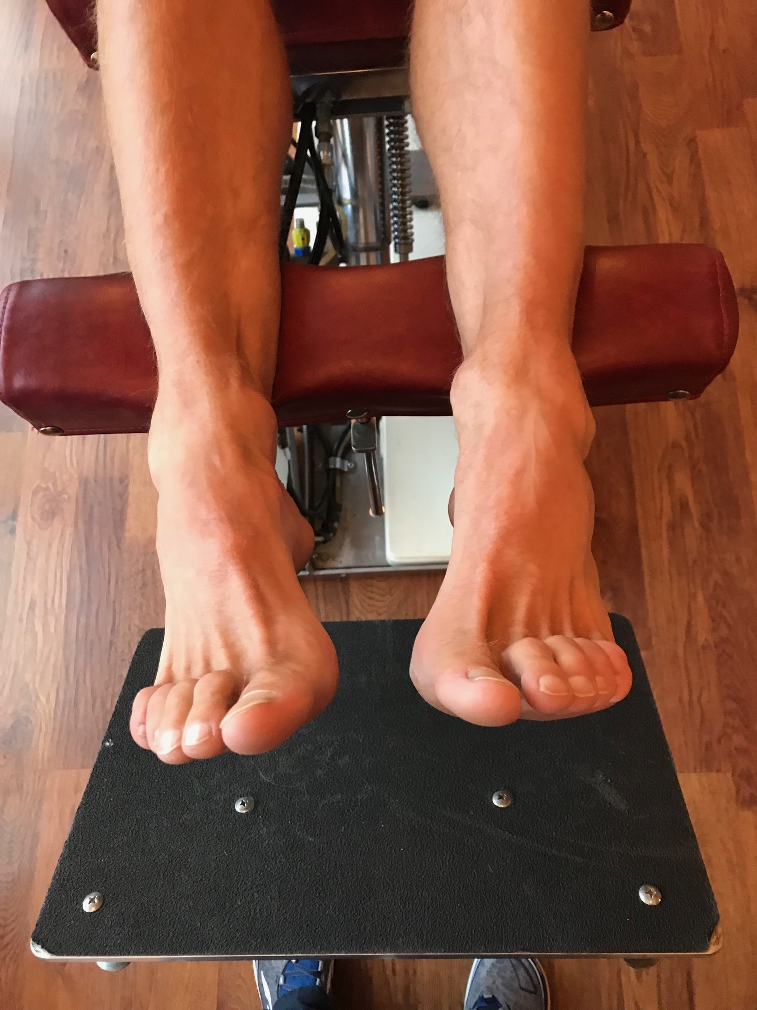
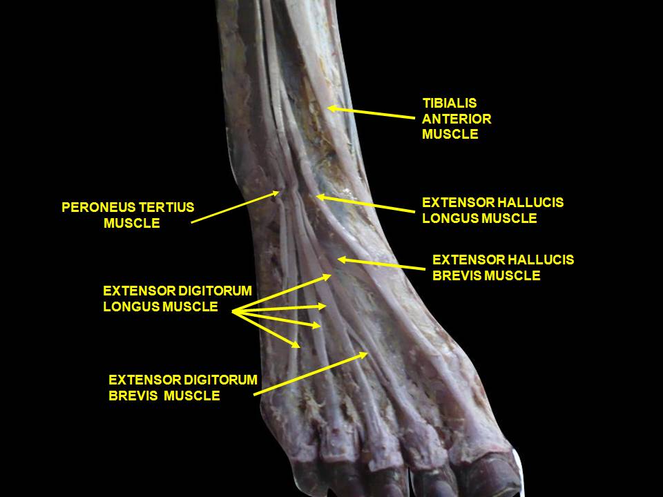
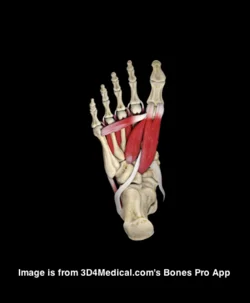
As you remember, the peroneii (3 heads) are on the outside of the lower leg (in a nice, easy to remember order of longus, brevis and tertius, from top to bottom) and help to stabilize the lateral ankle. The peroneus brevis and tertius dorsiflex and evert the foot while the peroneus longus plantarflexes and everts the foot. We discuss the peroneii more in depth here in this post. It then is probably no surprise to you that people with ankle issues, probably have some degree of peroneal dysfunction. Over the years the literature has supported notable peroneal dysfunction following even a single inversion sprain event. 

Functional ankle instability (FAI) is defined as “ the subjective feeling of ankle instability or recurrent, symptomatic ankle sprains (or both) due to proprioceptive and neuromuscular deficits." 

Arthrogenic muscle inhibition (AMI) is a neurological phenomenon where the muscles crossing a joint become "inhibited”, sometimes due to effusion (swelling) of the joint (as seen here) and that may or may not be the case with the ankle (see here), or it could be due to nociceptive input altering spindle output or possibly higher centers causing the decreased muscle activity. 

This paper (see abstract below) merely exemplifies both the peroneals and FAI as well as AMI.

Take home message?

Keep the peroneals strong with lots of balance work!                                                             


2009 May;37(5):982-8. doi: 10.1177/0363546508330147. Epub 2009 Mar 6.

Peroneal activation deficits in persons with functional ankle instability.

Palmieri-Smith RM, Hopkins JT, Brown TN.

Source

School of Kinesiology, University of Michigan, 401 Washtenaw Avenue, Ann Arbor, MI 48109, USA. riannp@umich.edu

Abstract

BACKGROUND:

Functional ankle instability (FAI) may be prevalent in as many as 40% of patients after acute lateral ankle sprain. Altered afference resulting from damaged mechanoreceptors after an ankle sprain may lead to reflex inhibition of surrounding joint musculature. This activation deficit, referred to as arthrogenic muscle inhibition (AMI), may be the underlying cause of FAI. Incomplete activation could prevent adequate control of the ankle joint, leading to repeated episodes of instability.

HYPOTHESIS:

Arthrogenic muscle inhibition is present in the peroneal musculature of functionally unstable ankles and is related to dynamic peroneal muscle activity.

RESULTS:

The FAI patients had larger peroneal H:M ratios in their nonpathological ankle (0.399 +/- 0.185) than in their pathological ankle (0.323 +/- 0.161) (P = .036), while no differences were noted between the ankles of the controls (0.442 +/- 0.176 and 0.425 +/- 0.180). The FAI patients also exhibited lower EMG after inversion perturbation in their pathological ankle (1.7 +/- 1.3) than in their uninjured ankle (EMG, 3.3 +/- 3.1) (P < .001), while no differences between legs were noted for controls (P > .05). No significant relationship was found between the peroneal H:M ratio and peroneal EMG (P > .05).

CONCLUSION:

Arthrogenic muscle inhibition is present in the peroneal musculature of persons with FAI but is not related to dynamic muscle activation as measured by peroneal EMG amplitude. Reversing AMI may not assist in protecting the ankle from further episodes of instability; however dynamic muscle activation (as measured by peroneal EMG amplitude) should be restored to maximize ankle stabilization. Dynamic peroneal activity is impaired in functionally unstable ankles, which may contribute to recurrent joint instability and may leave the ankle vulnerable to injurious loads.


The Beef on the EDL.....

We have long been promoting appropriate function of the long extensors of the toes  here, in our practices, our lectures, on Youtube, in our book......You get the idea. Lets take a closer look at this often weakened and overlooked muscle.

We remember that the EDL lies mostly in the superior and somewhat lateral part of the anterior compartment of the lower leg, comprising approximately the upper 2/3 from under the lateral tibial plateau and fibula, and from the interosseus membrane. It lies under the tibialis anterior, and the extensor hallucis longus lies below it. Its tendons pass inferiorly and travel under the extensor retinaculum and attaches to the base of the distal phalanges of toes 2-4. These muscles act from initial contact to loading response to help eccentrically lower the foot to the ground and ensure smooth heel rocker and most likely attenuate the speed of initial pronation as the talus glides anteriorly on the calcaneal facets and again from terminal stance through initial swing to provide compression of the metatarsal phalangeal and interphalangeal joints, to offset the long flexors (which are often overactive) and create clearance for the toes during swing.  

jhowardL.gif
jhowardR.gif

What does it look like when the long extensors don’t work so well? Have a look at the pedograph on the right (pair J howard r). what do we see? First we notice the lack of printing under the head of the 1st metatarsal and increased printing of the second metatrsal head. Looks like this individual has a forefoot supinatus, or possibly a forefoot varus (cannot get the head of the 1st metatarsal to the ground, and thus a weak medial tripod, possibly insufficient extensor hallucis brevis, peroneus longus, flexor digitorum brevis, or all of the above). Next we see increased printing of the distal phalanges of digits 2-4. Looks like the long flexors are dominant, which means the long extensors are inhibited. What about the lack of printing of the 5th toe? I thought the flexors were overactive? They are, but due to the supinatus, the foot is tipped to the inside and the 5th barely contacts the ground!

How do you fix this?

  • Help make a better foot tripod using the toe wave, tripod standing and extensor hallucis brevis exercises.
  • Make sure the articulations are mobile with joint mobilization, manipulation and massage.
  • How about dry needling and acupuncture to improve function?
  • Make sure the knee and hip are functioning appropriately.
  • Put them in footwear that will allow the foot to function better (a less rigid, less ramp delta shoe).
  • As a last resort, if they cannot make an adequate tripod because of lack of motivation, anatomical constraints or both, use a foot leveling orthotic.


Pain on the outside of one leg, inside of the other. 

Whenever you see this pattern of discomfort, compensation is almost always at play and it is your job to sort it out. 

This patient presents with with right sided discomfort lateral aspect of the right fibula and in the left calf medially. Pain does not interfere with sleep.  He is a side sleeper 6 to 8 hours. His shoulders can become numb; left shoulder bothers him more than right.

PAST HISTORY: L shoulder surgery, rotator cuff with residual adhesive capsulitis. 

GAIT AND CLINICAL EVALUATION: see video. reveals an increased foot progression angle on the right side. Diminished arm swing from the right side. A definite body lean to the right upon weight bearing at midstance on that side.

He has external tibial torsion bi-lat., right greater than left with a right short leg which appears to be at least partially femoral. Bi-lat. femoral retrotorsion is present. Internal rotation approx. 4 to 6 degrees on each side. He has an uncompensated forefoot varus on the right hand side, partially compensated on the left. In standing, he pronates more on the left side through the midfoot. Ankle dorsiflexion is 5 degrees on each side. 

trigger points in the peroneus longus, gastroc (medial) and soles. 

Weak long toe extensors and short toe flexors; weak toe abductors. 

pathomechanics in the talk crural articulation b/l, superior tip/fib articulation on the right, SI joints b/l

WHAT WE THINK:  

1.    This patient has a leg length discrepancy right sided which is affecting his walking mechanics. He supinates this extremity as can be seen on video, especially at terminal stance/pre swing (ie toe off),  in an attempt to lengthen it; as a result, he has peroneal tendonitis on the right (peroneus is a plantar flexor supinator and dorsiflexor/supinator; see post here). The left medial gastroc is tender most likely due to trying to attenuate the midfoot pronation on the left (as it fires in an attempt to invert the calcaneus and create more supination). see here for gastroc info

2.    Left shoulder:  Frozen shoulder/injury may be playing into this as well as it is altering arm swing.

WHAT WE DID INITIALLY (key in mind, there is ALWAYS MORE we can do):    

  •  build intrinsic strength in his foot in attempt to work on getting the first ray down to the ground; EHB, the lift/spread/reach exercises to perform.
  • address the leg length discrepancy with a 3 mm sole lift
  • address pathomechanics with mobilization and manipulation. 
  • improve proprioception: one leg balancing work
  • needled the peroneus longus brevis as well as medial gastroc and soles. 
  • follow up in 1 week to 10 days.

Pretty straight forward, eh? Look for this pattern in your clients and patients

tumblr_o7p469lND91qhko2so1_1280.jpg
tumblr_o7p469lND91qhko2so2_1280.jpg
tumblr_o7p469lND91qhko2so3_1280.jpg
tumblr_o7p469lND91qhko2so4_1280.jpg
tumblr_o7p469lND91qhko2so6_1280.jpg
tumblr_o7p469lND91qhko2so8_1280.jpg
tumblr_o7p469lND91qhko2so9_1280.jpg
tumblr_o7p469lND91qhko2so5_r1_1280.png

Pain on the outside of the leg? Could it be your orthotic? What you wear on your feet amplifies the effect of the orthotic.

This woman presented with right-sided pain on the outside of her leg after hiking approximately an hour. She noticed a prominence of the arch in her right orthotic. She hikes in a rigid Asolo boot ( see below). Remember that footwear amplifies the effect of an orthotic!

In the pictures below you can see the prominent arch. The orthotic has her “over corrected” so that she toes off in varus on that side. The rigid footwear makes the problem worse. The peroneus group is working hard (Especially the peroneus longus)  to try and get the first Ray down to the ground.

The “fix” was to soften the arch of the orthotic and grind some material out. Look at the pictures where the pen is pointing to see how some of the midsole material was taken out. Notice how I ground it somewhat medial to further soften the arch.

She felt better much better after this change and is now a “happy hiker” :-)

Thoughts on the adductor grouping to ponder. I found this while prepping for the dry needling course I am teaching this weekend and thought you may enjoy it. Though the primary actions of the addcutors are well established, secondary actions (whethe…

Thoughts on the adductor grouping to ponder. 

I found this while prepping for the dry needling course I am teaching this weekend and thought you may enjoy it. Though the primary actions of the addcutors are well established, secondary actions (whether they are acually internal or external rotators) remains to be elucidated.

Here is a nice abstract that supports the dynamic function of them as external rotators (eccentrically) during gait.

“Anatomical texts agree on most muscle actions, with a notable exception being the action of the adductors of the hip in the transverse plane. Some texts list an action of the adductor brevis (AB), adductor longus (AL), and/or adductor magnus (AM) as internal rotation, whereas others list an action of external rotation. The purpose of this article is to present a functional model in support of the action of external rotation. Transverse plane motion of the femur at the hip during normal gait is driven by subtalar joint motion during the loading response, terminal stance, and preswing phases. During the loading response, the subtalar joint pronates, and the talus adducts. This talar adduction results in the lower leg, and subsequently the femur, internally rotating. During terminal stance and preswing, the opposite occurs; the subtalar joint supinates as the talus abducts in response to forces generated from the lower extremity and in the forefoot. Electromyographic (EMG) studies indicate varied activity in the AB, AL, and AM during the loading response, terminal stance, and preswing phases of the gait cycle. A careful analysis of EMG activity and kinematics during gait suggests that, in the transverse plane, the adductors may be eccentrically controlling internal rotation of the femur at the hip during the loading response, rather than the previously reported role as concentric internal rotators. In addition, these muscles may also concentrically produce external rotation of the femur at the hip during terminal stance and preswing. Physical therapists should consider this important function of the hip adductors during gait when evaluating a patient and designing an intervention program. Anatomical texts should consider listing the concentric action of external rotation of the femur at the hip as one action of the AB, AL, and AM, particularly when starting from the anatomic position.”

Leighton RD. A functional model to describe the action of the adductor muscles at the hip in the transverse plane.Physiother Theory Pract. 2006 Nov;22(5):251-62.Leighton RD. A functional model to describe the action of the adductor muscles at the hip in the transverse plane.Physiother Theory Pract. 2006 Nov;22(5):251-62.

tumblr_nozmmaOxAI1qhko2so1_500.png
tumblr_nozmmaOxAI1qhko2so2_540.png

Ahh yes, the lumbricals. 

One of our favorite muscles. And here it is in a recent paper! This one is for all you fellow foot geeks : )

Perhaps the FDL (which fires slightly earlier than the FHL) and FHL (which fires slightly later and longer) at loading response, slowing pronation and setting the stage for lumbrical function from midstance to terminal stance/preswing (flexion at the metatarsal phalangeal joint (it would have to be eccentric, if you think about this from a closed chain perspective) and extension (actually compression) of the proximal interphalangeal joints.

“The first lumbrical arose as two muscle bellies from both the tendon of the FDL and the tendinous slip of the FHL in 83.3 %, and as one muscle belly from the tendon of the FDL or the tendinous slip of the FHL in 16.7 %. These two muscle bellies subsequently merged to form the muscle belly of the first lumbrical. The second lumbrical arose from the tendinous slips of the FHL for the second and third toes as well as the tendon of the FDL in all specimens. The third lumbrical arose from the tendinous slips of the FHL for the third and fourth toes in 69.7 %, and the fourth lumbrical arose from the tendinous slip of the FHL for the fourth toe in 18.2 %. Some deep muscle fibers of the fourth lumbrical arose from the tendinous slip of the FHL for the second toe in 4.5 %, for the third toe in 28.8 %, and for the fourth toe in 15.2 %.”

Hur MS1, Kim JH, Gil YC, Kim HJ, Lee KS. New insights into the origin of the lumbrical muscles of the foot: tendinous slip of the flexor hallucis longus muscle. Surg Radiol Anat. 2015 May 12. [Epub ahead of print]

tumblr_nhoqkqFCHJ1qhko2so2_r1_250.jpg
tumblr_nhoqkqFCHJ1qhko2so4_r1_250.jpg
tumblr_nhoqkqFCHJ1qhko2so1_r1_250.jpg
tumblr_nhoqkqFCHJ1qhko2so3_r1_250.png

Thinking on your feet. You have less than 20 minutes with this gentleman, as he has to leave to catch a plane. See how you did. 

Lateral foot pain and cowboy boots?

A 55 YO male patient presents with pain in his left foot area of the cuboid and tail of the fifth metatarsal.  He was told that he had a “locked cuboid” on this side by his chiropractor, who provided some treatment and temporary relief. There has been  no history of trauma and Most recently, he has been wearing cowboy boots and doing “a lot of walking” particularly when he was over in Europe and feels this was a precipitating factor.

Watching him walk in his cowboy boots, the rear foot and heel plate of the cowboy boot is worn into varus. Gait evaluation reveals his left foot to remain in supination (and thus in varus) throughout the entire gait cycle. 

Examination of the foot revealed loss of long axis extension at the metatarsophalangeal and interphalangeal articulations. The cuboid appeared to be moving appropriately. (to see why cuboid function is integral, see this post here. ) There was weakness in the peroneus brevis and peroneus longus musculature with reactive trigger points in the belly of each.  There is tenderness over the tail of the fifth metatarsal and the groove where the peroneal muscle travels through as well as in the peroneal tendon as it travels through here. 

So, what’s up?

This patient has peroneal tendonitis at the point around the foot as it goes around the tail of the fifth metatarsal. Discomfort is dull and achy in this area.  The cowboy boot is putting his foot in some degree of supination (plantar flexion, inversion adduction); this combined with the rear foot varus (from wear on the heel) is creating excessive load on the peroneus longus, which is trying to descend the 1st ray and create a stable medial tripod. Look at the pictures above and check out this post here

What did we do?

Temporarily, we created a valgus post on an insole for him.  This will push him onto his 1st metatarsal as he goes through  midstance into termiinal stance. He was asked to discontinue using the boot until we could get the heel resoled with a very slight valgus cant. We also treated with neuromuscular acupuncture over the peroneal group (GB 34, GB 35, GB 36 and a few Ashi points between GB34 and 35) circle the Dragon about the tail of fifth metatarsal, GB41 as well as the insertion of peroneus onto the base of the first metatarsal (approximately SP4).   We K-taped the peroneus longus to facilitate function of peroneus longus.  He was given peroneus longus (plantarflexion and eversion) and peroneus brevis (dorsiflexion and eversion) theraband exercises. 

How did you do? Easy peasy, right? If they were all only this straight forward….


The Gait Guys. teaching you to think on your feet and increasing your gait literacy with each and every post.