3 things

Its subtle, but hopefully you see these 3 things in this video.

I just LOVE the slow motion feature on my iPhone. It save me from having to drag the video into Quicktime, slow it down and rerecord it.

This gal has a healing left plantar plate lesion under the 2nd and 3rd mets. She has an anatomical leg length deficiency, short on the left, and bilateral internal tibial torsion, with no significant femoral version. Yes, there are plenty of other salient details, but this sketch will help.

  1. 1st if all, do you see how the pelvis on her left dips WAY more when she lands on the right? There is a small amount of coronal plane shift to the right as well. This often happens in gluteus medius insufficiency on the stance phase leg (right in this case), or quadratus lumborum (QL) deficiency on the swing phase leg (left in this case) or both. Yes, there are other things that can cause this and the list is numerous, but lets stick to these 2 for now. In this case it was her left QL driving the bus.

  2. Watch the left and right forefeet. can you see how she strikes more inverted on the left? this is a common finding, as the body often (but not always) tries to supinate the shorter extremity (dorsiflexion, eversion and adduction, remember?) in an attempt to “lengthen” it. Yes, there is usually anterior pelvic tilt accompanying it on the side, because I knew you were going to ask : )

  3. Look how her knees are OUTSIDE the saggital plane and remain there in her running stride. This is commonly seen in folks with internal tibial torsion and is one of the reasons that in our opinion, these folks should not be put medially posted, torsionally rigid, motion control shoes as this usually drive the knees FURTHER outside the saggital plane and can macerate the meniscus.

Yep, lots more we could talk about on this video, but in my opinion, 3 is a good number.

Dr Ivo Waerlop, one of The Gait Guys

#thegaitguys #gaitanalysis #footpain #gaitproblem #internaltibialtorsion #quadratuslumborum #footstrike

https://vimeo.com/329212767

A bit about the QL...


As we have said in previous posts,  though they can’t act independently we like to think to think of the QL as having two divisions. The lower division arises from the medial portion of the iliac crest and adjacent iliolumbar ligament and inserts onto the transverse processes of the lumbar vertebrae, in the coronal plane from lateral to medial and in the saggital plane from posterior to anterior. The upper division arises from the lumbar transverse processes of the upper 4 lumbar vertebrae and insert into the inferior border of the 12th rib, running in the coronal plane from medial to lateral and in the saggital plane from anterior to posterior; about half of the fascicles of this second division act on the twelfth rib and the rest act on the lumbar spine.

The QL is primarily a coronal plane stabilizer causing lateral bending to the ipsilateral side when the foot is planted as well as posterior rotation of the lumbar spine on the weight bearing side.   When acting unilaterally without the ipsilateral foot fixed on the ground, it can raise the ilia on the side of contraction. It is active during single limb support during stance phase of gait on the contralateral side (along with the external oblique) to elevate the ilium. This is coupled with the ipsilateral anterior fibers of the gluteus medius and minimus pulling the iliac crest toward the stable femur. Sahrmann states “the QL is optimally situated to provide control of lateral flexion to the opposite side via its eccentric contraction to provide control of the return from lateral flexion via its concentric contraction. The muscle is also positioned to play a role in the rotation that occurs between the pelvis and spine during walking”.

Acting bilaterally, it extends the lumbar spine, deepening the lordosis and acting to limit anterior shear of the vertebral bodies.

It is also able to stabilize the 12th rib during forced expiration, thus acting as an accessory muscle of respiration. This fixation is important when we need to superimpose pelvic movements upon it. Furthermore, it increased activation in response to increasing compression in static upright standing postures.

Here is a video of a low back screen we often use

The QP....What's the deal?

tumblr_mau352bxll1qhko2so1_540.png

Possibly heard of, rarely implicated and not often treated, this is one muscle you should consider taking a look at.

The quadratus plantae is generally considered to arise from two heads of differing and variable  fiber type composition, with the lateral head having slightly more Type 1 endurance fibers (1) The two heads are separated from each other by the long plantar ligament, though it can arise from from one (somewhat more common)  to 3 heads (very rare).  The attachments can be variable, The medial head is larger and more muscular, attached to the medial calcaneus, lateral aspect of the long plantar ligament and often from the plantar calcaneocuboid ligament (2);  the lateral head is smaller and more tendinous, attaching to the lateral border of the inferior surface of the calcaneus and the long plantar ligament.  The two portions join and end in a flattened band which inserts into the lateral, upper and under surfaces of the muscles, tendons or aponeurosis of predominantly the flexor digitorum longus and usually of the second and third, and sometimes fourth toes (2,3). 

Its action can be equally as variable. In addition to augmenting the pull of the long flexor tendons along the long axis of the foot and so that the 3rd and 4th toes do not curl under the foot, the tendinous slips of the FHL may distribute the load of the great toe to the second toe to the third or fourth toe in the forefoot, especially during toe-off (3).

look at the 4th and 5th digits trying to "crawl under the foot"

look at the 4th and 5th digits trying to "crawl under the foot"

The main attachment of the QP to the tendinous slips of the FHL may provide more efficient control of the long flexor tendons in comparison with that of the QP to the tendon of the FDL (3). EMG studies suggest it resists extension of the toes during the stance phase of locomotion, which serves to increase the stability of the foot. Additional EMG studies suggest it actually acts as a primary toe flexor in voluntary movements, being preferentially recruited over flexor digitorum longus and from comparative anatomical considerations it also seems likely that quadratus plantae may be an intrinsic evertor of the foot (4).

This muscle is a major player in gait and rehabilitation of this muscle should not be overlooked. I could only find one study looking at exercise activation of the QP (5) . It was examined along with the abductor hallucis, flexor digitorum brevis, abductor digiti minimi, flexor digiti minimi, adductor hallucis oblique, flexor hallucis brevis, interossei and lumbricals during rehabilitative the short-foot exercise, toes spread out, first-toe extension, second- to fifth-toes extension.

So, what else can you do?

  • you could ignore the muscle and hope it gets better. (in all likelihood it will worsen)
  • you could give them long flexor, toe scrunching towel-curling, marble-grasping exercises, like you see all over the internet…and give the flexor digitorum longus even more of a mechanical advantage, and make the problem worse
  • you could give them exercises to increase the function of the long extensors, which would increase the mechanical advantage of the quadratus plantae. like the shuffle walk; lift, spread and reach and tripod standing exercises
  • look north of the foot to see what might be causing the problem (loss of ankle rocker, insufficient gluteal activity, loss of internal rotation of the hip, etc) 

Check out the QP on your next foot pain patient, or whenever you see the toes trying to crawl under the foot. You may be surprised at your results. 


1. Schroeder KL, Rosser BW, Kim SY. Fiber type composition of the human quadratus plantae muscle: a comparison of the lateral and medial heads. J Foot Ankle Res. 2014 Dec 13;7(1):54. doi: 10.1186/s13047-014-0054-5. eCollection 2014.

2. Pretterklieber B1. Morphological characteristics and variations of the human quadratus plantae muscle. Ann Anat. 2017 Nov 21;216:9-22. doi: 10.1016/j.aanat.2017.10.006. [Epub ahead of print]

3. Hur MS, Kim JH, Woo JS, Choi BY, Kim HJ, Lee KS. An anatomic study of the quadratus plantae in relation to tendinous slips of the flexor hallucis longus for gait analysis. Clin Anat. 2011 Sep;24(6):768-73. doi: 10.1002/ca.21170.

4. Sooriakumaran P, Sivananthan S. Why does man have a quadratus plantae? A review of its comparative anatomy. Croat Med J. 2005 Feb;46(1):30-5.

5. Gooding TM, Feger MA, Hart JM, Hertel J. ntrinsic Foot Muscle Activation During Specific Exercises: A T2 Time Magnetic Resonance Imaging Study. J Athl Train. 2016 Aug;51(8):644-650. Epub 2016 Oct 3.

The Mighty Quadratus Femoris

Ishial tuberosity pain that looks like a hamstring but is not responding? Think QF.

We have always have found the quadratus femoris is one of, if not the, 1st hip muscle to become dysfunctional in hip pain patients. Perhaps it is due to it being the southern most stabilizer of the deep 6. Long known as an adductor, but also external rotator, we find it is employed eccentrically when the foot the planted and people rotate to the same side as weight bearing, or people take a “sudden stumble” while running. It often mimics an insertional hamstring strain with regards to location. We were happy to see it is getting some of the attention it deserves : )

When is a hamstring strain not a hamstring strain?We have always have found the quadratus femoris is one of, if not the, 1st hip muscle to become dysfunctional in hip pain patients. Perhaps it is due to it being the southern most stabilizer of the d…

When is a hamstring strain not a hamstring strain?

We have always have found the quadratus femoris is one of, if not the, 1st hip muscle to become dysfunctional in hip pain patients. Perhaps it is due to it being the southern most stabilizer of the deep 6. Long known as an adductor, but also external rotator, we find it is employed eccentrically when the foot the planted and people rotate to the same side as weight bearing, or people take a “sudden stumble” while running. It often mimics an insertional hamstring strain with regards to location. We were happy to see it is getting some of the attention it deserves : )


http://www.anatomy-physiotherapy.com/articles/musculoskeletal/lower-extremity/hip/1528-function-of-the-quadratus-femoris-and-obturator-externus

Go ahead and try this at home.remember last mondays post? (if not, click here). Here is one way of telling whether your (or someone else’s) vestibular system is working. It will also give you an idea of how some people compensate. Ready?
Stand up (b…

Go ahead and try this at home.

remember last mondays post? (if not, click here). Here is one way of telling whether your (or someone else’s) vestibular system is working. It will also give you an idea of how some people compensate.

Ready?

  • Stand up (barefoot or shoes does not matter).
  • place your hands resting on the top of your hips with your thumbs to the back (like your Mom used to, when you were in trouble). Your thumbs should be resting on your quadratus lumborum (QL) muscle.
  • tilt your HEAD to the LEFT
  • you should feel the muscle (ie the QL) under your RIGHT thumb contract
  • come back upright


repeat, but this time lean your BODY to the LEFT

  • same thing right? Now check the other side.


Everything OK? Everything fire as it should?

Now lets add another dimension.

  • slide your fingers down so they are just below the crest of the hip, resting above the greater trochanter (the bump on the side of your upper thigh). This should place your fingers on the middle fibers of the gluteus medius.
  • tilt your head (or body ) to the LEFT.
  • You should feel the LEFT gluteus medius and the RIGHT QL contract. These muscles should be paired neurologically. When walking, during stance phase on the LEFT: the LEFT gluteus medius helps to maintain the pelvis level, while the RIGHT QL, assists in hiking the RIGHT side.


If everything works OK, then your vestibulospinal spinal system is intact and your QL and gluteus medius seem to be firing and appropriately paired. If not? That is the subject for another post.

The Gait Guys. Helping you to understand the concepts of WHY compensations occur.

tumblr_mwzu9gTBzS1qhko2so1_1280.jpg
tumblr_mwzu9gTBzS1qhko2so2_400.gif
tumblr_mwzu9gTBzS1qhko2so3_r1_1280.jpg
tumblr_mwzu9gTBzS1qhko2so4_r1_1280.jpg

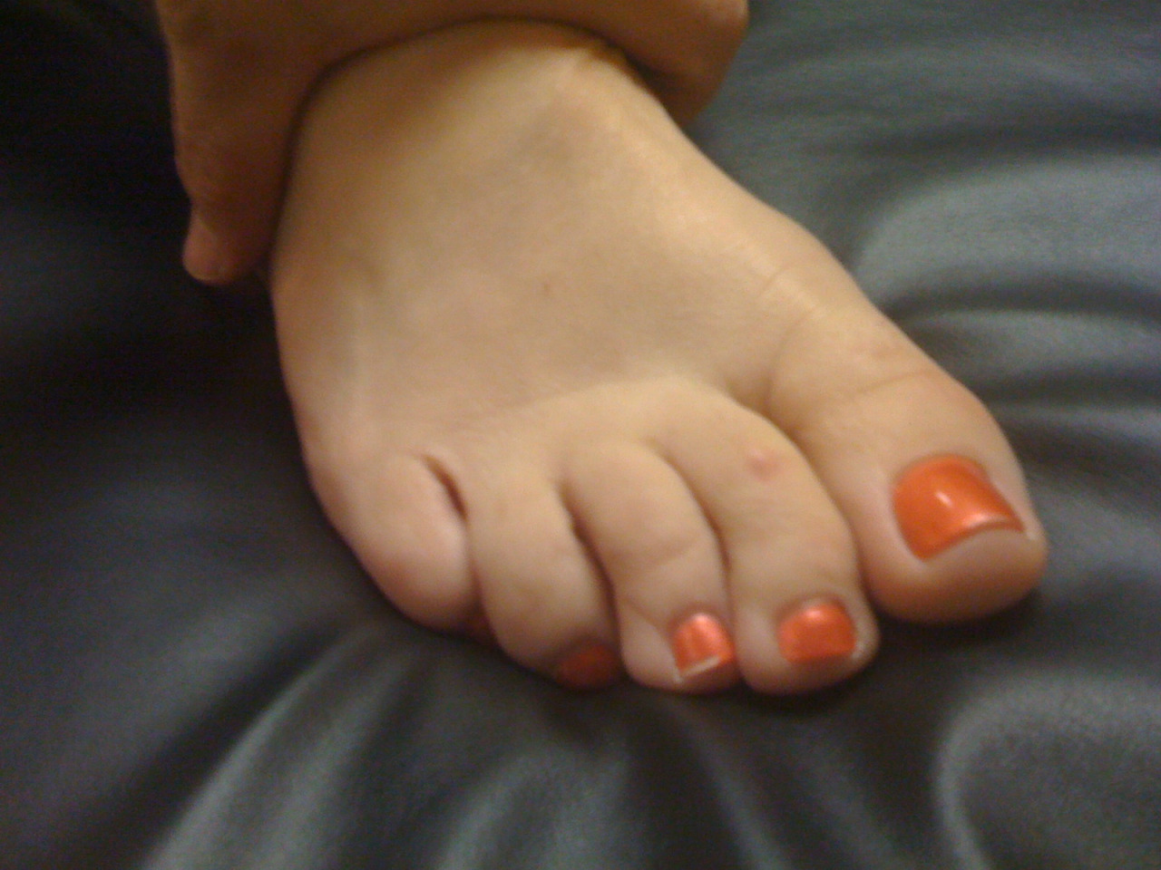
Is your 5th toe curled under ? What do you do when “this little piggy” can’t go wee wee wee all the way home.

Have a look at the 4 photos above.  You will see this curling of the lesser toes quite often in your practice, and when you know what it means it can help to guide your thinking, both from a diagnostic and treatment perspective.  

You should have noticed in the photos that the 4th and 5th toes curl under and are hyper-flexed, and this is at rest.  So, what does this mean ?

It means that the long flexors are overactive, the extensors are underactive, and the adduction pull of the long flexors is unopposed by the under appreciated quadratus plantae muscle.

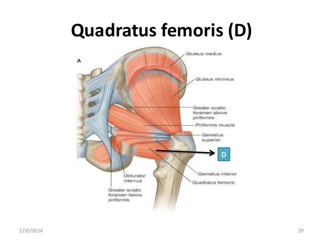
Look at the clinical drawing. The quadratus plantae has 2 heads, a medial head and a lateral head.  Being able to clinically test these two heads will give you much insight into the function of the foot and when you see these outer two toes curling under, as you see in the photo, you will always see weakness of the lateral head of the quadratus plantae.  

The quadratus plantae arises from two heads separated from each other by the long plantar ligament. The medial head is larger and more muscular, attached to the medial calcaneus;  the lateral head is smaller and more tendinous, attaching to the lateral border of the inferior surface of the calcaneus and the long plantar ligament.  The two portions join and end in a flattened band which inserts into the lateral, upper and under surfaces of the tendons of the flexor digitorum longus, usually the second, third, and fourth toes.

But this time, if you have studied the drawing, you should notice the oblique line of pull of the long flexors.  This should in fact create this undesirable curling effect of the lateral two toes since they are so far out on the oblique line of pull. However, if you look at the insertion of the lateral head of the quadratus plantae you should be able to conclude that this head is designed to offset this oblique pull of the outer two long flexor tendons.  The quadratus creates a posterior pull on the outer long flexor tendons ensuring that the curling effect (as seen in the photo) is nullified. Thus, we have a clinical presentation of a weak lateral head of the quadratus plantae (and probably a few others which we will not discuss here so as to not dilute the purpose of today’s post). Now you just have to figure out why it is weak or if there is a biomechanical reason for its insufficiency

  • is there a foot type presenting itself that makes it difficult for this muscle to create sufficient posterior pull to offset the tremendous leverage of the long flexors? Maybe a forefoot varus, which gives the flexor tendons a mechanical advantage or a forefoot valgus which puts the quadratus plantae at a mechanical disadvantage? (Taking our National Shoe Fit Certification Program will help you get closer to understanding many of these issues.)
  • Are their other anatomical variants like an increased forefoot width or bunions (medial or tailor’s)
  • is there excessive rear or midfoot pronation?
  • Shoe choice problem ?

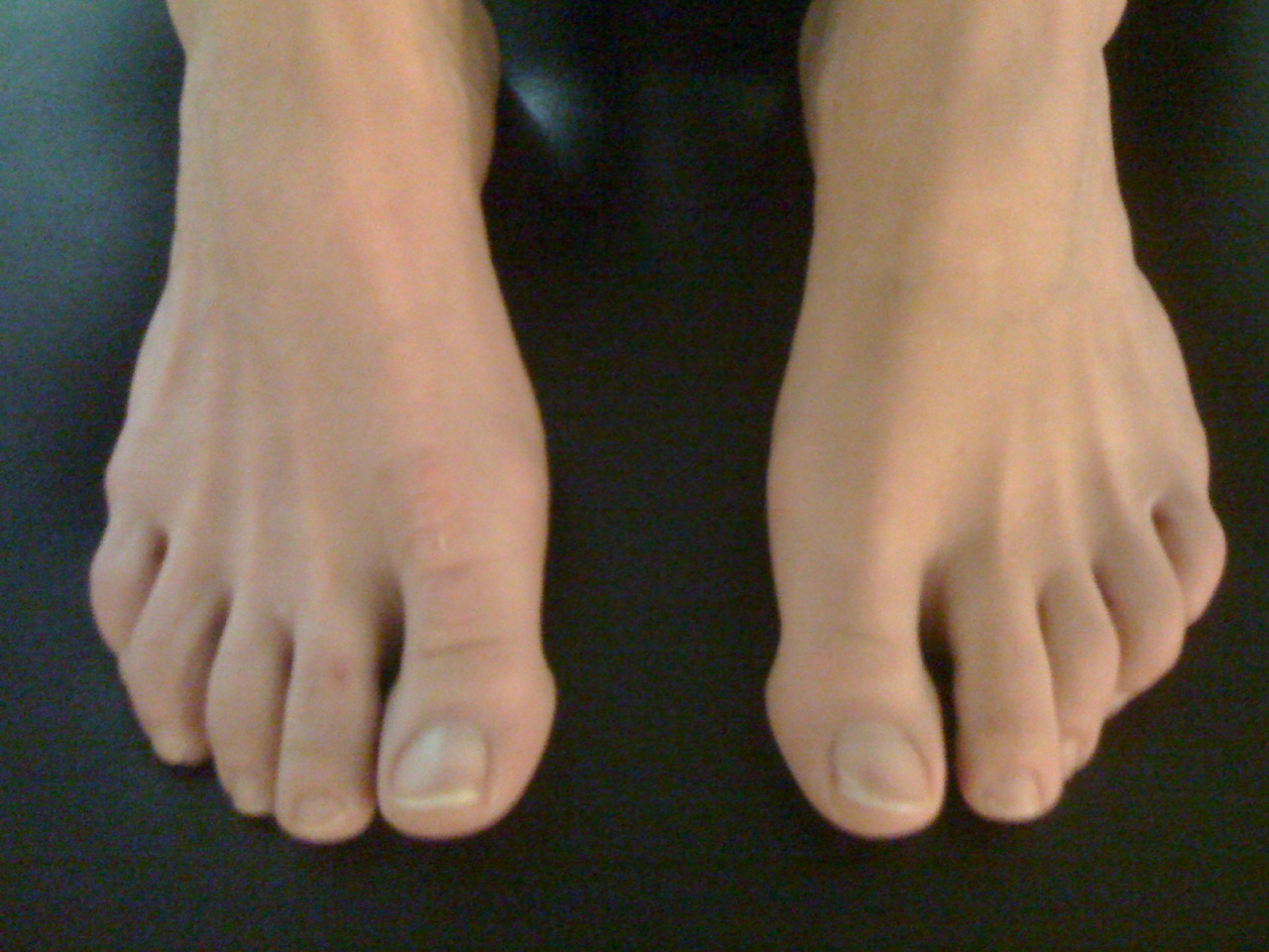
Some folks do have adequate function of the quadratus plantae. Note the lovely feet in the last picture … .  they must have strong lateral quadratus plantae and abductors of the lateral foot and toes ! And, they have great toe separation, thus great intrinsic interossei muscles, and nice flat toes (great balance between flexors and extensors).

So, what do you do?

  • you could do a surgery, amputate or fuse some of the joints to make them look better. Extreme for a problem like this
  • you could ignore the issue and hope it goes away. (in all likelihood it will worsen)
  • you could give them long flexor, toe scrunching Towel-curling, marble-grasping exercises , like you see all over the internet…and give the flexor digitorum longus even more of a mechanical advantage, and make the problem worse
  • you could give them exercises to increase the function of the long extensors, which would increase the mechanical advantage of the quadratus plantae. like the shuffle walk; lift, spread and reach and tripod standing exercises (hmm…sounding better)
  • be a real clinician and in addition to looking at the foot, look north of the foot to see what might be causing the problem (loss of ankle rocker, insufficient gluteal activity, loss of internal rotation of the hip, etc) Hmmm; sounding like a good idea too…

The Gait Guys. Hammering it home, day after day, about the importance of gait and giving you clues to be a better _________ (insert athlete, coach, trainer, clinician, shoe fitter, rehab specialist…).