Kinetic chain transfer.

Anyone would be silly to disagree with this.
We go into some deeper reasoning back in this older blog post (https://tmblr.co/ZrRYjxTJ6zw9) looking at arm swing and leg swing and pairing of pelvis and shoulder posturing and how clean pelvis function parlays into upper body function in softball pitching.

“Proper utilization of the kinetic chain allows for efficient kinetic energy transfer from the proximal segments to the distal segments. Dysfunction at a proximal segment may lead to altered energy transfer and dysfunction at more distal segments,”

Lower body conditioning may cut upper body injury risk in softball. -Hank Black

http://lermagazine.com/special-section/pediatric-clinical-news/lower-body-conditioning-may-cut-upper-body-injury-risk-in-softball

Gait and Autism spectrum disorder (ASD).

Gait and autism spectrum disorder (ASD):
“ … overall findings of the studies conducted in the area are inconclusive … however, some results suggest an emerging pattern. The current perspective on gait patterns in children with ASD is that there are a number of deviations present in terms of temporospatial, kinematic, and kinetic parameters and that gait, along with other movement pattern changes, may be used to allow for earlier diagnosis of ASD. There is, however, some consensus regarding the involvement of the cerebellum and basal ganglia in children with ASD and the relationship with observed motor deficits. ” - Kindregan et al

http://www.hindawi.com/journals/aurt/2015/741480/

Gait and the lower visual field.

Gait and the eyes. We forget about the eyes. If you have vision issues, your gait may change.
Gaze during adaptive gait involving obstacle crossing is typically directed two or more steps ahead where as visual information of the “in the moment” swinging lower-limb and its relative position during the task is available in the lower visual field. This study determined exactly when visual information is utilised to control/update lead-limb swing trajectory during obstacle negotiation.
In this study, when the lower visual field was blocked out the foot-placement distance and toe-clearance became significantly increased, suggesting the brain overcorrecting for safety. A logical assumption. “These findings suggest that lower visual field input is typically used in an online manner to control/update final foot-placement, and that without such control, uncertainty regarding foot placement causes toe-clearance to be increased.”

http://www.ncbi.nlm.nih.gov/pubmed/22424759

Base of support and chronic ankle sprains

We spoke at length about “base of support” and how if you have a narrow base of support, you have a small comfort zone of balance in single leg tasking (walking, running, sports etc). If you have a narrow base of support, you will wish and attempt to put the center of your body mass over the foot at all times…….hence the cross over gait often times. Increasing balance ability will help to increase base of support and hence help with reducing cross over gait (narrow step width gait and running) tendencies.
* This study here showed that a SINGLE episode of single 30-minute training session involving kicking a ball while standing on 1 foot promoted changes in postural-control strategies in individuals with chronic ankle instabilty(CAI).
Does this translate to the assumption that “CAI clients will have a narrow step width, narrow base of support, and a cross over gait”? No, but if you are thinking that way, we want you on our team.

Changes in Postural Control After a Ball-Kicking Balance Exercise in Individuals With Chronic Ankle Instability. Marcio Jose dos Santos PhD, Josilene Conceição PT, MSc, Felipe Gustavo Schaefer de Araújo, Gilmar Moraes SantosPhD, John Keighley PhD

tumblr_o92viq6Er51qhko2so1_1280.jpg
tumblr_o92viq6Er51qhko2so2_1280.jpg
tumblr_o92viq6Er51qhko2so3_1280.jpg

Holy Leg Length discrepancy!

These pix come to us from one of our brethren, Dr Scott Tesoro in Carbondale of a 73 yr old golfer with mild LBP and a  L knee replacement three yrs ago. He has a VERY short R leg (close to an inch).

What you are seeing is he ultimate compensation for a short leg. Note how he takes the shorter side and supinates it (to the max!). You can see the external rotation of the lower leg and thigh to go along with it. If you look carefully and extrapolate how his left leg would look “neutral”, you can see he has internal tibial torsion on this (right) side as well. He has some increased midfoot pronation on the right compared to the left, but not an excessive amount.

A full length sole lift would probably be in order, as well as potentially addressing some of his compensations. Wow, what a great set of pictures !

Sounds like a bad ideaOrthotics, can be useful adjunct to care. They can be used to give people biomechanics that they do not have while you were trying to improve them and help to make up for ranges of motion which do not seem attainable.From the g…

Sounds like a bad idea

Orthotics, can be useful adjunct to care. They can be used to give people biomechanics that they do not have while you were trying to improve them and help to make up for ranges of motion which do not seem attainable.

From the gate cycle we know that after initial contact and loading response the calcaneus should start to evert. The calcaneus will continue to evert until it encounters something (like the lateral heel counter of the shoe). At mid stance it should be fully everted and as the opposite leg comes in to swing, begin to invert. The lateral heel counter assists in the inversion/supination process.

To our knowledge, flip-flops, even if they have an increased arch, do not have a lateral heel counter and therefore will promote further lateral excursion of the calcaneus while the medial longitudinal arch is collapsing  (i.e.: midfoot pronation). Go ahead and place your foot into inversion and see what happens to your heel. It’s slides laterally.

It’s also well-established that flip-flops, through flexion of the distal toes and engagement of the long flexor tendons, inhibits ankle rocker. It is often necessary to engage these muscles to keep the flip-flop from coming off. Lack of ankle rocker usually will inhibit hip extension and that can cause a constellation of problems.

Though engagement of the long flexors of the toes will have a partial anti-pronatory effect, this is not enough to counter the excessive heel  eversion which is happening.

We generally do not think the flip-flops are a great idea and telling someone that it’s “OK” to wear flip-flops as long as it has appropriate arch support, is silly.

So here is somewhat of a controversial subject.Perhaps, though not discussed in this article, activating more axial extensors (vestbulospinal pathways, things like your erector spinae) could be somewhat protective, in that it could, at least theoret…

So here is somewhat of a controversial subject.

Perhaps, though not discussed in this article, activating more axial extensors (vestbulospinal pathways, things like your erector spinae) could be somewhat protective, in that it could, at least theoretically, help to normalize flexor/extensor ratios in the lower extremity. 

We see flexor dominance (increased corticospinal activity) in many cases of lower extremity problems causing an imbalance. Perhaps activating extensors the lower extremity (tibialis interior, extensor digitorum longest, etc.) could explain, in part, some of these (controversial) results.

We’re not recommending or condoning taking up smoking to preserve your knees. This is merely food for thought in the ever-changing landscape of clinical application.


http://lermagazine.com/cover_story/smoking-knee-oa-from-clinical-controversy-to-therapeutic-possibility

Pain on the outside of one leg, inside of the other. 

Whenever you see this pattern of discomfort, compensation is almost always at play and it is your job to sort it out. 

This patient presents with with right sided discomfort lateral aspect of the right fibula and in the left calf medially. Pain does not interfere with sleep.  He is a side sleeper 6 to 8 hours. His shoulders can become numb; left shoulder bothers him more than right.

PAST HISTORY: L shoulder surgery, rotator cuff with residual adhesive capsulitis. 

GAIT AND CLINICAL EVALUATION: see video. reveals an increased foot progression angle on the right side. Diminished arm swing from the right side. A definite body lean to the right upon weight bearing at midstance on that side.

He has external tibial torsion bi-lat., right greater than left with a right short leg which appears to be at least partially femoral. Bi-lat. femoral retrotorsion is present. Internal rotation approx. 4 to 6 degrees on each side. He has an uncompensated forefoot varus on the right hand side, partially compensated on the left. In standing, he pronates more on the left side through the midfoot. Ankle dorsiflexion is 5 degrees on each side. 

trigger points in the peroneus longus, gastroc (medial) and soles. 

Weak long toe extensors and short toe flexors; weak toe abductors. 

pathomechanics in the talk crural articulation b/l, superior tip/fib articulation on the right, SI joints b/l

WHAT WE THINK:  

1.    This patient has a leg length discrepancy right sided which is affecting his walking mechanics. He supinates this extremity as can be seen on video, especially at terminal stance/pre swing (ie toe off),  in an attempt to lengthen it; as a result, he has peroneal tendonitis on the right (peroneus is a plantar flexor supinator and dorsiflexor/supinator; see post here). The left medial gastroc is tender most likely due to trying to attenuate the midfoot pronation on the left (as it fires in an attempt to invert the calcaneus and create more supination). see here for gastroc info

2.    Left shoulder:  Frozen shoulder/injury may be playing into this as well as it is altering arm swing.

WHAT WE DID INITIALLY (key in mind, there is ALWAYS MORE we can do):    

  •  build intrinsic strength in his foot in attempt to work on getting the first ray down to the ground; EHB, the lift/spread/reach exercises to perform.
  • address the leg length discrepancy with a 3 mm sole lift
  • address pathomechanics with mobilization and manipulation. 
  • improve proprioception: one leg balancing work
  • needled the peroneus longus brevis as well as medial gastroc and soles. 
  • follow up in 1 week to 10 days.

Pretty straight forward, eh? Look for this pattern in your clients and patients

Ankle sprains and the reorganization of the sensorimotor system

“Our subjects with unilateral chronic ankle sprains had weaker hip abduction strength and less plantar-flexion range of motion on the involved sides. Clinicians should consider exercises to increase hip abduction strength when developing rehabilitation programs for patients with ankle sprains.”-Friel et al

Awhile back we wrote about the principle that if the hip abductors are weak, the leg will posture more adducted (ie, cross over type pattern) and this places the foot more directly below the body midline plumb, this will posture the foot in inversion and thus at greater risk for future inversion sprains.  This sets up the vicious cycle of hip abductor weakness, frontal plane drift of pelvis, inversion of the foot and more ankle sprain risks/events.  The cycle must be broken. The hip must be addressed. That lateral chain must be restored all the way up from the foot.  

Another newer study by Bowker discusses the somatosensory feedback necessary for postural adjustments, walking, and running stating that they may be hampered by a decrease in soleus spinal reflex excitability.  The study adds more validity to what we are all growing to know more clearly, that the central nervous system via supraspinal circuitry plays deeply into chronic ankle instability (CAI). The studies suggest that CAI may be more about coordination and control of dynamic stabilizers and changes in the motor neuron excitability rather than the function of static stabilizers.

“A successful reorganization of the sensorimotor system after an initial ankle sprain is the critical point when individuals suffer chronic ankle instability or become copers [individuals who do not develop chronic instability after an ankle sprain] who break the cycle of recurrent injuries and disabilities seen in CAI,” Masafumi Terada, PhD

According to LER and the Terada work, 

The slow-twitch fibers in the soleus muscle are mostly innervated by small alpha motoneurons, Terada explained, so the study findings suggest that some people may restore their ability to reflexively recruit alpha motoneurons after ankle injury, and some may not.

“Therapeutic interventions that can increase the H-reflex in the soleus may help to break the cycle of recurrent injuries and disabilities seen in CAI,” he said. “Lower-intensity transcutaneous electrical stimulation, joint manipulations, and reflex conditioning protocols may be effective in increasing the soleus spinal excitability.”

The Gait Guys


Reference:

CAI and the CNS: Excitability may influence instability. Larry Hand

http://lermagazine.com/news/in-the-moment-sports-medicine/cai-and-the-cns-excitability-may-influence-instability

Taken from original source:

Bowker S, Terada, M, Thomas AC, et al. Neural excitability and joint laxity in chronic ankle instability, coper, and control groups. J Athl Train 2016 Apr 11. [Epub ahead of print]

J Athl Train. 2006; 41(1): 74–78.PMCID: PMC1421486Ipsilateral Hip Abductor Weakness After Inversion Ankle SprainKaren Friel,Nancy McLean,Christine Myers, and Maria Caceres
http://www.ncbi.nlm.nih.gov/pmc/articles/PMC1421486/

Can you guess why this person has left-sided plantar fasciitis?This question probably seem somewhat rhetorical. Take a good look at these pedographs which provide us some excellent clues.First of all,  note how much pressure there is over the metata…

Can you guess why this person has left-sided plantar fasciitis?

This question probably seem somewhat rhetorical. Take a good look at these pedographs which provide us some excellent clues.

First of all,  note how much pressure there is over the metatarsal heads. This is usually a clue that people are lacking ankle rocker and pressuring these heads as the leg cantilevers forward.  This person definitely have a difficult time getting the first metatarsal head down to the ground.

Notice the overall size of the left foot compared to the right (right one is splayed or longer). This is due to keeping the foot and somewhat of a supinated posture to prevent excessive tension on the plantar fascia.

The increase splay of the right foot indicates more mid foot pronation and if you look carefully there is slightly more printing at the medial longitudinal arch. This is contributing to the clawing of the second third and fourth toes on the right. Stand up, overpronate your right foot and notice how your center of gravity (and me) move medially.The toes will often clench in an attempt to create stability.

The patient’s pain is mostly at the medial and lateral calcaneal facets, and within the substance of the quadratus plantae with weakness of that muscle and the extensor digitorum longus. She has 5° ankle dorsiflexion left and 10 degrees on the right and hip extension which is similar.

The lack of ankle rocker and hip extension or causing her to pronate through her midfoot, Tensioning are plantar fascia at the insertion. The problem is worse on the left and therefore that is where the symptoms are.

Pedographs can be useful tool in the diagnostic process and provide clues as to biomechanical faults in the gait cycle.

tumblr_o84av4iR3i1qhko2so2_250.jpg
tumblr_o84av4iR3i1qhko2so1_1280.png

Shoe Drop and its effects on the shoe itself

An interesting, free, full text article we ran across entitled: “THE EFFECT OF WALKING IN FOOTWEAR WITH VARYING HEELSOLE DIFFERENTIALS ON SHANK & FOOT SEGMENT KINEMATICS”

Not exactly a page turner but some important pearls to glean here.

CONCLUSION: During 0-50% gait cycle stance phase shank kinematics do not change with changes in Heel Sole Differential. Actual foot angles do change, increasing with increasing Heel Sole Differential of footwear and by the angle of pitch of the footwear.

In other words, the kinematics (read: flex and physical characteristics) of the shank (the platform that the shoe is built on) do not change with increased ramp delta (ie: “drop”, from heel to toe), but foot kinematics (ie: how the foot moves) DOES change.

http://www.oandp.org/publications/jop/2016/2016-020.pdf

tumblr_o864jbYF2R1qhko2so1_1280.jpg
tumblr_o864jbYF2R1qhko2so2_1280.jpg
tumblr_o864jbYF2R1qhko2so3_1280.jpg
tumblr_o864jbYF2R1qhko2so4_1280.jpg

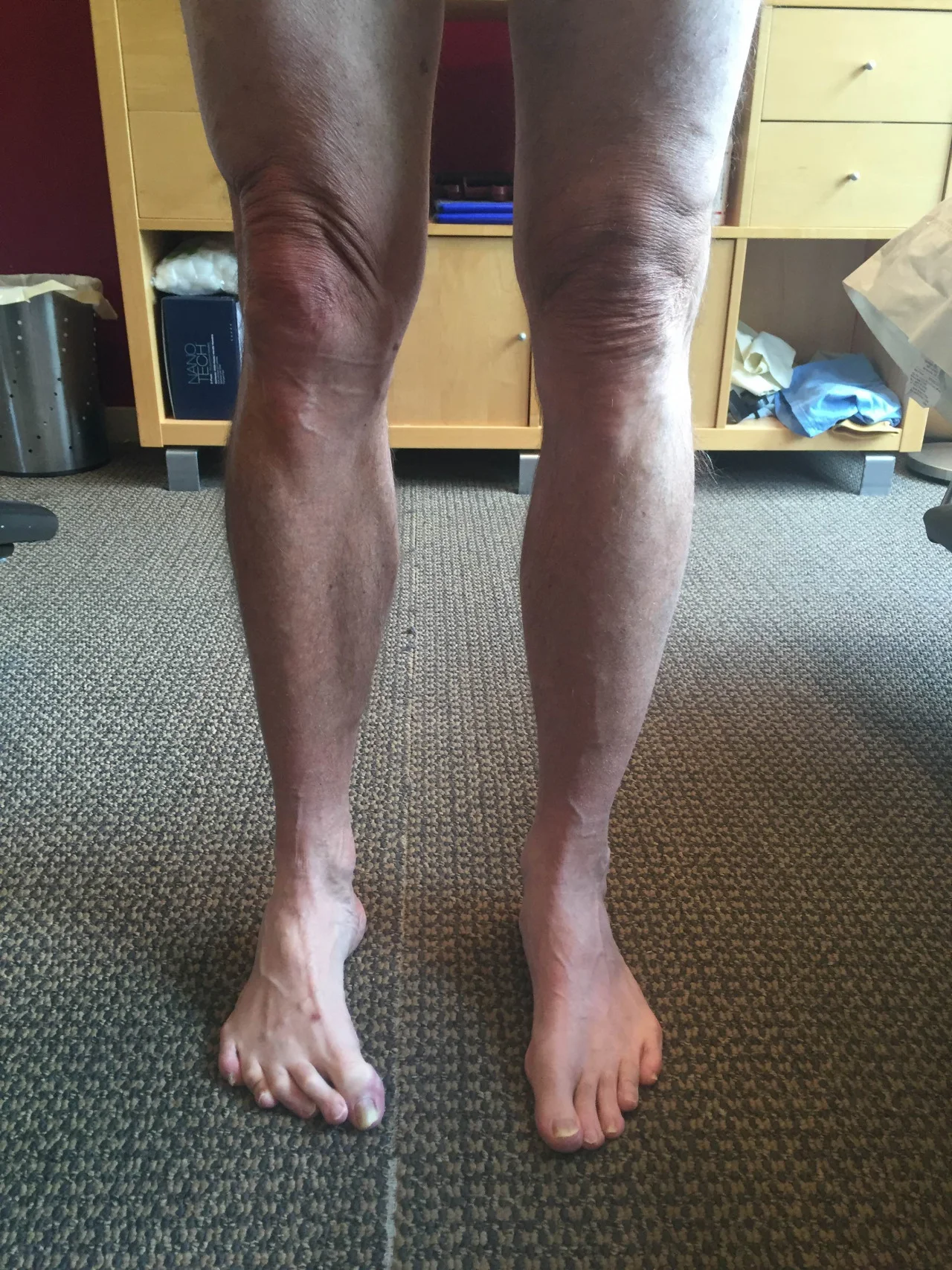
Short leg and mottling of the skin

Have you ever heard of Klippel-Trenaunay Syndrome? I hadn’t either, until I had a patient come in with low back pain and a gait issue and said she had it.

Evidently, in 1900, noted French physicians Klippel and Trenaunay first described a syndrome in 2 patients presenting with a port-wine stain and varicosities of an extremity associated with hypertrophy of the affected limb’s bony and soft tissue. Klippel-Trenaunay-Weber syndrome (KTWS) is characterized by a triad of port-wine stain, varicose veins, and bony and soft tissue hypertrophy involving an extremity (1).

Most cases KTWS are sporadic, although a few cases in the literature report an autosomal dominant pattern of inheritance (2). There is no racial predilection, even distribution between males and females and presents at birth or during early childhood (3). It generally affects a single extremity, although cases of multiple affected limbs have been reported. The leg is the most common site followed by the arms, the trunk, and rarely the head and the neck(4).

This patient had a history of low back pain with a recent epidural steroid injection. Exam highlights included a R sided leg length discrepancy approximately 5mm (tibial and femoral). Pelvic tilt to the right (for LLD) with anterior rotation of that side of the pelvis, posterior on the opposite side (counter clockwise pelvic distortion pattern). Lumbar flexion off 60/90 with all motion occurring in the lumbar spine (ie: no hip hinge), extension 20/30, lateral bending 30/45 BL with pain ipsilateral. Decreased low back endurance of <50 seconds in extension.

Right lower extremity was smaller (appeared hypoplastic) than left and had multiple discolorations in the skin (see pictures). L sided Q angle > R (12 vs 8 degrees). Less internal rotation of the right lower extremity compared to left, but with normal limits. Gait revealed a shift and hike to the right during stance phase with an increased arm swing on the right. Foot intrinsics were weak (lumbricals, EDL, FDB, dorsal intrerossei)

She walked in a pair of Chaco sandals with allowed much greater calcaneal eversion bilaterally R > L.

MRI revealed paraspinal marbling at the lower part of the lumbar spine, improving as you move rostrally. Small disc herniations at L3/4, 4/5, 5/S1, which did not effect the exiting nerve roots. Degenerative changes in the lumbar facet joints. There was no radiographic evidence of instability.

Impression:
It seems that she did not have enough intrinsic for the strength to stop calcaneal eversion in her Chaco’s and therefore this was causing increased foot pronation. This, combined with her leg length discrepancy, was contributing to increasing the lordosis in her lumbar spine, causing facet joint irritation. This was compounded by weakness and lack of endurance of the lumbar paraspinal musculature. The effects of the Klippel-Trenaunay Syndrome are evident with the IPO plasticity of the right lower extremity and accompanying musculoskeletal abnormalities.

What did we do?

  • Gave her endurance exercises for the lumbar spine.
  • Gave her propriosensorv exercises for the lumbar spine
  • Recommended she continue with the 5 mm sole lift.
  • Advised getting rid of the Chaco sandals as they allow too much calcaneal eversion and sticking to a shoe that has a stronger/larger heel counter.
  • acupuncture to improve circulation and proprioception as well as muscular function
  • we will monitor weekly for the next 4 to 6 weeks.

All in all, and interesting use with a little twist (not a torsion, of course!) : )


1. http://reference.medscape.com/article/1084257-overview
2. Ceballos-Quintal JM, Pinto-Escalante D, Castillo-Zapata I. A new case of Klippel-Trenaunay-Weber (KTW) syndrome: evidence of autosomal dominant inheritance. Am J Med Genet. 1996 Jun 14. 63(3):426-7.
3. Sung HM, Chung HY, Lee SJ, Lee JM, Huh S, Lee JW, et al. Clinical Experience of the Klippel-Trenaunay Syndrome. Arch Plast Surg. 2015 Sep. 42 (5):552-8.
4. http://reference.medscape.com/article/1084257-clinical

tumblr_ni6lv4HU1v1qhko2so2_r1_250.png
tumblr_ni6lv4HU1v1qhko2so1_500.jpg

Riding the inside edge of the sandal. Mystery hunting with Dr. Allen.

You can see it in the photo above, the heel is a third of the way off the sandal. (there are 2 photos provided today, find the arrow and tab to see both)

You either have it or have seen it. It is frustrating as hell if you have it. Your heel rides on only half of your flip flop or summer sandals. You do not notice it in shoes, only in sandals, typically ones without a back or back strap.  This is because the heel has no controlling factors to keep it confined on the rear of the  sandal sole. But there is a reason this happens to some, but not everyone. It is best you read on, this isn’t as simple as it might seem. 

These clients have restricted ankle rocker (dorsiflexion), restricted hip extension and/or adductor twist (if your reference is the direction the heel is moving towards). I could even make a biomechanical case that a hallux limitus could result in the same scenario. So what happens is that as the heel lifts and adducts it does not rise directly vertically off the sandal, it spins off medially from the “adductor twist” event. This event is largely from a torque effect on the limb from the impaired sagittal mechanics as described above, manifesting  at the moment of premature heel rise resulting in an slightly externally rotating limb (adducting heel). The sandal eventually departs the ground after the heel has risen, but the sandal will rise posturing slightly more laterally ( you can clearly see this on the swing leg foot in the air, the sandal remains laterally postured). Thus, on the very next step, the sandal is not entirely reoriented with its rear foot under the heel, and the event repeats itself. The sandal is slightly more lateral at the rear foot, but to the wearer, we believe it is our heel that is more medial because that is the way it appears on the rear of the sandal or flip flop.  Optical illusion, kind of… . . a resultant biomechanical illusion is more like it.

You will also see this one all over the map during the winter months in teenagers who swear by their Uggs and other similar footwear, as you can see in the 2nd photo above. This is not an Ugg or flip flop problem though, this is often a biomechanical foot challenge that is not met by a supportive heel counter and may be a product of excessive rear foot eversion as well.  This does not translate to a “stable” enough shoe or boot, that is not what this is about. This is about a rearfoot that moves to its biomechanical happy place as a result of poor or unclean limb and foot biomechanics and because the foot wear does not have a firm stable and controlling heel counter.  The heel counter has several functions, it grabs the heel during heel rise so that the shoe goes with the foot, it give the everting rearfoot/heel something to press against, and as we have suggested today, it helps to keep the rearfoot centered over the shoe platform.  To be clear however, the necessary overuse and gripping of the long toe flexors to keep flip flops and backless sandals on our feet during the late stance and swing phases of gait, clearly magnifies these biomechanical aberrations that bring on the “half heel on, half heel off” syndrome.

There you have it. Another solution to a mystery in life that plagues millions of folks. 

Dr. Shawn Allen, mystery hunter, and one of the gait guys.

Treadmills, motorized or nonmotorized can have some pitfalls. Here are seven of our biggest concerns.

More on non motorized treads from Mike Reinold which came to my attention via Scott Tesoro (thanks!).

1. Watch out for how much ankle dorsiflexion(and great toe extension) your client has to be able to take advantage of the “curve”

2. The treadmill, whether motorized or not, is constantly moving, opposite the direction of travel. With the foot on the ground, this provides a constant rate of change of length of the gastroc/soleus (ie, it is putting it through a slow stretch); so, once the muscle is activated, it contracts for a longer period of time because of the treadmill putting a slow stretch on the gastroc and soleus.

3. The moving deck also has a tendency to put the ankle in dorsiflexion ( see point number one) initiating a stretch reflex in the tricep surae (gastroc/soleus) facilitating toe off through here and pushing you through the gait cycle, rather than pulling you through (with your hip extensors).

4. Likewise, the treadmill pulls the hip into extension and places a pull on the anterior hip musculature, especially the hip flexors including the rectus femoris, iliopsoas and iliacus. This causes a slow stretch of the muscle, activating the muscle spindles (Ia afferents) and causing a mm contraction (ie the stretch reflex). This acts to inhibit the posterior compartment of hip  extensors through reciprocal inhibition, especially the glute max, making it difficult to fire them.

5. Because the deck is moving, the knee is brought into extension, with stretch of the hamstrings, the quads become reciprocally inhibited.

6.  the moving deck forces you to flex the thigh forward for the next footstrike (ie footstance), firing the RF, IP and Iliacus, and reciprocally inhibit the g max

7. If your core isn’t engaged, the pull of the rectus femoris and iliopsoas/iliacus pulls the ilia and pelvis into extension (ie increases the lordosis) and you reciprocally inhibit the erectors and increase reliance on the multifidus and rotatores, which have short lever arms and are supposed to be more proprioceptive in function.

We are not saying they are bad and in fact, we tend to like self-propelled models more than motorized ones  and agree with many of the points made. We are just saying that treadmills are not the same as walking on a flat surface and approximate but do not simulate actual gait.

Podcast 107: Unilateral Training: Warping the Nervous System

Plus: Changing an existing orthotic to make it work, Meniscal tear truths, Shoe Insole truths, Plantar Pressures

Show Sponsors:

softscience.com
Altrarunning.com

Other Gait Guys stuff

A. Podcast links:

http://traffic.libsyn.com/thegaitguys/pod_107f.mp3

http://thegaitguys.libsyn.com/episode-107-0

B. iTunes link:
https://itunes.apple.com/us/podcast/the-gait-guys-podcast/id559864138

C. Gait Guys online /download store (National Shoe Fit Certification & more !)
http://store.payloadz.com/results/results.aspx?m=80204

D. other web based Gait Guys lectures:
Monthly lectures at : www.onlinece.com type in Dr. Waerlop or Dr. Allen, ”Biomechanics”

-Our Book: Pedographs and Gait Analysis and Clinical Case Studies
Electronic copies available here:

-Amazon/Kindle:
http://www.amazon.com/Pedographs-Gait-Analysis-Clinical-Studies-ebook/dp/B00AC18M3E

-Barnes and Noble / Nook Reader:
http://www.barnesandnoble.com/w/pedographs-and-gait-analysis-ivo-waerlop-and-shawn-allen/1112754833?ean=9781466953895

https://itunes.apple.com/us/book/pedographs-and-gait-analysis/id554516085?mt=11

-Hardcopy available from our publisher:
http://bookstore.trafford.com/Products/SKU-000155825/Pedographs-and-Gait-Analysis.aspx

________________________

Show Notes:

Running helps mice slow cancer growth
https://www.sciencedaily.com/releases/2016/02/160216142825.htm

The future of Wearables
http://readwrite.com/2016/02/19/future-of-wearables

mensicus surgery is dead ?
http://www.regenexx.com/should-i-have-meniscus-surgery/#

Why you should be training your CNS
http://www.outsideonline.com/2055066/cross-educate-your-body#article-2055066

The business of insoles
http://www.outsideonline.com/2057156/business-insoles-support-system-or-super-rip

Altered plantar pressures
http://link.springer.com/article/10.1007%2Fs00167-016-4015-3

The diaphragm and chronic ankle instability.

I have been treating the global manifestations of unaddressed chronic ankle sprains for decades now. I am never unsurprised to find frontal plane hip weakness and dysfunction of the same side obliques , shoulder and spinal stabilizers. Here is one more piece of proof that unaddressed ankles are monster problems, slowly eroding the stability of the system.
But, shame on those who attempt to simplify this, just correcting the breathing and throwing some corrective spinal stability work at this problem. This approach will fail, repeatedly. At some point the ankle has to be addressed and the impaired supra spinal programming. Gait will have to be retrained as well, forget to do this and your efforts will be muted.
-Dr. Allen

“Previous investigations have identified impaired trunk and postural stability in individuals with chronic ankle instability (CAI). The diaphragm muscle contributes to trunk and postural stability by modulating the intra-abdominal pressure. A potential mechanism that could help to explain trunk and postural stability deficits may be related to altered diaphragm function due to supraspinal sensorimotor changes with CAI.”

Reference:

Diaphragm Contractility in Individuals with Chronic Ankle Instability.

Terada, Masafumi; Kosik, Kyle B.; McCann, Ryan S.; Gribble, Phillip A.  Medicine & Science in Sports & Exercise:

http://journals.lww.com/acsm-msse/Abstract/publishahead/Diaphragm_Contractility_in_Individuals_with.97497.aspx

tumblr_o80wf8tU9u1qhko2so1_400.jpg
tumblr_o80wf8tU9u1qhko2so2_1280.jpg

Do you know where your rocker is?

At 1st pass, some articles may seem like a sleeper, but there can be some great clinical pearls to be had. I recently ran across one of these. It was a presentation from the  42nd annual American Academy of Orthotists and Prosthetists meeting in Orlando, March 2016 entitled “ Shifting Position of Shoe Heel Rocker Affects Ankle Mechanics During Gait”. The title caught my eye.

They looked at ankle kinematics while keeping the toe portion of rocker constant at 63% of foot length, angled at 25 degrees and shifting the base of a rockered shoe from 1cm behind the medial malleolus, directly under it and 1cm anterior to it. Knee and hip kinematics did not differ significantly, however ankle range of motion did.

The more forward the ankle rocker, the less plantarflexion but more ankle dorsiflexion at midstance. So, the question begs, why do we care? Lets explore that further…

  • Think about the “average” heel rocker in a shoe. It largely has to do with the length of the heel and heel flare (base) of the shoe. The further back this is (ie; the more “flare”) the more plantar flexion at heel strike and less ankle dorsiflexion (and thus ankle rocker, as described HERE) you will see. Since loss of ankle dorsiflexion (ie: rocker) usually means a loss of hip extension (since these 2 things should be relatively equal during gait (see here), and that combination can be responsible for a whole host of problems that we talk about here on the blog all the time. Picking a shoe with a heel rocker based further forward (having less of a flare) would stand to promote more ankle dorsiflexion.
  • Having a shoe with a greater amount of “drop” from heel to toe (ie: ramp delta) is going to have the same effect. It will move the calcaneus forward with respect to the heel of the shoe and effectively move the rocker posteriorly.
  • Lastly, look a the shape of the outsole of the shoe. The toe drop is usually clear to see, but does it have a heel rocker (see the picture above)?

These are  a few examples of what to look for in a clients shoe when examining theirs or making a recommendation, depending on whether you are trying to improve or decrease ankle rocker. We can’t think of why you would want to decrease ankle rocker, but with conditions like rigid hallux limitus, where the person has limited or no dorsiflexion of the great toe, you may want to employ a rockered sole shoe. We would recommend one with the rocker set more forward.

Walking in sync makes enemies seem less scary

Walking in sync makes enemies seem less scary.
“When men match each others’ steps, purported criminals seem less physically formidable, a new study shows. The results, published August 27 in Biology Letters, suggest that matched movements in men may foster fighting alliances, a behavior seen in apes and some dolphins and whales.”
What this does not discuss, but that which we do all over our blog, is whether they are talking about in-phase or out-of-phase synchrony. This is an important distinction, though not likely when it comes to the topic at hand here. We discussed this and many other things on podcast 74.

http://thegaitguys.tumblr.com/post/96787952409/podcast-74-cross-fit-more-on-squatting-and-hip

https://www.sciencenews.org/blog/science-ticker/walking-sync-makes-enemies-seem-less-scary

tumblr_o7p469lND91qhko2so1_1280.jpg
tumblr_o7p469lND91qhko2so2_1280.jpg
tumblr_o7p469lND91qhko2so3_1280.jpg
tumblr_o7p469lND91qhko2so4_1280.jpg
tumblr_o7p469lND91qhko2so6_1280.jpg
tumblr_o7p469lND91qhko2so8_1280.jpg
tumblr_o7p469lND91qhko2so9_1280.jpg
tumblr_o7p469lND91qhko2so5_r1_1280.png

Pain on the outside of the leg? Could it be your orthotic? What you wear on your feet amplifies the effect of the orthotic.

This woman presented with right-sided pain on the outside of her leg after hiking approximately an hour. She noticed a prominence of the arch in her right orthotic. She hikes in a rigid Asolo boot ( see below). Remember that footwear amplifies the effect of an orthotic!

In the pictures below you can see the prominent arch. The orthotic has her “over corrected” so that she toes off in varus on that side. The rigid footwear makes the problem worse. The peroneus group is working hard (Especially the peroneus longus)  to try and get the first Ray down to the ground.

The “fix” was to soften the arch of the orthotic and grind some material out. Look at the pictures where the pen is pointing to see how some of the midsole material was taken out. Notice how I ground it somewhat medial to further soften the arch.

She felt better much better after this change and is now a “happy hiker” :-)