Peroneus brevis is a more effective evertor than peroneus longus

"A primary function of the peroneus longus and peroneus brevis is to provide the eversion moment necessary to balance the opposing inversion moments. "
The peronei have to be rehabilitated when injured, and they have to be strong to effectively control that rear and midfoot and work in a balance fashion. This is not a simple task and this will take some specific focused efforts, in our experience. On example we would strongly suggest would be to put far more focus on loaded weight bearing peroneal challenges in various heel heights rather that waste time with non-weight bearing band/theraloop work, it just cannot replicate the loaded rear/mid/forefoot.

Foot and Ankle. 2004 Apr;25(4):242-6.Peroneus brevis is a more effective evertor than peroneus longus.Otis JC1, Deland JT, Lee S, Gordon J.

https://www.ncbi.nlm.nih.gov/pubmed/15132932

Abnormal Forefoot loading creates fatigue ?

How is your medial-lateral foot and ankle stability ? In many injured folks it is an issue and needs to be investigated. Remember, skill, endurance, strength IN THAT ORDER ! Today, Fatigue is the topic du jour !
If you are in a heeled shoe of any degree (greater heels suggests greater risk) , and this likely pertains to increased heel dropped running shoes we would propose, fatigue of the frontal plane is risky business. 
“EMG measurements from habitual high-heeled shoe wearers demonstrated an imbalance of gastrocnemius lateralis versus gastrocnemius medialis activity in fatigue conditions, which correlated with abnormal lateral shifts in the foot-ground or shoe-ground COP of these women.”
Some of this is from the natural foot architecture, a blog post on this very topic is in order we think !
“The results demonstrated accelerated fatigue of the peroneus longus muscle in marching conditions … . EMG analysis further revealed substantial fatigue of the pre-tibial and triceps surae muscles during intensive marching”
Don’t forget to assess fatigue gait brethren ! Motor patterns, skill, strength are all great, but what about fatigue ?! Test for it and you will find it ! 
And, think about this if you are a forefoot strike runner, for obvious reasons, the heels is similarly in a raised posture.

Dr. Shawn Allen

Gait Posture. 2002 Feb;15(1):56-63.
Analysis of muscular fatigue and foot stability during high-heeled gait.
Gefen A

Med Biol Eng Comput. 2002 May;40(3):302-10.
Biomechanical analysis of fatigue-related foot injury mechanisms in athletes and recruits during intensive marching.
Gefen A1.

Pain on the outside of one leg, inside of the other. 

Whenever you see this pattern of discomfort, compensation is almost always at play and it is your job to sort it out. 

This patient presents with with right sided discomfort lateral aspect of the right fibula and in the left calf medially. Pain does not interfere with sleep.  He is a side sleeper 6 to 8 hours. His shoulders can become numb; left shoulder bothers him more than right.

PAST HISTORY: L shoulder surgery, rotator cuff with residual adhesive capsulitis. 

GAIT AND CLINICAL EVALUATION: see video. reveals an increased foot progression angle on the right side. Diminished arm swing from the right side. A definite body lean to the right upon weight bearing at midstance on that side.

He has external tibial torsion bi-lat., right greater than left with a right short leg which appears to be at least partially femoral. Bi-lat. femoral retrotorsion is present. Internal rotation approx. 4 to 6 degrees on each side. He has an uncompensated forefoot varus on the right hand side, partially compensated on the left. In standing, he pronates more on the left side through the midfoot. Ankle dorsiflexion is 5 degrees on each side. 

trigger points in the peroneus longus, gastroc (medial) and soles. 

Weak long toe extensors and short toe flexors; weak toe abductors. 

pathomechanics in the talk crural articulation b/l, superior tip/fib articulation on the right, SI joints b/l

WHAT WE THINK:  

1.    This patient has a leg length discrepancy right sided which is affecting his walking mechanics. He supinates this extremity as can be seen on video, especially at terminal stance/pre swing (ie toe off),  in an attempt to lengthen it; as a result, he has peroneal tendonitis on the right (peroneus is a plantar flexor supinator and dorsiflexor/supinator; see post here). The left medial gastroc is tender most likely due to trying to attenuate the midfoot pronation on the left (as it fires in an attempt to invert the calcaneus and create more supination). see here for gastroc info

2.    Left shoulder:  Frozen shoulder/injury may be playing into this as well as it is altering arm swing.

WHAT WE DID INITIALLY (key in mind, there is ALWAYS MORE we can do):    

  •  build intrinsic strength in his foot in attempt to work on getting the first ray down to the ground; EHB, the lift/spread/reach exercises to perform.
  • address the leg length discrepancy with a 3 mm sole lift
  • address pathomechanics with mobilization and manipulation. 
  • improve proprioception: one leg balancing work
  • needled the peroneus longus brevis as well as medial gastroc and soles. 
  • follow up in 1 week to 10 days.

Pretty straight forward, eh? Look for this pattern in your clients and patients

tumblr_o7p469lND91qhko2so1_1280.jpg
tumblr_o7p469lND91qhko2so2_1280.jpg
tumblr_o7p469lND91qhko2so3_1280.jpg
tumblr_o7p469lND91qhko2so4_1280.jpg
tumblr_o7p469lND91qhko2so6_1280.jpg
tumblr_o7p469lND91qhko2so8_1280.jpg
tumblr_o7p469lND91qhko2so9_1280.jpg
tumblr_o7p469lND91qhko2so5_r1_1280.png

Pain on the outside of the leg? Could it be your orthotic? What you wear on your feet amplifies the effect of the orthotic.

This woman presented with right-sided pain on the outside of her leg after hiking approximately an hour. She noticed a prominence of the arch in her right orthotic. She hikes in a rigid Asolo boot ( see below). Remember that footwear amplifies the effect of an orthotic!

In the pictures below you can see the prominent arch. The orthotic has her “over corrected” so that she toes off in varus on that side. The rigid footwear makes the problem worse. The peroneus group is working hard (Especially the peroneus longus)  to try and get the first Ray down to the ground.

The “fix” was to soften the arch of the orthotic and grind some material out. Look at the pictures where the pen is pointing to see how some of the midsole material was taken out. Notice how I ground it somewhat medial to further soften the arch.

She felt better much better after this change and is now a “happy hiker” :-)

Dry Needling and Proprioception. What a great combination. Since dry needling and proprioception both have such profound effects on muscle tone, why not combine them to treat chronic ankle instability? We do all the time and here is a FREE FULL TEXT…

Dry Needling and Proprioception. What a great combination.

Since dry needling and proprioception both have such profound effects on muscle tone, why not combine them to treat chronic ankle instability? We do all the time and here is a FREE FULL TEXT article that ties the two together nicely!

And what better to muscle to use than the peroneii? These babies help control valgus/varus motions of the foot and influence plantar and dorsiflexion AND the longus descends the 1st ray. We call that a triple win!

“This study provides evidence that the inclusion of TrP-DN within the lateral peroneus muscle into a proprioceptive/strengthening exercise program resulted in better outcomes in pain and function 1 month after the end of the therapy in individuals with ankle instability. Our results may anticipate that the benefits of adding TrP-DN in the lateral peroneus muscle for the management of ankle instability are clinically relevant as large between-groups effect sizes were observed in all the outcomes.”

link to full text
http://www.ncbi.nlm.nih.gov/pmc/articles/PMC4430654/

photo from this past weekends Dry Needling Seminar: working on the dorsal interossei

tumblr_nhoqkqFCHJ1qhko2so2_r1_250.jpg
tumblr_nhoqkqFCHJ1qhko2so4_r1_250.jpg
tumblr_nhoqkqFCHJ1qhko2so1_r1_250.jpg
tumblr_nhoqkqFCHJ1qhko2so3_r1_250.png

Thinking on your feet. You have less than 20 minutes with this gentleman, as he has to leave to catch a plane. See how you did. 

Lateral foot pain and cowboy boots?

A 55 YO male patient presents with pain in his left foot area of the cuboid and tail of the fifth metatarsal.  He was told that he had a “locked cuboid” on this side by his chiropractor, who provided some treatment and temporary relief. There has been  no history of trauma and Most recently, he has been wearing cowboy boots and doing “a lot of walking” particularly when he was over in Europe and feels this was a precipitating factor.

Watching him walk in his cowboy boots, the rear foot and heel plate of the cowboy boot is worn into varus. Gait evaluation reveals his left foot to remain in supination (and thus in varus) throughout the entire gait cycle. 

Examination of the foot revealed loss of long axis extension at the metatarsophalangeal and interphalangeal articulations. The cuboid appeared to be moving appropriately. (to see why cuboid function is integral, see this post here. ) There was weakness in the peroneus brevis and peroneus longus musculature with reactive trigger points in the belly of each.  There is tenderness over the tail of the fifth metatarsal and the groove where the peroneal muscle travels through as well as in the peroneal tendon as it travels through here. 

So, what’s up?

This patient has peroneal tendonitis at the point around the foot as it goes around the tail of the fifth metatarsal. Discomfort is dull and achy in this area.  The cowboy boot is putting his foot in some degree of supination (plantar flexion, inversion adduction); this combined with the rear foot varus (from wear on the heel) is creating excessive load on the peroneus longus, which is trying to descend the 1st ray and create a stable medial tripod. Look at the pictures above and check out this post here

What did we do?

Temporarily, we created a valgus post on an insole for him.  This will push him onto his 1st metatarsal as he goes through  midstance into termiinal stance. He was asked to discontinue using the boot until we could get the heel resoled with a very slight valgus cant. We also treated with neuromuscular acupuncture over the peroneal group (GB 34, GB 35, GB 36 and a few Ashi points between GB34 and 35) circle the Dragon about the tail of fifth metatarsal, GB41 as well as the insertion of peroneus onto the base of the first metatarsal (approximately SP4).   We K-taped the peroneus longus to facilitate function of peroneus longus.  He was given peroneus longus (plantarflexion and eversion) and peroneus brevis (dorsiflexion and eversion) theraband exercises. 

How did you do? Easy peasy, right? If they were all only this straight forward….


The Gait Guys. teaching you to think on your feet and increasing your gait literacy with each and every post. 


tumblr_my9vb37Ozy1qhko2so1_1280.jpg
tumblr_my9vb37Ozy1qhko2so2_1280.jpg
tumblr_my9vb37Ozy1qhko2so3_540.png

And what have we here?

The above is a pedograph, a simple, effective pressure map of the foot as someone is walking across an inked grid. For more info on pedographs, click here.

Did you note the increased ink present under the great toe bilaterally? What could be causing this? If you look carefully, you will note that it is at the base of the proximal phalynx of the great toe. This could be none other than the tendon of the flexor hallucis brevis!

This bad boy arises from the medial part of the under surface of the cuoid and the adjacent 3rd cunieform, with a small slip from the tendon of the tibialis posterior. As it travels forward it splits into two parts, which are inserted into the medial and lateral sides of the base of the proximal phalanyx of the great toe. There is a sesamoid bone present in each tendon, which offers the FHB a mechanical advantage when flexing the toe.  The medial portion is blends with the abductor hallucis and the lateral portion blends with the adductor hallucis.

Had the increased printing on the pedograph been more distal, it most likely would have been due to increased action of the flexor hallucis longus.   Had it been more proximal (under the head of the 1st metatarsal) it would have been due to the peroneus longus.

Cool, eh?

Reading pedographs and making you a sharper clinician/coach/trainer/sales person is just one of the many skills we try to teach here on the blog. Keep up the great work!

The Gait Guys

tumblr_m0lqunwlic1qhko2so1_1280.jpg
tumblr_m0lqunwlic1qhko2so2_1280.jpg

The Sole Truth and Nothing but the Truth

Thicker soles mean more muscle activity. Nothing new here. We have posted on the fallacy of increased cushioning and decreased impact many times. Here is another supporting study.


Here are part of the results:Compared to the barefoot condition, there is an increase in the magnitude of muscle contraction on wearing shoes, which further increases with thickening shoe soles.”

and the conclusion...“Footwear with increasing shoe sole thickness evokes a correspondingly stronger protective eversion response from the peroneus longus to counter the increasing moment at the ankle-subtalar joint complex following sudden foot inversion. Hence, fashion footwear with thicker sole is likely to increase the risk of lateral ligament injury of the ankle when such protective response is overwhelmed. Similarly, the clinicians need to be cautious regarding the amount of shoe raise that they could provide for patients with limb length discrepancy without any detrimental untoward side effects.”


We remember the peroneus longus attaches from the upper, lateral fibula, traveling down the fibular shaft, around the lateral malleolus and attaching to the base of the 1st  1st metatarsal and lateral cunieform. It fires from just prior to heel strike to terminal stance, assisting in eversion of the foot and cuboid,  locking the lateral column of the foot during supination, and plantar flexes the 1st ray (brings the medial tripod down to the ground). More sole = More activity = More potential for injury

more on peroneus here


The Gait Guys. Bringing you the science of shoes and the impact on gait, every day.


http://www.ncbi.nlm.nih.gov/pubmed/22017890

Foot Ankle Surg. 2011 Dec;17(4):218-23. Epub 2010 Sep 17.

The influence of shoe sole’s varying thickness on lower limb muscle activity.

Source

Institute of Motion Analysis & Research, Department of Orthopaedic & Trauma Surgery, TORT Centre, Ninewells Hospital & Medical School, University of Dundee, Dundee, DD1 9SY, Scotland, UK.

Abstract

BACKGROUND:

The lateral ligament injury of the ankle is acknowledged to be the most common ankle injury sustained in sport. Increased peroneus longus muscle contraction in the shod population has already been documented. This study aimed to quantify the effect of shoe sole’s varying thickness on peroneus longus muscle activity.

METHODS:

Electromyographic recordings of the peroneus longus muscle activity following unanticipated inversion of the foot from 0° to 20° in a two-footplate tilting platform were collected from 38 healthy participants. The four test conditions were: barefoot, standard shoe, and shoes with 2.5 cm and 5 cm sole adaptation respectively.

RESULTS:

Compared to the barefoot condition, there is an increase in the magnitude of muscle contraction on wearing shoes, which further increases with thickening shoe soles. The peroneus longus was responding earlier in the shod conditions when compared to the barefoot, although the results were variable within the three shod conditions.

CONCLUSION:

Footwear with increasing shoe sole thickness evokes a correspondingly stronger protective eversion response from the peroneus longus to counter the increasing moment at the ankle-subtalar joint complex following sudden foot inversion. Hence, fashion footwear with thicker sole is likely to increase the risk of lateral ligament injury of the ankle when such protective response is overwhelmed. Similarly, the clinicians need to be cautious regarding the amount of shoe raise that they could provide for patients with limb length discrepancy without any detrimental untoward side effects.

Copyright © 2010. Published by Elsevier Ltd.

tumblr_lznqh0u6g31qhko2so1_r1_400.jpg
tumblr_lznqh0u6g31qhko2so5_r1_400.jpg
tumblr_lznqh0u6g31qhko2so6_r1_400.jpg
tumblr_lznqh0u6g31qhko2so7_r1_400.jpg

A Case for “Reverse Engineering”

You have often heard us say: “think of muscle function from a closed kinetic chain perspective”. In other words, the muscle(in the case of gait) working from the foot (or ground) up. Here is a study exemplifying this with the tibialis anterior and peroneus longus.

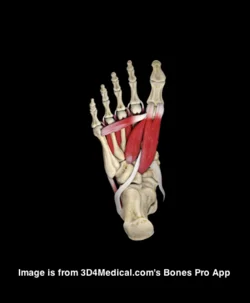
We remember the tibialis anterior is usually the most prominent muscle on the anterior leg (see 1st 2 pictures above). It has two origins, the lateral tibial condyle and the upper lateral surface of the tibia; it inserts on the medial surface of the 1st (medial) cuneiform and proximal part of base of the first metatarsal of the foot. It is a dorsiflexor and inverter of the foot in open chain (ie before and at heel strike/initial contact), and a dorsi flexor and inverter of the foot (or it brings the tibia medially and everts the foot relative to the tibia) from loading response to midstance. It also helps to maintain the medial longitudinal arch up to this point, and assist in decelerating pronation (along with its antagonist to dorsiflexion, the tibialis posterior). It takes a break at midstance and then contracts again at terminal stance (to counter act its antagonist, the peroneus longus). When running, it remains active through midstance to help pull the leg forward over the foot.

The authors have this to say: “Tibialis anterior restrained rearfoot plantarflexion from heel contact to 10% stance, and eversion between 10% stance and footflat.”

No surprises here.

Now the peroneus longus: “Activity in peroneus longus was consistent with its role in causing eversion after heel contact, then as a stabiliser of the forefoot after heel rise. ”

The peroneus (or fibularis) longus arises from the head and upper two-thirds of the lateral surface of the fibula, from the deep surface of the fascia, and from the intermuscular septa between it and the muscles on the front and back of the leg; occasionally also by a few fibers from the lateral condyle of the tibia.  You can see from it attachments that it can influence the entire upper lateral leg.

It’s tendon runs down the fibular shaft, wraps around the lateral malleolus, travels obliquely under the foot, crossing the lateral cubiod (which it everts after midstance to help with supination) crosses the sole of the foot obliquely, and inserts into the lateral side of the base of the first metatarsal and lateral aspect of the 1st cunieform.  

It acts from just prior to heel strike to limit excessive rearfoot inversion, through midstance to decelerate subtalar pronation and assists in stabilization of the midfoot articulations, and into terminal stance and pre swing to lock the lateral column of the foot for toe off and plantar flex the 1st ray (creating a good foot tripod), allowing dorsal posterior shift of the 1st metatarsal-phalangeal joint axis (necessary for dorsiflexion of the hallux (big toe)).

The peroneus brevis arises from the lower two-thirds of the lateral surface of the shaft of the fibua and from the intermuscular septa separating it from the adjacent muscles on the front and back of the leg. Again. lots of influence here.

It travels behind the lateral malleolus (and in front of the peroneus longus) and inserts into the tuberosity at the base of the fifth metatarsal.

It acts in a similar timeframe as the longus, copressing the tarsals to provide midfoot stabilization, and a significant eversion moment of the foot (helping to push you on to the base of the 1st metatarsal).

You can see how the peroneii could work together also to produce a small plantar flexion moment of the ankle and lateral movement of the lower leg. Because of their route around the lateral malleolus, they also can internally rotate the tibia (from a closed chain perspective; remember the tibia SHOULD be extenally rotation at this this point) so it can act to dampen or attenuate supination. This is also supported by the study:

“Activity in peroneus brevis suggested a role in restraining lateral rotation of the leg over the foot, late in stance.”

We are definitely the Uber Gait Nerds of the internet. We are bending your mind around the foot (rather than the foot around your mind).

Ivo and Shawn

Abstract

This study examined stance phase foot kinematics, kinetics and electromyographic (EMG) activity of extrinsic muscles of 18 healthy males. Three-dimensional kinematic and kinetic data were obtained via video analysis of surface markers and a force plate. Ankle joint moments are described about orthogonal axes in a segmental coordinate system. Kinematic data comprise rearfoot and forefoot motion, described about axes of a joint coordinate system, and medial longitudinal arch height. Surface EMG was obtained for tibialis anterior, soleus, gastocnemius medialis and lateralis, peroneus longus and peroneus brevis and extensor digitorum longus. It was concluded that the demands on the controlling muscles are greatest prior to foot flat and after heel rise. Tibialis anterior restrained rearfoot plantarflexion from heel contact to 10% stance, and eversion between 10% stance and footflat. Activity in peroneus longus was consistent with its role in causing eversion after heel contact, then as a stabiliser of the forefoot after heel rise. Activity in peroneus brevis suggested a role in restraining lateral rotation of the leg over the foot, late in stance.

Foot Ankle Int. 2001 Jan;22(1):31-41.

Extrinsic muscle activity, foot motion and ankle joint moments during the stance phase of walking.

School of Physiotherapy, Faculty of Health Sciences, Lidcombe, NSW, Australia. a.hunt@cchs.usyd.edu.au

Erratum in

  • Foot Ankle Int 2001 Jul;22(7):543.