You might think your shoe is doing more to control motion of your foot than it is actually doing.

You might think your shoe is doing more to control motion of your foot than it is actually doing.

"The measurement of rearfoot kinematics by placing reflective markers on the shoe heel assumes its motion is identical to the foot’s motion."
The results of this study revealed that "calcaneal frontal plane ROM was significantly greater than neutral and support shoe heel ROM. Calcaneus ROM was also significantly greater than shoe heel ROM in the transverse and sagittal planes. No change in tibial transverse plane ROM was observed."

It is easy to underestimate the calcaneal ROM across all planes of motion. Motion is going to occur somewhere, hopefully you can help your client control the excessive ROMs that are occurring and causing their symptoms. But just do not think that a shoe is going to markedly help, it might, but let your interventions and your client's feedback on pain lead you.

Calcaneus range of motion underestimated by markers on running shoe heel.
Ryan S. Alcantara'Correspondence information about the author Ryan S. AlcantaraEmail the author Ryan S. Alcantara
, Matthieu B. Trudeau, Eric S. Rohr
Human Performance Laboratory, Brooks Running Company, 3400 Stone Way N, Suite 500, Seattle, WA 98103 United States

Premature heel rise: Part 2

VIDEO: an atypical case of Premature heel rise. A follow up video for yesterdays discussion on the topic.

You should easily see premature heel rise here in this video. We will discuss this case at length with other video projections on our Patreon site next week, if you wish to dive further.

But here you should see, lets focus on the right limb, premature heel rise (again, stick with just watching the right foot/leg). This is, in-part, because this person does not achieve adequate hip extension, you should clearly be able to see that. Loss of terminal hip extension means premature heel rise, no exceptions. Train your eye to see this, you do not need expensive video software to see this.

So, Why inadequate hip extension? Well, just look at the amount of right knee flexion going into terminal stance, it is still heavily flexed and this forces them to prematurely heel rise, avoiding terminal hip extension, and prematurely load the forefoot. Without a knee that extends sufficiently, the hip cannot extend sufficiently, and thus premature heel rise is inevitable. And, trying to solve this issue down at the foot/ankle level is foolish in this case. Stretching this calf day after day until aliens come visit earth will still not be enough stretch time to fix this premature heel rise (ie. get that heel to stay down longer). There is a good reason why this is happening in this person, and it is a neurologic one, one we will discuss on the Patreon site for our Patrons. And, the reason does not matter for the concept I am teaching here today.

For today, you need to be able to see premature heel rise, and know all of the issues behind it, including causes, so that you can direct your phyiscial examination to solve your client's puzzle.
I have included yesterday's post below so you can review and bring this further together.
This is the kind of stuff we will do at Dr. Allen's Friday night Gait Lab, over some beverages. A unique, clinically curious and hungry 25 people need only apply. If you want to get to the next level of your human movement game, this is a way to get there.

Yesterday's post: We know that early/premature heel rise (PHR) leads to premature loading of the forefoot.
We know that premature heel rise (PHR) speeds us through many of the timely mechanical events that need and should occur for to get to safe and effective toe off during walking and running gaits.
This is why there are so many variables that need to be assessed and checked before instituting care to address the premature heel rise, because many times the problem is not even near the heel.
Consider, examine, assess (this is not an exhaustive list either) of causes of PHR
-short calf complex
-short quad (limits hip extension)
- short hip flexors
-anterior pelvis tilt as one's deviated norm posture
- prolonged or excessive rearfoot inversion
-lack of appropriate pronation (sustained supination)
-hallux limitus, rigidus
- weak anterior compartment lower leg
-lack of hip extension/weak glutes
-knee flexion contracture
- neurologic (toe walking gait from youth)
-painful achilles tendon mechanism
- loss of ankle rocker (which has its own long list)
. . . . to name a few

This is why you need to examine your clients, even after a gait analysis. Because, as we like to say, what you see is not your clients gait problem, it is their work around to other mechanical deficits.
After all, telling someone they just need to lengthen/stretch their calf to keep that heel down longer is utterly foolish.

*want to learn more about this stuff, you can join the upcoming Dr. Allen, Friday night Gait Lab series that he will be having in his office one Friday a month, in his Chicagoland office. Stay tuned for that notice. I will take only 25 people per session. We will dive into videos, cases, concepts, white-board rabbit holes, and enjoy some beverages and learn together. Stay tuned. The first 25 to pay and sign up are in !

Shawn Allen, the other gait guy

#gait, #gaitproblems, #gaitanalysis, #heelrise, #PHR, #prematureheelrise, #achilles, #achillestendinitis, #anklerocker, #heelrocker, #forefootpain, #halluxlimitus, #halluxrigidus, #heelpain

Premature heel rise: Part 1

IMG_1603.jpg

We know that early/premature heel rise (PHR) leads to premature loading of the forefoot.
We know that premature heel rise (PHR) speeds us through many of the timely mechanical events that need and should occur for to get to safe and effective toe off during walking and running gaits.
This is why there are so many variables that need to be assessed and checked before instituting care to address the premature heel rise, because many times the problem is not even near the heel.
Consider, examine, assess (this is not an exhaustive list either) of causes of PHR
-short calf complex
-short quad (limits hip extension)
- short hip flexors
-anterior pelvis tilt as one's deviated norm posture
- prolonged or excessive rearfoot inversion
-lack of appropriate pronation (sustained supination)
-hallux limitus, rigidus
- weak anterior compartment lower leg
-lack of hip extension/weak glutes
-knee flexion contracture
- neurologic (toe walking gait from youth)
-painful achilles tendon mechanism
- loss of ankle rocker (which has its own long list)
. . . . to name a few

This is why you need to examine your clients, even after a gait analysis. Because, as we like to say, what you see is not your clients gait problem, it is their work around to other mechanical deficits.
After all, telling someone they just need to lengthen/stretch their calf to keep that heel down longer is utterly foolish.

*want to learn more about this stuff, you can join the upcoming Dr. Allen, Friday night Gait Lab series that he will be having in his office one Friday a month, in his Chicagoland office. Stay tuned for that notice. I will take only 25 people per session. We will dive into videos, cases, concepts, white-board rabbit holes, and enjoy some beverages and learn together. Stay tuned. The first 25 to pay and sign up are in !

Shawn Allen, the other gait guy

#gait, #gaitproblems, #gaitanalysis, #heelrise, #PHR, #prematureheelrise, #achilles, #achillestendinitis, #anklerocker, #heelrocker, #forefootpain, #halluxlimitus, #halluxrigidus, #heelpain

A video primer on foot biomechanics.

Rewind Video Friday.
If you ever were unclear on how the sesamoids, 1st MET and FBH (flexor hallucis brevis) and others party together, this video will help you get up to speed.

As we begin the process of generating new videos, we came across this little gem from 8 years ago. Who is this younger punk ? Its Dr. Allen, showing some foot skills and sharing knowledge, stuff that will serve you well as we move forward with new videos.

https://www.youtube.com/watch?v=TyRE9dReVTE

Neuroma! Triple Threat....

Can you guess why this patient is developing a neuroma on the left foot, between the 3rd and 4th metatarsals?

IMG_6220.jpg
IMG_6218.jpg
IMG_6219.jpg

This gal presented to the office with pain in the left foot, in the area she points to as being between the 3rd and 4th metatarsals. It has been coming on over time and has become much worse this spring with hiking long distances, especially in narrower shoes. It is relieved by rest and made worse with activity.

Note the following:

  • She has an anatomical short leg on the left (tibial)

  • internal tibial torsion on the left

  • left forefoot adductus (see the post link below if you need a refresher)

Lets think about this.

The anatomical short leg on the left is causing this foot to remain in relative supination compared the right and causes her to bear weight laterally on the foot.

The internal tibial torsion has a similar effect, decreasing the progression angle and again causing her to bear weight laterally on the foot, compressing the metatarsals together.

We have discussed forefoot adductus before here on the blog. Again, because of the metararsal varus angle, it alters the forces traveling through the foot, pushing the metatarsals together and irritating the nerve root sheath, causing hypertrophy of the epineurium and the beginnings of a neuroma.

In this patients case, these things are additive, causing what I like to a call the “triple threat”.

So, what do we do?

  • give her shoes/sandals with a wider toe box

  • work on foot mobility, especially in descending the 1st ray on the left

  • work on foot intrinsic strength, particularly the long extensors

  • treat the area of inflammation with acupuncture

Dr Ivo Waerlop, one of The Gait Guys

#forefootadductus #metatarsusadductus #neuroma #gaitanalysis #thegaitguys #internaltibialtorsion

Hip flexors do not initiate hip flexon.

We have been saying it in writing and podcasts for years, the hip flexors are limb swing phase PERPETUATORS, not initiators of hip flexion.
It is the elastic response discussed below and the changing of the pelvis obliquity (from posterior positioning to anterior) via the abdominal wall acting on the pelvis-hip interval in conjunction with the stance phase hip musculature that drives hip flexion.
The next time you go after the psoas as a culprit in your meanderings for solutions, because that is what is all over the internet, think bigger, smarter, deeper.

"These experiments also showed that the trailing leg is brought forward during the swing phase without activity in the flexor muscles about the hip joint. This was verified by the absence of EMG activity in the iliacus muscle measured by intramuscular wire electrodes. Instead the strong ligaments restricting hip joint extension are stretched during the first half of the swing phase thereby storing elastic energy, which is released during the last half of the stance phase and accelerating the leg into the swing phase. This is considered an important energy conserving feature of human walking."

Dan Med J. 2014 Apr;61(4):B4823.
Contributions to the understanding of gait control.
Simonsen EB1.

#thegaitguys, #hip, #hipflexors, #pelvismechanics, #swingphase, #gait, #gaitanalysis, #gaitproblems

Knee braces and long legs?

Knee brace fixed at a zero to 5 degree flexion angle, creating a long leg? 

We know that the knee is supposed to flex during stance phase, usually around 20-25 degrees (depending on speed and weight, increases in bot increases the flexion requirement) to create dampening from vertical oscillation of the pelvis. What happens if they cannot flex? This creates a virtual "long leg" on that side this will usually result in:

  • Increased vertical translation of the pelvis upward on the braced side and

  • A compensation to make up for this "long leg; circumduction in this case,  but it could be any of the other compensations that we have talked about in posts here on the blog. 


work arounds? They are tough as each can create their own set of problems

  • allow more flexion in the knee on the braced side (not always possible)

  • place a full length sole lift on the opposite side to make up for the difference

  • use crutches

  • use a skateboard : )

we are sure you have some as well that you would LOVE to share with us

Dr Ivo Waerlop, one of The Gait Guys

#shortleg #LLD #compensations #legbrace #gaitproblem #thegaitguys


K ShamaeiGS SawickiAM Dollar Estimation of quasi-stiffness and propulsive work of the human ankle in the stance phase of walking - PloS one, 2013 - journals.plos.org

MORAIS FILHO, Mauro César; REIS, Renata Albertin dos  and  KAWAMURA, Cátia Myuki.Evaluation of ankle and knee movement pattern during maturation of normal gait. Acta ortop. bras. [online]. 2010, vol.18, n.1 [cited  2019-04-25], pp.23-25.

Where do you want to load your foot in relation to your center of mass ?

Screen Shot 2018-11-16 at 6.00.30 PM.png

Who do you want to be ? The guy loading his head over his foot
(narrow step width), or the gal loading the head and COM inside the foot (less narrow step width) ?
It is not hard to guess who is gonna be faster and more powerful from these photos. The lady is stacking the knee over the foot, the hip over the knee and stabilizing the hip and pelvis sufficiently and durably to keep the pelvis level for the next powerful loading step, and the other is flexion collapsing into the stance phase knee, insufficiently loading the hip and thus dropping the opposite side pelvis. He is not stacking the joints, there is a pending cross over (look at the swing leg knee approaching midline with barely any knee spacing, thus guaranteeing a cross over step or at the very least a very narrow step width.)
Sure, some one is going to say one is a distance runner and the other is a sprinter. Yes, and our point is that the sprinter is not head-over-foot, the one with all the highly suspect flaws is head over foot ! Wider step width means more glutes. Go ahead, walk around right now with a very narrow step width and see how little efficient glute contraction you get, then walk with a notably wider step width, and you will see wider means more glutes. Keep your COM moving forward, not oscillating back and forth sideways over each stance foot, that is a power leak.

Screen Shot 2018-11-16 at 6.00.11 PM.png

The distance runner is showing sloppy in technique. Say what you want, but one of these runners is weak and very likely at greater risk for injury, the other is strong and durable, and likely at less risk for injury.
If you ask us, but what do we know . . . .
So, again, was ask . . . . which one do you want to be ?

Dual tasking and neurocognitive decline.

Your holiday homework . . . . look for the gait clues Ivo and Shawn have talked about this year (*see below)

Dual tasking and neurocognitive decline.
Mild cognitive impairment (MCI) is considered a predementia state associated with a 10-fold increased risk of progression to dementia. Dual tasking during gait may help predict neurocognitive decline.

So, When you are around aging family this holiday season, pay close attention to them when moving about around them. Dual tasking during gait should not be difficult for most healthy folks, but if you add in things that the aging population are challenged with (things like physical weaknesses, mild vestibular challenges, visual challenges , mild neuropathy, cold feet, proprioceptive losses) and then throw in some dual tasking (talking, carrying bags) we can often bring out predictors of future decline.
Remember, falls in the elderly are huge predictors of near term morbidity.

* Look for the clues during dual tasking or during intimidating situations (ie, crossing a busy street), look for things like slowing of gait, wider or narrower step width, shorter steps, frustration, confusion, reaching for support (grapping your hand or arm), stopping, shuffling, arresting of talk to negotiate an area, etc.

"A dual-task gait test evaluating the cognitive-motor interface may predict dementia progression in older adults with MCI (mild cognitive impairment)."

Association of Dual-Task Gait With Incident Dementia in Mild Cognitive Impairment
Results From the Gait and Brain Study. Manuel M. Montero-Odasso et al.
JAMA Neurol. 2017 Jul; 74(7): 857–865.

Circumducting gait , at the ankle level ?


We often circumduct a leg to get around a clearance problem. Sometimes the clearance problem is the leg length itself, and sometimes it is a foot clearance issue, one that doesn't dorsiflex/toe extend enough.

This is what the foot clearance circumduction strategy looks like (more clear on the left foot). It is a heavy peroneal, tib anterior (more lateral belly, interosseous) and lesser toe extensor strategy. The foot clearly dorsiflexes and everts the rear and fore foot during early swing. It is not until just before heel strike that the tib anterior seem to jump in to do its primary job of dorsiflexion AND inversion.
Finding out why a client is circumducting this way is the key. It could be from the opposite hip abductors being weak, and it could be poor abdominal control on the same side, or it could be down in the foot (perhaps extensor hallucis/big toe extensor) and of highest suspect is a weak or motor pattern delayed tib anterior. Bad lazy habits can happen around trivial weakness, and then can mushroom into other bigger things.

Your exam will help you.
Seeing a problem in someones gait is not their problem, it is their strategy to get around the parts that are not working well.

shawn allen, the other gait guy

#gait, #gaitproblems, #gaitanalysis, #anklerocker, #ankledorsiflexion, #shinsplints, #swingphase, #thegaitguys, #circumductinggait

The “Standing on Glass” Static Foot/Pedograph... PART 2

The “Standing on Glass” Static Foot/Pedograph... PART 2
We hope you find this case presentation dialogue interesting.

Screen Shot 2019-01-13 at 7.51.15 PM.png

* note: This is a static assessment dialogue. One cannot, and must not, make clinical decisions from a static assessment. As in all assessments, information is taken in, digested and them MUST be confirmed, denied and/or at the very least, folded into a functional and clinically relevant assessment of the client before the findings are accepted, dismissed and acted upon.

Here is the case . . .

Part 2: “Standing on Glass” Static Foot/Pedograph Assessment

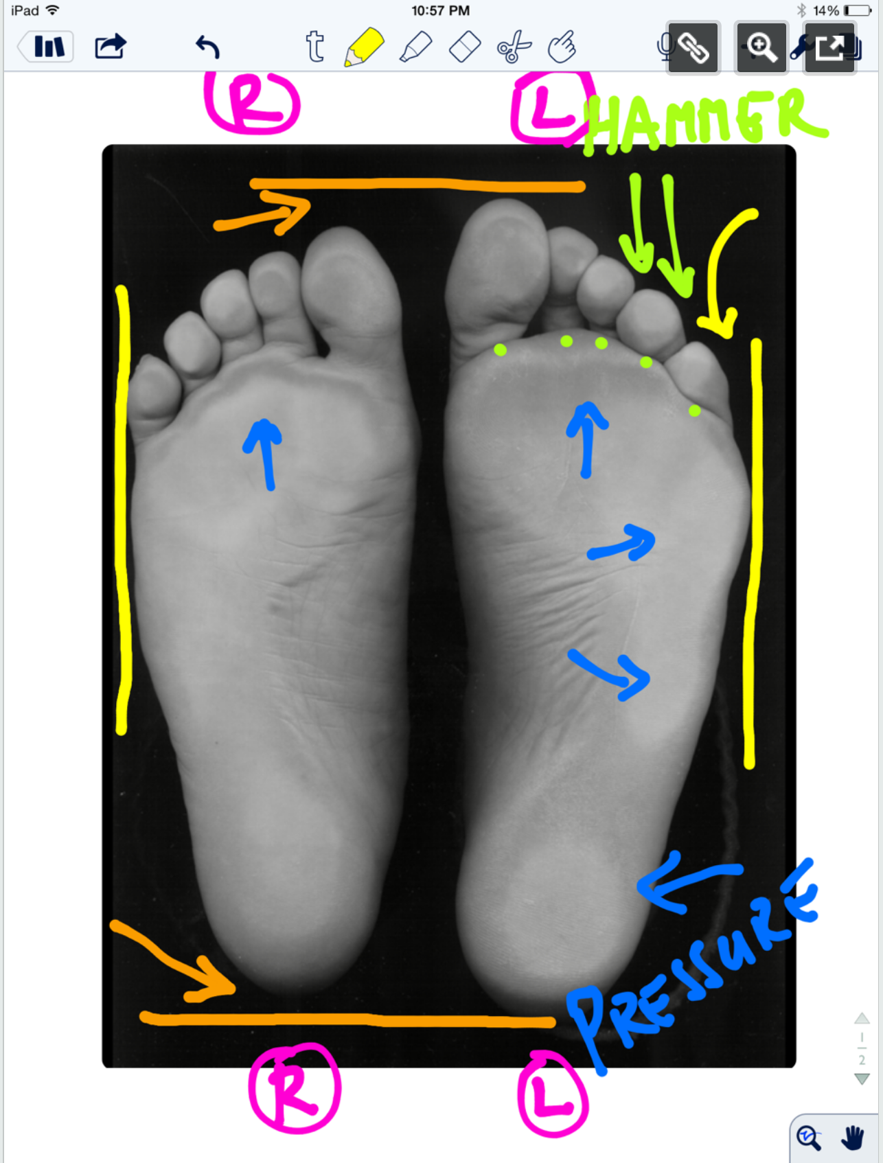
* note (see warning at bottom): This is a static assessment dialogue. One cannot, and must not, make clinical decisions from a static assessment. The right and left sides are indicated by the R and L circled in pink. There are 4 photos here today.

Blue lines: Last time we evaluated possible ideas on the ORANGE lines here, it would be to your advantage to start there.

We can see a few noteworthy things here in these photos. We have contrast-adjusted the photo so the pressure areas (BLUE) are more clearly noted. There appears to be more forefoot pressure on the right foot (the right foot is on the readers left), and more rearfoot pressure on the left (not only compare the whiteness factor but look at the displacement of the calcaneal fat pad (pink brackets). There is also noticeably more lateral forefoot pressure on the left. There is also more 3-5 hammering/flexion dominance pressure on the left. The metatarsal fat pad positioning (LIME DOTS represent the distal boundary) is intimately tied in with the proper lumbrical muscle function (link) and migrates forward toward the toes when the flexors/extensors and lumbricals are imbalanced. We can see this fat pad shift here (LIME DOTS). The 3-5 toes are clearly hammering via flexor dominance (LIME ARROWS), this is easily noted by visual absence of the toe shafts, we only see the toe pads. Now if you remember your anatomy, the long flexors of the toes (FDL) come across the foot at an angle (see photo). It is a major function of the lateral head of the Quadratus plantae (LQP) to reorient the pull of those lesser toe flexors to pull more towards the heel rather than on an angle. One can see that in the pressure photos that this muscle may be suspicious of weakness because the toes are crammed together and moving towards the big toe because of the change in FDL pull vector (YELLOW LINES). They are especially crowding out the 2nd toe as one can see, but this can also be from weakness in the big toe, a topic for another time. One can easily see that these component weaknesses have allowed the metatarsal fat pad to migrate forward. All of this, plus the lateral shift weight bearing has widened the forefoot on the left, go ahead, measure it. So, is this person merely weight bearing laterally because they are supinating ? Well, if you read yesterday’s blog post we postulated thoughts on this foot possibly being the pronated one because of its increased heel-toe and heel-ball length. So which is it ? A pronated yet lateral weight bearing foot or a normal foot with more lateral weight bearing because of the local foot weaknesses we just discussed ? Or is it something else ? Is the problem higher up, meaning, are they left lateral weight bearing shift because of a left drifted pelvis from weak glute medius/abdominal obliques ? Only a competent clinical examination will enlighten us.

Is the compensation top-down or bottom up, or both in a feedback cycle trying to find sufficient stability and mobility ? These are all viable possibilities and you must have these things flowing freely through your head during the clinical examination as you rule in/rule out your hands-on findings. Remember, just going by a screen to drive prescription exercises from what you see on the movement screen is not going to necessarily fix the problem, it could in fact lead one to drive a deeper compensation pattern.

Remember this critical fact. After an injury or a long standing problem, muscles and motor patterns jobs are to stabilize and manage loads (stability and mobility) for adequate and necessary movement. Injuries leave a mark on the system as a whole because adaptation was necessary during the initial healing phase. This usually spills over during the early movement re-introduction phase, particularly if movement is reintroduced too early or too aggressively. Plasticity is the culprit. Just because the injury has come and gone does not mean that new patterns of skill, endurance, strength (S.E.S -our favorite mnemonic), stability and mobility were not subsequently built onto the apparently trivial remnants of the injury. There is nothing trivial if it is abnormal. The forces must, and will, play out somewhere in the body and this is often where pain or injury occurs but it is rarely where the underlying problem lives.

Come back tomorrow. We will try to bring this whole thing together, but remember, it will just be a theory for without an exam one cannot prove which issues are true culprits and which are compensations. Remember, what you see is often the compensatory illusion, it is the person moving with the parts that are working and compensating not the parts that are on vacation. See you tomorrow friends !

Shawn and ivo, the gait guys

* note: This is a static assessment dialogue. One cannot, and must not, make clinical decisions from a static assessment. As in all assessments, information is taken in, digested and then MUST be confirmed, denied and/or at the very least, folded into a functional and clinically relevant assessment of the client before the findings are accepted, dismissed and acted upon. As we always say, a gait analysis or static pedograph-type assessment (standing force plate) is never enough to make decisions on treatment to resolve problems and injuries. What is seen and represented on either are the client’s strategies around clinical problems or compensations. Today’s photo and blog post are an exercise in critical clinical thinking to get the juices flowing and to get the observer thinking about the client’s presentation and to help open up the field to questions the observer should be entertaining. The big questions should be, “why do i see this, what could be causing these observances ?”right foot supinated ? or more rear and lateral foot……avoiding pronation ?


The “Standing on Glass” Static Foot/Pedograph Assessment: Part 1

Screen Shot 2019-01-13 at 7.42.41 PM.png

The “Standing on Glass” Static Foot/Pedograph... PART 1
We hope you find this case presentation dialogue interesting.

* note: This is a static assessment dialogue. One cannot, and must not, make clinical decisions from a static assessment. As in all assessments, information is taken in, digested and them MUST be confirmed, denied and/or at the very least, folded into a functional and clinically relevant assessment of the client before the findings are accepted, dismissed and acted upon.

The “Standing on Glass” Static Foot/Pedograph Assessment: Part 1

* note: This is a static assessment dialogue. One cannot, and must not, make clinical decisions from a static assessment. As in all assessments, information is taken in, digested and them MUST be confirmed, denied and/or at the very least, folded into a functional and clinically relevant assessment of the client before the findings are accepted, dismissed and acted upon. As we always say, a gait analysis or pedograph-type assessment is never enough to make decisions on treatment to resolve problems and injuries. What is seen and represented on either are the client’s strategies around clinical problems or compensations. Today’s photo and blog post are an exercise in critical clinical thinking to get the juices flowing and to get the observer thinking about the client’s presentation and to help open up the field to questions the observer should be entertaining. The big questions should be, “why do i see this, what could be causing these observances ?”

* note the right and left sides by the R and L circled in pink.

ORANGE lines: The right foot appears to be shorter, or is it that the left is longer (see the lines and arrows drawing your attention to these differences)? A shorter foot could be represented by a supinated foot (if you raise the arch via the windlass mechanism you will shorten the foot distance between the rear and forefoot). A longer foot could be represented by a more pronated foot. Is that what we have here ? There is no way to know, this is a static presentation of a client standing on glass. What we should remember is that the goal is always to get the pelvis square and level. If an anatomically or functionally short leg is present, the short leg side MAY supinate to raise the mortise and somewhat lengthen the leg. In that same client, they may try to meet the process part way by pronating the other foot to functionally “shorten” that leg. Is that what is happening here ? So, does this client have a shorter right leg ? Longer left ? Do you see a plunking down heavily onto the right foot in gait ? Remember, what you see is their compensation. Perhaps the right foot is supinating, and thus working harder at the bottom end of the limb (via more supination), to make up for a weak right glute failing to eccentrically control the internal spin of the leg during stance phase ? OR, perhaps the left foot is pronating more to drive more internal rotation on the left limb because there is a restricted left internal hip rotation from the top ? Is the compensation top-down or bottom up ? These are all viable possibilities and you must have these things flowing freely through your head during the clinical examination as you rule in/rule out your hands-on findings. Remember, just going by a FMS-type screen to drive prescription exercises from what you see on a movement screen is not going to necessarily fix the problem, it could in fact lead one to drive a deeper compensation pattern. You can be sure that Gray Cook’s turbo charged brain is juggling all of these issues (and more !) when he sees a screen impairment, although we are not speaking for him here.

Remember this critical fact. After an injury or a long standing problem, muscles and motor patterns jobs are to stabilize and manage loads (stability and mobility) for adequate and necessary movement. Injuries leave a mark on the system as a whole because adaptation was necessary during the initial healing phase. This usually spills over during the early movement re-introduction phase, particularly if movement is reintroduced too early or too aggressively. Plasticity is the culprit. Just because the injury has come and gone does not mean that new patterns of skill, endurance, strength (S.E.S -our favorite mnemonic), stability and mobility were not subsequently built onto the apparently trivial remnants of the injury. There is nothing trivial if it is abnormal. The forces must, and will, play out somewhere in the body and this is often where pain or injury occurs but it is rarely where the underlying problem lives.

Come back tomorrow, where we will open your mind into the yellow, pink, blue and lime markings on the photo. Are the hammering toes (lime) on the left a clue ? How about the width of the feet (yellow) ? The posturing differences of the 5th toe to the lateral foot border ? What about the static plantar pressure differences from side to side (blue)? Maybe, just maybe, we can bring a logical clinical assumption together and then a few clinical exam methods to confirm or dis-confirm our working diagnostic assumption. See you tomorrow friends !

Shawn and ivo, the gait guys

Here is the case link.......

https://thegaitguys.tumblr.com/post/99409232289/the-standing-on-glass-static-footpedograph?fbclid=IwAR3gd3d81Gwt3ywAB7BcTwXqST2Z_5nmieODzSb8rJQYBcJFhTs6rS_9auA

Forefoot running, achilles loads & gait retraining

tag/key words: gait, gaitproblems, gaitanalysis, forefootrunning, forefootstrike, achilles, heelstrike, elastography, thegaitguys, microvascularity, rockeredshoes, HOKA, metarocker, gaitretraining,

Links to find the podcast:
Look for us on iTunes, Google Play, Podbean, PlayerFM and more.
Just Google "the gait guys podcast".

Our Websites:
www.thegaitguys.com
Find Exclusive content at: https://www.patreon.com/thegaitguys
doctorallen.co
summitchiroandrehab.com
shawnallen.net

Our website is all you need to remember. Everything you want, need and wish for is right there on the site.
Interested in our stuff ? Want to buy some of our lectures or our National Shoe Fit program? Click here (thegaitguys.com or thegaitguys.tumblr.com) and you will come to our websites. In the tabs, you will find tabs for STORE, SEMINARS, BOOK etc. We also lecture every 3rd Wednesday of the month on onlineCE.com. We have an extensive catalogued library of our courses there, you can take them any time for a nominal fee (~$20).

Our podcast is on iTunes and just about every other podcast harbor site, just google "the gait guys podcast", you will find us.

Where to find us, the podcast Links:

iTunes page:
https://itunes.apple.com/us/podcast/the-gait-guys-podcast/id559864138?mt=2

Google Play:
https://play.google.com/music/m/Icdfyphojzy3drj2tsxaxuadiue?t=The_Gait_Guys_Podcast

Direct download URL: http://traffic.libsyn.com/thegaitguys/pod_150.2_-_42719_5.00_PM.mp3

Permalink URL: http://thegaitguys.libsyn.com/forefoot-running-achilles-loads-gait-retraining

Libsyn Directory URL: http://directory.libsyn.com/episode/index/id/9555122

Show notes:

Ultrasound elastographic assessment of plantar fascia in runners using rearfoot strike and forefoot strike. Tony Lin-WeiChen et al
https://www.sciencedirect.com/science/article/pii/S0021929019302775
J Sci Med Sport. 2015 Mar;18(2):133-8. doi: 10.1016/j.jsams.2014.02.008. Epub 2014 Feb 14.

Rocker shoes reduce Achilles tendon load in running and walking in patients with chronic Achilles tendinopathy.. Sobhani S et al
https://www.ncbi.nlm.nih.gov/pubmed/24636129/


The increase in muscle force after 4 weeks of strength training is mediated by adaptations in motor unit recruitment and rate coding. Alessandro Del Vecchio et al
https://physoc.onlinelibrary.wiley.com/doi/10.1113/JP277250


Learning new gait patterns is enhanced by specificity of training rather than progression of task difficulty. ChandramouliKrishnan et al
https://www.sciencedirect.com/science/article/pii/S0021929019301927


The microvascular volume of the Achilles tendon is increased in patients with tendinopathy at rest and after a 1-hour treadmill run. Pingel J et al
Am J Sports Med. 2013 Oct;41(10):2400-8. doi: 10.1177/0363546513498988. Epub 2013 Aug 12.
https://www.ncbi.nlm.nih.gov/pubmed/23940204/

*** Our PODcast disclaimer:
This podcast is for general informational purposes only. It does not constitute the practice of medicine, nursing, rehab, treatment, therapy recommendations or anything of the sort. This podcast should not replace proper medical advise that should only be attained through proper medical channels that would entail a full medical and/or biomechanical physical examination and/or appropriate diagnostic testing. No doctor-patient relationship is formed by listening to this podcast or any information gleaned from our writings or social media work.
The use of this information and the materials linked to the podcast is taken at the users own risk. This podcast and the content shared is not intended to replace or be a substitute for appropriate professional medical advise diagnosis or treatment. Users should not disregard or delay obtaining medical advice for any condition they have and should seek the advice and assistance from their providers for any such conditions.

Top end heel raises. The top end might matter.

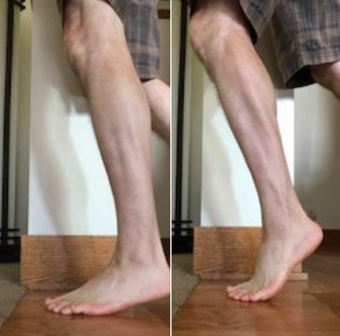
Screen Shot 2019-04-19 at 6.34.09 PM.png

Thought experiment . . .
If top end posterior compartment (loosely, the calf complex) strength is lacking, then heel rise may not be optimized to transfer body mass forward sufficiently and effectively.
This lack of forward progression, fails to move the body mass sufficiently forward enough to reduce the external moment arms and optimize the internal moment arms to take maximal advantage of the calf complex (I am talking about moment arms between the grounded 1st MTP joint and ankle mortise & ankle mortise and achilles tendon).
These are rough thoughts today gang, letting you inside our heads and how we juggle multiple parameters when we are struggling to solve a client's problems.

In the lower heel rise photo, The body mass does not progress forward enough over the grounded first MTP joint at the big toe (during gait, the heel doesn’t just rise up, the axis of the ankle joint moves both up and forward).
In this case, the foot may not be fully rigid in a supinated position to benefit from joint closed-packed positions. Thus, the foot may be more pliable and one might suppose that if not adequately supinated, they are inadequately still too much relatively pronated. This might put more load into the tibialis posterior and other soft tissue mechanical loading scenarios that are less optimally suited to do this job. Over time, might this lead to pathology? Likely.
Thus, when running on a weaker posterior mechanism (often found unilaterally) the higher up posterior chains might be overburdened, the tendon loads and loading response of the achilles, tibialis posterior, and long flexors will be most likely altered, likely negatively, the naturally occurring foot locking mechanisms might be less optimal than desired, subtalar and forefoot loading might be premature (ie. sesamoid malpositioning for one, as a simple example), etc etc. Loading a foot(the mid and forefoot) into heel rise that is still somewhat pronated creates a different moment arm around the subtalar joint axis (that moves through the 1st metatarsal), than a foot that is more supinated.

Now, put these ideas into the 2 photos from yesterday where one might be loading the forefoot laterally or more medially, and now make the top end strength more in one of those scenarios. Is it any wonder why so many struggle with posteiror mechanism tendonopathies ? There are so many parameters to consider and examine. And, if not examined in great detail, the key lacking parameter can be missed.
Hence, just forcing calf strength loading is too simple a solution, there is a needle in that haystack that upset the client's apple cart, it is the job of the clinician to find it and remedy it.

Today, looking into the research and finding some interesting things that are spurring some thoughts.

Shawn Allen, one of the gait guys

You are mostly likely not getting to your big toe at push-off if you are doing this.

Screen Shot 2019-04-08 at 4.20.41 PM.png

You are mostly likely not getting to your big toe at push-off if you are doing this. And, if you have a painful big toe, you will do it as well. Oh, and Head-over-foot related, yup. Read on . . .


Yes, the cross over gait. Yes, when you are into a cross over gait you are most certainly head over foot. And that is most likely not a good thing.


If you are not closer to stacking the hip over the knee, and knee over the foot (like in the photo "SUI" bib runner) you are not likely getting to much of your big toe at terminal stance loading, when you could be getting more power at push off.
Said another way, if you are attacking the ground with the feet closer together, as if you are running on a line (as in the photo) you are going to be more on the outside of the foot (note the lateral foot contact), show a similar wear/loading pattern as in these shoes, and hardly load the medial foot tripod effectively.
Go ahead, walk around your office or home right now . . . . with a very narrow step width and see how little you can load into the bit toe-medial foot tripod (note how little effective glute engagement you get as well). Then, walk with a wider step width, note the easier more effective big toe-medial tripod loading, and, not the glutes come into play.

Screen Shot 2019-04-08 at 4.20.52 PM.png

Thus, head over foot/cross over gait is foolish for effective gait. You have a big toe, don't you wish to use it ? One has to find that balance between an economical step width that still allows an effective toe off event in walking and running. A very narrow cross over-style gait does not afford us this.
So, should it be any surprise to any of us that someone with pain in the big toe or medial tripod complex will choose a narrow step width to avoid the painful loading ? No, no surprise there at all.
We have been writing about the cross over gait for 10 years, bringing little pieces of research to the forefront to prove our theories on it as the research presents itself. We first brought it to you with our 3 part video series here. Search our blog, type in "cross over gait" into the search box on the site www.thegaitguys.com and get a LARGE coffee before hand, you are going to be reading for several hours.

https://www.youtube.com/watch?v=LG-xLi2m5Rc
https://www.youtube.com/watch?v=WptxNrj2gCo
https://www.youtube.com/watch?v=oJ6ewQ8YUAA

Screen Shot 2019-04-08 at 4.21.31 PM.png
Screen Shot 2019-04-08 at 4.21.23 PM.png

Support for visual gait analysis... with respect to leg length discrepancies


image credit: https://pixabay.com/photos/eye-blue-eyelashes-vision-make-up-691269/

image credit: https://pixabay.com/photos/eye-blue-eyelashes-vision-make-up-691269/

We talk about leg length discrepancies all the time here on the blog and sometimes, how small discrepancies cause changes in peoples biomechanics. The gold standard for measuring a leg length difference is full length lower extremity X ray, but this presents a problem due to the ionizing radiation, accessibility as well as impracticality of X rays every person with a suspected difference.

We have talked about different compensations as to how to get around a leg length discrepancies. Last week we actually did a tell a seminar on this entire subject. Your patient or client needs to “create clearance” for the longer leg side. This can be accomplished in many ways.

The 5 most common strategies (keep in mind there are many more) are:

  • lean the torso to the short leg side (essentially hip adduction of the longer side)

  • hike the torso on the long leg side

  • circumduct the longer lower extremity

  • increase plantar flexion of the calf of the short leg side

  • increase hip and knee flexion on the longer leg side

And that is exactly what this study found. They looked at kinematics in people with anatomical leg length discrepancies and found that hip adduction as well as increased hip and knee flexion were 2 variables that were consistent in folks with anatomical differences and suggest these variables are a useful screening tool.

Paying attention to how people move and looking for asymmetries. In our opinion, that’s the name of the game : )

Dr Ivo Waerlop, on of The Gait Guys

Zeitoune GNadal JBatista LAMetsavaht LMoraes APLeporace G.Prediction of mild anatomical leg length discrepancy based on gait kinematics and linear regression model. Gait Posture. 2019 Jan;67:117-121. doi: 10.1016/j.gaitpost.2018.09.027. Epub 2018 Sep 29.

#LLD #leglengthdifference #leglengthinequality #visualgaitanalysis #thegaitguys #gaitanalysis

Pronation anyone? Coupling? Walking Speed? How do they relate?

We have talked many times here on TGG about pronation, supination, overpronation, asymmetrical pronation, and more.

When most people think of pronation, they think of midfoot pronation, or pronation about the subtalar or transverse tarsal joints. Pronation can actually occur about any articulation or bone, but with respect to the foot, we like to think of rearfoot (ie. talo-calcaneal), midfoot (talo-navicular) and forefoot (transverse tarsal). The question is why does this matter?

Pronation, with respect to the foot, is defined as a combination of eversion, abduction and dorsiflexion (see picture attached) which results in flattening of the planter vault encompassing the medial and lateral longitudinal arches. In a normal gait cycle, this begins at initial contact (heel strike) and terminates at midstance, lasting no more than 25% of the gait cycle.

In a perfect biomechanical world, shortly following initial contact with the ground, the calcaneus should evert 4-8 degrees, largely because the body of the calcaneus is lateral to the longitudinal axis of the tibia. This results in plantar flexion, adduction and eversion of the talus on the calcaneus, as it slides anteriorly. At this point, there should be dorsiflexion of the transverse tarsal (calcaneo-cuboid and talo-navicular joints). Due to the tight fit of the ankle mortise and its unique shape, the tibial rotates internally (medially). This translates up the kinetic chain and causes internal rotation of the femur, which causes subsequent nutation of the pelvis and extension of the lumbar spine. This should occur in the lower kinetic chain through the 1st half of stance phase. The sequence should reverse after the midpoint of midstance, causing supination and creating a rigid lever for forward propulsion.

Pronation, along with knee and hip flexion, allow for shock absorption during throughout the 1st half of stance phase. Pronation allows for the calcaneo-cuboid and talo-navicular joint axes to be parallel making the foot into a mobile adaptor so it can contour to irregular surfaces, like our hunter gatherer forefathers used to walk on before we paved the planet. Problems arise when the foot either under pronates (7 degrees valgus results in internal tibial rotation), resulting in poor shock absorption or over pronates (> 8 degrees or remains in pronation for greater than 50% of stance phase).

This paper talks about how foot and ankle pathologies have effects on other articulations in the foot. They looked at stance phase of gait in 14 people without pathology at 3 different walking speeds. they found:

coupling relationships between rear foot inversion and hallux plantar flexion and rear foot eversion with hallux dorsiflexion.... When the rear foot everts (as it does as discussed above) during pronation from initial contact to mid stance , the hallux should be extending AND when the rear foot everts, as it should from mid stance to terminal stance/pre swing, the hallux should be plantar flexing to get the 1st ray down to the ground

medial (internal) rotation of there leg was accompanied by mid foot collapse (read pronation) and lateral (external) rotation with mid foot elevation (read supination).... Because of the shape of the talar dome and shape of the talo calcaneal facet joints, the talus plantar flexes, everts and adducts from initial contact to mid stance, and dorsiflexes, inverts and adducts from mid stance to terminal stance/ pre swing

walking speed significantly influenced these coupling relationships....meaning that the faster we go, the faster these things must happen and the greater degree that the surrounding musculature and associated cortical control mechanisms must act

So, when these relationships are compromised, problems (or more often, compensations) ensue. Think about these relationships and the kinetics and kinematics the next time you are studying someones gait.

Dr Ivo Waerlop, one of The Gait Guys

Dubbeldam R1, Nester C, Nene AV, Hermens HJ, Buurke JH. Kinematic coupling relationships exist between non-adjacent segments of the foot and ankle of healthy subjects.Kinematic coupling relationships exist between non-adjacent segments of the foot and ankle of healthy subjects.Gait Posture. 2013 Feb;37(2):159-64. doi: 10.1016/j.gaitpost.2012.06.033. Epub 2012 Aug 27

cool video on our blog to go with this post:

https://www.thegaitguys.com/thedailyblog/2018/8/7/what-do-you-know-about-pronation-and-supination

#gaitanalysis #thegaitguys #pronation #couplingrelationshipsandgait #pronation

Case Studies in Gait Analysis: Focus on the Short Leg (online video class)

Case Studies in Gait Analysis: Focus on the Short Leg
*link is below

*this is the online Continuing education class we did last week, for those of you who could not get to the Wednesday evening class.
*our entire catalogue of lectures and seminars are all here on this site for CE/CEU

Case Studies in Gait Analysis: Focus on the Short Leg
- Review anatomical vs functional short leg
-Review the kinematics and kinetics of the short leg during the gait cycle
-View and discuss case studies looking at functional and anatomical short legs
-Predict pathomechanics that will arise from a short leg
-Propose remedies for the gait abnormalities seen

Link: https://chirocredit.com/course/Chiropractic_Doctor/Biomechanics_211

Screen Shot 2019-04-22 at 1.20.16 PM.png

The hip flexors do not pull the leg forward during swing (mostly).

The hip flexors are not responsible for pulling/flexing the swing leg forward in gait or running. The psoas is a mere swing phase perpetuator, not an initiator.
For about 2 decades we have been saying in our lectures, posts and podcasts that it is the reduction of the obliquity of the pelvis during gait from various other tissues and biomechanical events that causes leg swing, meaning the trail leg is brought forward in swing largely by the abdominal muscle linkage to the pelvis (and other loaded tissues) that is responsible for forward swing of the leg. It is not the hip flexor group that does this hip flexion action. Thus it could be considered foolish to train the hip flexors to be the primary swing drivers. Here is another supporting piece of research.

"These experiments also showed that the trailing leg is brought forward during the swing phase without activity in the flexor muscles about the hip joint. This was verified by the absence of EMG activity in the iliacus muscle measured by intramuscular wire electrodes. Instead the strong ligaments restricting hip joint extension are stretched during the first half of the swing phase thereby storing elastic energy, which is released during the last half of the stance phase and accelerating the leg into the swing phase. This is considered an important energy conserving feature of human walking. "

Dan Med J. 2014 Apr;61(4):B4823.
Contributions to the understanding of gait control.
Simonsen EB1.