Can a cross over occur on one side of the body ? Sure, this case is a perfect example. The heavy lateral shoe wear on the left is a huge clue. But remember, what you see is not the problem, it is the result of their problem(s).
. . . a talented marathoner came into our office complaining of a long standing deep posterior right hip pain and an equally longstanding left chronic lateral ankle and foot pain. The ankle had been treated regularly for chronic peroneal tendonitis with various manual therapy modalities and yet the right hip seemed to be left out of the equation in terms of treatment.
After taking a detailed history this runner unknowingly pretty much told us they had all the qualifications of ischial-femoral impingement (IFI). What they did not realize was that they had a cross over gait style that was a significant contributor to the clinical problem.
Here is a nice rewind case for your Friday read.
____________________
link: https://thegaitguys.tumblr.com/post/116468620969/what-ischial-femoral-impingement-might-look-like
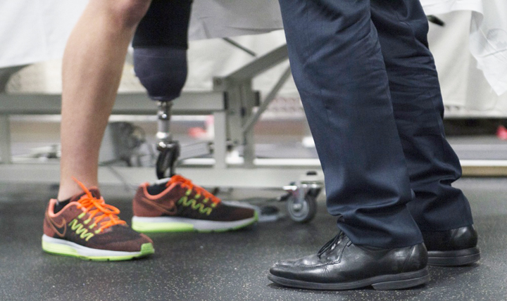
What ischial-femoral impingement might look like as aberrant shoe wear.
A few weeks ago we wrote an article on ischial-femoral impingement. For you to best understand today’s blog post you really should go back and review this interesting clinical phenomenon, here is the link to that piece.
Three weeks ago a talented marathoner came into our office complaining of a long standing deep posterior right hip pain and an equally longstanding left chronic lateral ankle and foot pain. The ankle had been treated regularly for chronic peroneal tendonitis with various manual therapy modalities and yet the right hip seems to be left out of the equation in terms of treatment.
After taking a detailed history this runner unknowingly pretty much told us they had all the qualifications of ischial-femoral impingement (IFI). What they did not realize was that they had a cross over gait style that was a significant contributor to the clinical problem.
Lets now have a look at the shoe wear patterns above. On the left shoe, (a shoe we love, New Balance Fresh Foam (find your next pair at NewBalance Chicago)) we see that the entry zone or crash zone of rear foot impact is heavily worn, especially laterally. Heavy entry zone wear can be from several things, but one thing we always check for and assume until proven otherwise is a cross over gait. It can also just be from excessive rearfoot inversion at foot strike but when excessive there is usually a reason for it, especially when unilaterally as seen here. This foot is not stacking under the knee and hip, it is striking more midline to a plumb line dropped from the hip joint. This creates a steep medial angle of attack. The question is why ? Well, in the history the right hip pain began first and then the left ankle pain, so one should at least consider a compensatory timeline, that being the foot is a compensation in the gait cycle from the painful hip.
This client on examination tested pretty obviously for a right frontal plane drift, meaning when the right foot impacts there is not enough lateral line support to hold the hip/pelvis over the foot and so the pelvis drifts laterally to the right in this case. This can be fought by inverting the foot. This is a strategy to try and stop the lateral drift. In this case, excessive wear is seen on the entire lateral side of the right shoe to represent this compensation. Changing this clients foot wear, shoe, orthotic is not fixing the problem, in fact it is impairing their ability to compensate and could create more problems, and even another deeper layer of compensation. Again, the inverted/supinated right foot moves the weightbearing line laterally, by moving the foot’s center of pressure from within the confines of the foot tripod towards the lateral border of the foot tripod, in attempt to restack the loading over the laterally drifted hip (hence the right lateral shoe wear pattern). Unfortunately this does not solve the reason for the lateral drifted pelvis. That solution has to come from improved stablization of the hip, pelvis and core and since they tested weak on the right side abdominals, gluteus medius, gluteus max and other accessory lateral stabilizers, work must be done there. Interestingly, this runner is stuck into a vicious cycle. The lateral drift to the right is allowing the left hemi-pelvis to dip and this is challenging rotational control of the stance limb and it is causing ischial-femoral impingement (suspecting of the quadratus femoris). It was clear on examination that there was impairment of the quadratus femoris and obturator externus upon detailed testing and deep palpation was pin point tender over these structures. Resistance to rotational challenges to the limb, especially iso and eccentric internal rotational challenges, were very poor when it came to coordination, endurance and certainly strength.
Remember, when you are spending time going sideways (right frontal plane drift), you are not spending time moving forwards. This could cause an early right departure and force and early left stance engagement. But it goes deeper than that in this case. Here, the right frontally drifting pelvis will pull the left swing leg across the midline with it, creating a left cross over gait. This will make more sense if you watch our popular video here. Link
So, when this left swing leg is forced into the cross over gait variant, it will force a strong lateral heel strike, as evidenced on the left shoe wear. This is a compensation to what is going on in the right side body mechanics.
Can a cross over occur on one side of the body ? Sure, this case is a perfect example.
Can a cross over gait on the left in this case, cause a vicious cycle and in itself create an environment whereby a right ischio-femoral impingment occurs ? Sure, neuronal plasticity can be a bitch, it can work in your favor, and against you.
This is not a tough case, if you have seen the beast before and you recognize all of its parameters. If you have not seen the beast before, this case is a nightmare with all these pieces (deep buttock pain, impingement, frontal drift, cross over, strange shoe wear pattern, opposite ankle peroneal pain etc). Do you have to get this right every time with a bulls eye diagnosis and remedy? Heck no, we flounder every day with new things and variants of old. Sometimes the layers of compensations are so deep that it takes weeks before a recognizable layer presents itself. Patience on both the client and the doctor are necessary.
So what we have here is a fairly classic shoe wear pattern of a right laterally drifting pelvis and a cross over left leg. In this case it was from a weak right core and pelvis drift creating an environment for the generation of a right ischial-femoral impingement syndrome, driving a left peroneal tendonopathy scenario from the ensuing left cross over gait.
Remember, don’t fix your clients shoe wear pattern and certainly do not make shoe recommendations from what you see in their shoe wear pattern. Recommending a different shoe to fix this clients problem is a mistake. As is prescribing an orthotic, different foot bed, adding wedges and postings to the shoe or foot bed can also be mistake. Define the source of the problem, before you go start tinkering around with the bottom of the kinetic chain. Want more ? Try taking our National Shoe fit program to get deeper into this kind of stuff.
We were lucky enough to get this runner’s problem spot on. After many failed attempts by others, this case was 50-75% resolved in one session with the right homework and a great understanding by the runner of their problem. They fully engaged themselves in the understanding of the problem and what they needed to be aware of in their walking and running gait. They were diligent with their homework and understood how it would help the presentation. They presented again to the clinic this week for a focused session to drive the problem further out of town and they are now on their way to the Boston Marathon with a smile and tools to fix the problem. There is a little more fine tuning to do here, but we can wait until they return from Boston.
Good luck in Boston everyone !
We hope this case helps you help someone else, that is the point after all.
Shawn and Ivo, the gait guys