More subtle clues..LLD's

IMG_5518.jpg
IMG_5515.jpg
IMG_5521.jpg

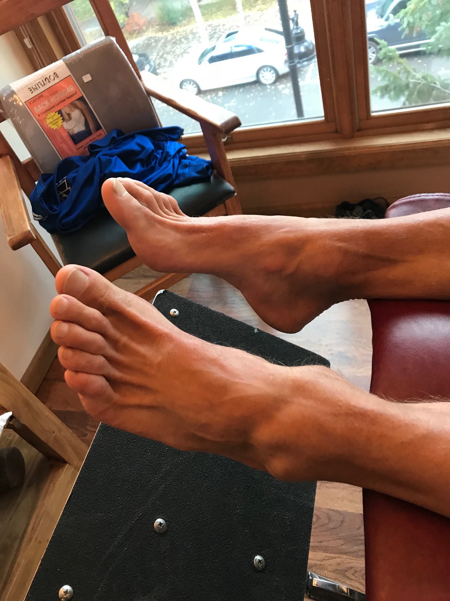
This gentleman presented to the office with left-sided knee pain at the medial collateral ligament following a cutting injury, moving from right to left with the left foot planted. As you can see, he has an anatomical leg length discrepancy with tibial and probable femoral length deficiencies on the left side. Can you see the subtle, increased tone of the long flexors of the toes on the left hand side as it evidenced by the increased prominence of the long extensor tendons to a greater degree on the shorter side? This is a common compensation seen in true leg length discrepancies with clawing of the toes in attempt to create stability on the shorter leg side. Often times, the progression angle on the shorter side will be increased as well.

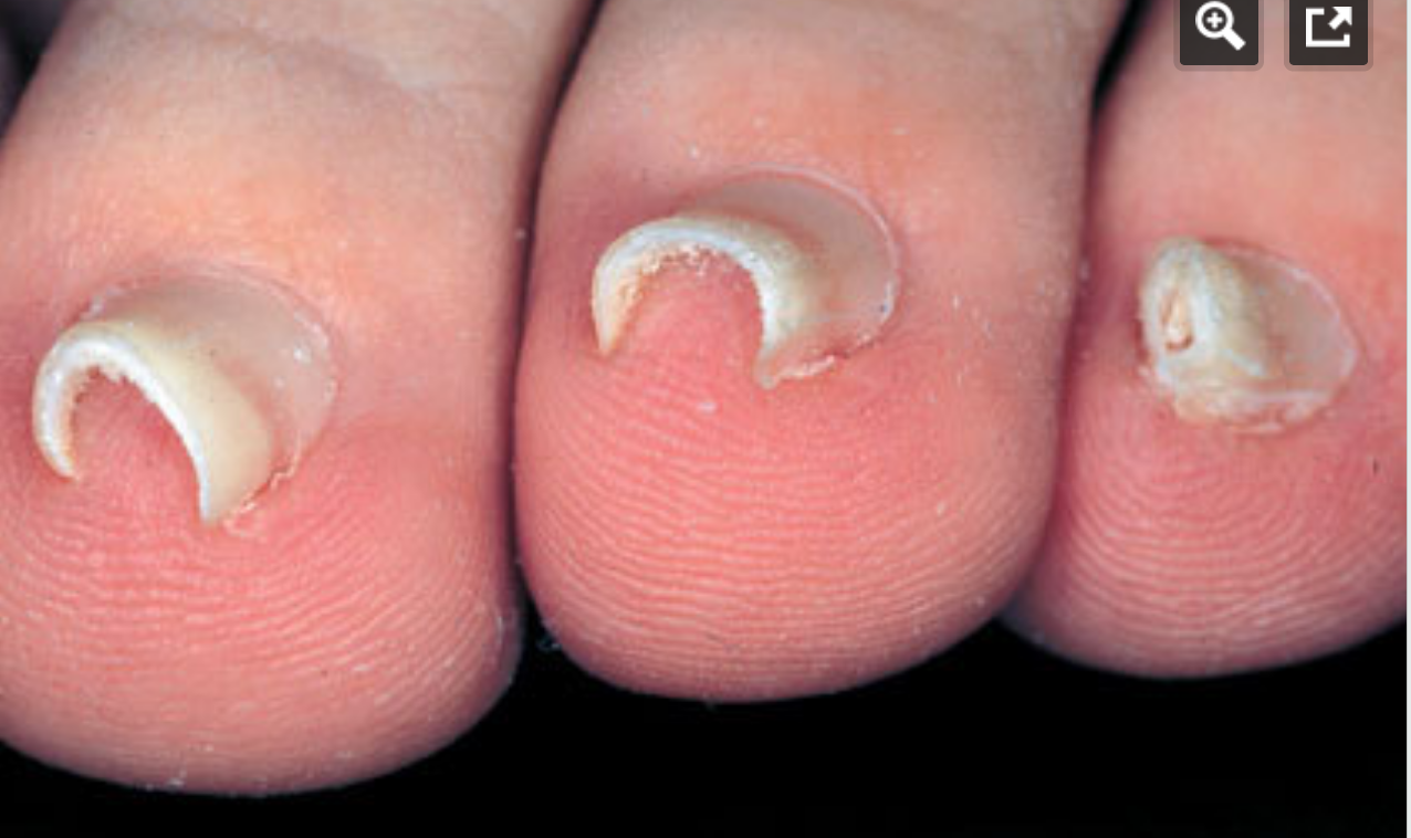
Pincer toe nails.

Screen Shot 2018-11-11 at 8.47.23 AM.png

Pincer Toe nails: You've seen them; did you know what they were and how they got that way? Or, did you dismiss them?

We think Hitomi’s hypothesis is correct. Here is why (this is paraphrased from our blog post on subungal hematomas and our revolutionary thinking on why they occur and it seems to fit well with pincer nail formation as well).

… when the skin is pulled at a differential rate over the distal phalange (from gripping of the toes rather than downward pressing through the toe pad) there will be a net lifting response of the nail from its bed as the skin is drawn forward of the backward drawn phalange (there is a NET movement of skin forward thus lifting the nail from its bedding). For an at-home example of this, put your hand AND fingers flat on a table top. Now activate JUST your distal long finger flexors so that only the tip of the fingers are in contact with the table top (there will be a small lifting of the fingers). There should be minimal flexion of the distal fingers at this point. Note the spreading and flattening of the nail. Now, without letting the finger tip-skin contact point move at all from the table, go ahead and increase your long flexor tone/pull fairly aggressively. You are in essence trying to pull the finger backward into flexion while leaving the skin pad in the same place on the table. Feel the pressure building under the distal tip of the finger nail as the skin is RELATIVELY drawn forward.] This is fat pad and skin being drawn forward (relative to the phalange bone being drawn backward) into the apex of the nail. Could this be magnifying the curvature of the nail and not offsetting the “automatic curving and shrinkage” function of the nail ? We think it is quite possible.

We have more to say on this topic, the above is just an excerpt of our blog post. More here, in the link below

https://thegaitguys.tumblr.com/post/127638788139/pincher-nails-who-knew-written-by-dr-shawn?fbclid=IwAR06ol516n9WF2Qh5TadlKd8esXrH5pVviycT_7QiMeScL0UJ3H9r1FF_OQ

Do you have dorsal (top) foot pain? Think you are tying your shoes too tightly?

Do you have dorsal (top) foot pain, at the peak of the arch? Think you are tying your shoes too tightly and that is the cause? Do you have pain over the dorsal or plantar mid foot on heel rise or jumping/landing or going up stairs ?

Just because you raise your heel and load the ball of the foot does not necessarily mean you have adequately plantarflexed the 1st metatarsal and loaded it soundly/stable with the medial tarsal bone. Heel rise, and thus loading onto the medial foot tripod, must be met with ample, stable, durable, 1st metatarsal plantarflexion and the associated medial tarsal bones. Also, without this, loading of the sesamoids properly cannot occur, and pain may ensue.

The first ray complex can be delicate in people who are symptomatic. In some people who do not have a good tibialis posterior-peroneus sling mechanism working harmoniously, in conjunction with a competent arch tripod complex to achieve a compentent arch complex (ie, EDL, EHL, tib anterior and some of the other foot intrinsics) this tarsometatarsal interval can become painful and instead of the 1st ray complex being stable and plantarflexing as the heel departs and the 1st ray begins taking load, it may not do so in a stable plantarflexed posturing. In some people it can momentarily dorsiflex as the arch subtly collapses (when it should be stable and supinated in heel rise).

"Subtle hypermobility of the first tarsometatarsal joint can occur concomitantly with other pathologies and may be difficult to diagnose. Peroneus Longus muscle might influence stability of this joint. Collapse of the medial longitudinal arch is common in flatfoot deformity and the muscle might also play a role in correcting Meary's angle."-Duallert et al

Soon, I hope to show you a video of how to watch for this problem, how to train it properly, how we do it in my office.
Dr. Allen

https://www.ncbi.nlm.nih.gov/pubmed/27015031

Clin Biomech (Bristol, Avon). 2016 May;34:7-11. doi: 10.1016/j.clinbiomech.2016.03.001. Epub 2016 Mar 10.

The influence of the Peroneus Longus muscle on the foot under axial loading: A CT evaluated dynamic cadaveric model study.

Dullaert K1, Hagen J2, Klos K3, Gueorguiev B4, Lenz M5, Richards RG6, Simons P7.

Your center of mass in relation to foot strike position.

For those arm swing/pulsers/ COM and head over foot folks consider some more research below.
Let the CNS drive the show, it is what it is there for . . . The leg motor patterns are dominant, the arms are passive and "shape" and influence the leg swing as a balance and ballast effect. As we discuss in an upcoming podcast, to cross the arms in a pumping motion across the midline of the body means one has to have compromised scapular mechanics (mostly protraction) to afford that much humeral adduction. This means we are forcing thoracic rotation as well. This means we are reversing what we know is more true, that "arm motion is driven passively by rotation of the thorax (Pontzer et al., 2009), an idea which is supported by shoulder muscle EMG data" (and not thoracic rotation by arm swing). Why would we try to create more unnatural axial spin through the spine when we are actually trying to move forward in the sagittal plane? Why would we try to force more rotation through the spine when the function of the thoracopelvic canister (ie. the core) is to stabilize rotational /angluar momentum? Hmmmm, things to ponder.

"Previous modelling studies have clearly shown that motion of the arms effectively counterbalances the angular momentum of the lower extremities during running (Hamner & Delp, 2013; Hamner et al., 2010). It has further been suggested that arm motion is driven passively by rotation of the thorax (Pontzer et al., 2009), an idea which is supported by shoulder muscle EMG data, consistent with the shoulders as spring-like linkages (Ballesteros, Buchthal, & Rosenfalck, 1965). Our data are con- sistent with this idea, showing motion of the thorax to be in the opposite direction to that of the swinging leg. Pontzer et al. (2009) also suggested that motion of the thorax is driven passively by motion of the pelvis. However, our data shows that the thorax reaches its peak angular velocity earlier than the pelvis, indicating that thorax motion is not completely passively driven by pelvic movements."

-S.J. Preece et al. / Human Movement Science 45 (2016) 110–118

Fatigue matters. Today's article looks at pre and post exercise fatigue and how, on EMG, our body changes.

Photo credit: pixabay.com

Photo credit: pixabay.com

Even for those of us who do (and should) know better, "the problem is, we are all often knee deep into compensations before we are aware of it, so most of us are always working on adding strength and endurance into our compensations without even knowing it. Our workouts layer things deeper. Yes, almost all of us are on this bus. Don't deny it. The next time you feel that tightness in your shoulder, or in your hip, or feel that tightness or soreness on one side of the low back, or one side of the neck, stop, and ask yourself that honest question. Again, you are on the bus with the rest of us."

We have spent much time discussing our order of things when intervening between a person and what ails them. Namely, our order is to first restore proper skill and patterning, then add endurance (move well often), and then add load, namely strength, power, force, explosive movements and the like. So, Skill, Endurance, Strength. This is a neurologic order, there is good reason for the necessity of this order. We have spend many an hour listening to Dr. Ivo explain why the CNS dictates this is the order with good reason. Cheat this order and you lay down neuroplastic patterns that are anything but what you want for your client. Enough said.

Today we introduce and article that the looks at the lumbo-pelvic-hip complex, a very complicated area, subject to large multi-planar movements and distortions (and hence, large complex multi-planar compensations). We must have good skill, endurance and strength in controlling this massive area safely, meaning, to avoid developing cheating compensatory patterns to negotiate around our days and activities and sports. The problem is, we are often knee deep into compensations before we are aware of it, so most of us are always working on adding strength and endurance into our compensations without even knowing it. Yes, almost all of us are on this bus. Don't deny it. The next time you feel that tightness in your shoulder, or in your hip, or feel that tightness or soreness on one side of the low back, or one side of the neck, stop, and ask yourself that honest question. Again, you are on the bus with the rest of us.

Today's article looks at pre and post exercise fatigue and how, on EMG, our body changes. Now keep in mind, and I will remind you of this again at the end of today's writing, keep in mind of the asymmetries, poor-skill, poor-endurance and poor strength in some areas that pre-exist, before even starting into our exercises. Imagine, assume, that these were there in all of this study's subjects, even prior to the exercise challenge. You should now fully grasp how layered things get for our clients.

Here is what the article said,

"fatigue may affect muscle recruitment, active muscle stiffness and trunk kinematics, compromising trunk stability".-Chang et al.

"The purpose of this study was to compare trunk muscle activation patterns, and trunk and lower extremity kinematics during walking gait before and after exercise. Surface electrodes were placed over the rectus abdominis, external oblique, erector spinae, gluteus medius, vastus lateralis, and vastus medialis of twenty-five healthy indviduals."

"The amplitude increased in the rectus abdominis during loading, midstance , terminal stance, and late swing after exercise. Amplitude also increased during swing phase in the erector spinae, vastus lateralis, and vastus medialis after exercise. There was less trunk and hip rotation from initial contact to midstance after exercise. Neuromuscular fatigue significantly influenced the activation patterns of superficial musculature and kinematics of the lumbo-pelvic-hip complex during walking. Increased muscle activation with decreased movement in a fatigued state may represent an effort to increase trunk stiffness to protect lumbo-pelvic-hip structures from overload."-Chang et al

What we found particularly notable was that they found less trunk and hip rotation from initial contact to midstance after exercise. And that, "neuromuscular fatigue significantly influenced the activation patterns of superficial musculature and kinematics of the lumbo-pelvic-hip complex during walking". As they concluded, increased muscle activation with decreased movement in a fatigued state plausibly indicates an effort to increase trunk stiffness as a protective measure. Translation, a protective compensation.

Here is what we have to say about that: do not leave the problem on the table and merely train your client around this. Resolve the underlying problem. The underlying problem may not, and likely will not, come out in a "functional screen". What will come out in the screen is how they are moving about with this existing compensation pattern(s). The screen shows WHAT they are doing with their limitations, not WHY Dive keep dear brethren. This is what it is all about, taking the time and diving deep. Find the "why".

So, as promised, here I am again, reminding you to keep in mind of the asymmetries, poor-skill, poor-endurance and poor strength in some areas that pre-exist, before even starting into our exercises. Imagine, assume, that these were there in all of this study's subjects, even prior to the exercise challenge. You should now fully grasp how layered things get for our clients.This is what can make, "helping someone get well", a difficult challenge, even on a good day.

*Muscle activation patterns of the lumbo-pelvic-hip complex during walking gait before and after exercise. Chang M1, Slater LV2, Corbett RO1, Hart JM1, Hertel J1.

Photo credit: pixabay.com Thank you for making such beautiful photos like this available for free use. Gorgeous photography !

How does hallux valgus and bunion formation cause toe hammering?

Screen Shot 2018-11-13 at 4.36.12 PM.png

Photo: you need to recognize this predictable pattern.

When the hallux begins to become incompetent, from perhaps pain, hallux rigidus, hallux limitus, bunion and in this case hallux valgus with bunion (in this case rotational instability) when this incompetence kicks in, we must find stability elsewhere. One will often, unconsciously, begin to increase the flexor tone and pressure to try and find stability since one cannot get it sufficiently from the hallux anymore.

Screen Shot 2018-11-16 at 6.28.27 PM.png


Here you see the tenting up of the 2nd toe, from increased long flexor activity (FDL) over time.
And here is the interesting thing you will notice, over time, the 3rd toe will start the same strategy, then the 4th. We see this often. It is not set in stone, but we see it a lot.
Notice it in your clients feet. Teach them why they are getting hammer toes, flexible ones at first, and possibly rigid ones over time. Hammer toes can have many causes, this is just one.
Solution: find a way to help your client re-find better hallux and medial forefoot stability to halt the progression.

How hallux valgus and bunions can affect the shoe toe box space.

Bunions and hallux valgus can change the toe box volume and shoe choice, so be careful, don't be fooled.
This photo shoes how a change in the forefoot width and length can be a result of a bunion or hallux valgus. Notice both feet are aligned the same, but the length of the foot is different in the hallux valgus foot.

Screen Shot 2018-11-16 at 6.23.31 PM.png


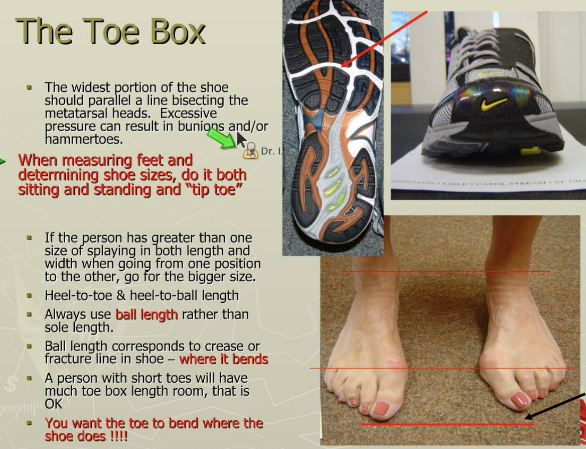
The old Brannock device use to help us all see this more clearly. You may recall that the device measured "heel to toe" (True foot length) and ALSO "heel to ball" length (the functional length and more important one. This length measured heel to the metatarsophlanageal joint line. This concept is important to know because we want the shoe "break point" or "bend point" at the forefoot to occur where the foot bends. Not all shoes have the flex lines (the creases on the bottom of the shoe were it is most likely to bend) in the same place, there is no standard. And if your client has shorter toes, longer toes or a long or short "heel to ball" length they man needs some help from a knowledgeable person like yourself making sure that their current forefoot complaints are not from a mis-fitted shoe.
Bottom line, the "heel to ball" length of a foot is far more important than the global foot length "heel to toe". So stop judging your shoe fit by pinching the front of the shoes to "make sure you have plenty of room"! Doh ! Face palm !

Because despite what many of the "experts" online are saying, that being "shoes don't matter". The fact is "sometimes they do". Period.

WAnt to learn this stuff? Got our website and buy the National Shoe Fit program. Hours of deep shoe, anatomy and biomechanics fun with ivo and shawn, in your own home over the holidays ! Give yourself the "gift" of ivo and shawn this year ! LOL

And for all of you who joined us last night on onlineCE.com for the 55 minute condensed nuclear version of the 3+ hours shoe fit program, we hope you have recovered with a good nights sleep !

GHS (generalized hypermobility syndrome) and foot loading.

GHS: Generalized Hypermobility Syndrome

Screen Shot 2018-11-16 at 6.20.23 PM.png

We have all seen these types of clients/patients. They have joints that have more than ample full range. It is easy for them to hyperextend their elbows and knees. They can fold over and touch their toes, everything seems lax and flexible. What they need is help gaining more control of their joints. But what about their gait ? Albeit a focal study, finally someone has looked at how these people interface the ground,

The forefoot region received higher loading in GHS clients.
So what could this mean? Does it mean they have challenges transitioning from rearfoot to forefoot? Does it mean their center of pressure is more foreward biased ? Does it mean they have to impart heavier loads through the forefoot during gait to feel stable? Are they premature heel raisers thus showing the forefoot bias increase? There are many questions here, too broad for this study, but they are the keys to understanding how the GHS body interprets movement. Regardless, it is highly suspect that these clients dominate their gait with the calf muscle complex doing plenty of extra work. We suspect they will be toe clenchers/grippers ( ie, have increased toe flexor dominance) and this can have long term impact on things like metatarsal loads, neuroma formation, bunions, lumbrical weakness, fat pad displacement, hammer toes and many other related issues that occur with premature or excessive forefoot loading. They may even have a little of that vertical bouncy gait we often discuss. We will keep our eyes open for this stuff and keep you in the loop.

J Back Musculoskelet Rehabil. 2018 Nov 2. doi: 10.3233/BMR-170973. [Epub ahead of print]
Generalized hypermobility syndrome (GHS) alters dynamic plantar pressure characteristics.
Simsek IE1, Elvan A1, Selmani M2, Cakiroglu MA2, Kirmizi M2, Angin S1, Bayraktar BA3.

Hip muscles may be smaller in unilateral OA.

Here, some "low quality evidence that specific hip muscles are smaller in unilateral hip OA".
Does this translate to osteoarthritis leads to muscular atrophy and thus weakness, or does this translate to muscle atrophy and weakness leads to OA ? Chicken or the egg ?
Sure, there is the whole trauma thought process, but that is not the discussion today.

Regardless, some possible implications here for targeted interventions in hip OA. This is why so many with arthritic joints improve with pointed, well thought out and target strengthening.

Muscle size and composition in people with articular hip pathology: a systematic review with meta-analysis

P.R. LawrensonEmail the author P.R. Lawrenson, K.M. CrossleyEmail the author K.M. Crossley, B.T. VicenzinoEmail the author B.T. Vicenzino, P.W. HodgesEmail the author P.W. Hodges, G. JamesEmail the author G. James, K.J. CroftEmail the author K.J. Croft, M.G. KingEmail the author M.G. King, A.I. Semciw

https://www.oarsijournal.com/article/S1063-4584(18)31512-7/fulltext?fbclid=IwAR2DTYxb2Azz4Df4I2Wy7CHst7egAg_x32DDH8DDfCbnAtYNf8h8iqlOQag

Adding strength to compensations and asymmetry.

FootNotes, with The Gait Guys.

Screen Shot 2018-11-13 at 4.36.12 PM.png

The CNS runs the show. Compensations are real, they are a calculated response, they are meaningful adaptive protective behaviors. Adding strength to a compensation makes them even more real, plastic, permanent. Fix the problem. Adding random strength is juvenile thinking. Letting one's client load/train/lift when in pain is juvenile (read below). Once we realize adding load to the problem can be helpful or hurtful, we are on the right path, we are winning. But it takes a deep understanding of how to add load safely, wisely, so that our client can benefit. We must try to understand adaptive behaviors, we must try to understand why our client's CNS made the choices it did.
Now, imagine a client with ankle pain, and resultant ankle dorsiflexion/ankle rocker loss. Now, imagine what their gait will look like as well with that premature heel rise and everything that adapts from that premature heel rise. Now, read below and understand one way how the CNS adapts. Why? So that the next time one chooses a stretch, mob, flossing, etc to gain a range of motion, without any additional meaningful measures, hopefully they will realize they are likely not addressing the deeper problem. Pushing a range of motion is not the same as safely re-earning a range of motion. Far from it. -Dr. Allen

*Effect of Achilles tendinosis on the agonist, synergist and antagonist muscles. Chang and Kulig

"In addition to the altered control system, the present study also observed an adaptive behaviour, as illustrated by the activity of agonist, synergist and antagonist muscles. This was seen during single-legged hopping, where the contribution from the triceps surae muscle to the plantar flexors was decreased and the co-contraction from the tibialis anterior muscle was also decreased on the involved side in individuals with Achilles tendinosis. This may be attributed to the protective mechanism shielding the already injured tendon from further injury or even rupture (Lund et al. 1991)."- Chang and Kulig

J Physiol. 2015 Aug 1; 593(Pt 15): 3373–3387.
Published online 2015 Jun 30. doi: [10.1113/JP270220]
PMCID: PMC4553058
PMID: 26046962
The neuromechanical adaptations to Achilles tendinosis
Yu-Jen Chang and Kornelia Kulig

Forget face recognition, gait recognition is here.

Gait recognition. Forget face recognition, your gait has its own neuroplastic pattern you have developed over the years. It is subconscious and like we repeatedly say, your gait is a representation of the parts that are working, not working and injuries you have compensated around. Sure, you can change your gait, but it is going to take retraining over 10+ weeks to adopt a new gait pattern and gait ID.

Remember this as well, coaching a gait form change does not fix anything, you are just teaching a new pattern on their existing patterns of ability and compensation. This is our main problem with basing diagnosis and corrective homework off of just a gait evaluation, they tell you nothing of what is wrong with your client, they only tell you what your client is presently doing.

https://abcnews.go.com/amp/Technology/wireStory/chinese-gait-recognition-tech-ids-people-walk-58988215?__twitter_impression=true&fbclid=IwAR2SvPXCBBya_P84THHtc6hdwvdjV47xn6JjFbRhaWZ_GCojarFxSrnnE_E

This could happen to you if your ankle dorsiflexors get weak.

This could happen to you if your ankle dorsiflexors get weak.

This (read below) is actually a normal compensation. Building strength on this compensation without resolving the anterior compartment weakness can lead to knee pain (or other things). It is why, again, we say that asymmetry and especially asymmetrical compensations, can matter in your client.

"muscle strength asymmetry in the ankle joint may lead to counterbalancing muscle strengthening of the knee joint to maintain the center of body mass."

"It is not only the balance between the agonist and antagonist muscles, but also the balance between the left and the right, and between the proximal joint and distal joint in a weight-bearing position that are involved during exercise. As seen in the university players in this study, the weakening of dorsiflexors due to muscle strength asymmetry in the right ankle joint is thought to cause the strengthening of the extension muscles of the knee joint in order to maintain the center of body movement and stability." - Kyoungkyu et al.

What The Gait Guys have to say about this:

As that foot is approaching the ground, and begins loading, the anterior compartment may not skillfully allow a forefoot loading response and may not protect that anterior ankle mortise joint let alone appropriate and skillful pronation. Ankle dorsiflexion loading may occur too fast and uncontrolled leading to sudden undesirable knee flexion (too much, too fast, too long). After all, closed chain ankle dorsiflexion and knee flexion are deeply paired loading movements. One way to slow down this increased amount of forward tibial progression (ankle dorsiflexion and knee flexion), is to increase the activity of the quadriceps/knee extensors. The compensation of increased knee extension loading will offset the increased knee flexion resulting in improved control the ankle dorisflexion. It is a reasonable compensation, but not one you want to stick around for long or unaddressed. If subtle, and left over time, perhaps even when subclinically presenting, knee pain from increased shear/compression or other mechanical results, may be the first presentation when the true problem is down in the lower region.

Remember, this is about controlling gait and thus center of body mass. This is about not falling over. This is about the central nervous system making necessary adaptations to protect the entire organism, which sometimes means doing anything so as not to fall. Now, just to make this more complicated and fun, imagine that this is a fairly common problem and compensation ON ONE SIDE of the body. So now imagine, the changes in arm swings, maladaptive loading onto the other side, pelvis asymmetry and the list goes on.

Of course, one could just globally add strength to this client, such as more squats, lunges or more deadlifts (instead of a focal intervention at the anterior ankle structures) thus adding another layer of compensation, and perhaps making them feel better, for the time being. Essentially, adding global strength to asymmetry. But make no mistake, this problem is likely to sit here and percolate under the surface with more possible compensations, subtle movement alterations and adaptations, until someone addresses the problem. And remember this, if one does those things long enough, those become a client's new adaptive neuroplastic patterns. Strength first is not always a good first intervention.

J Phys Ther Sci. 2016 Apr; 28(4): 1289–1293.
Effects of muscle strength asymmetry between left and right on isokinetic strength of the knee and ankle joints depending on athletic performance level
Kyoungkyu Jeon, PhD,1 Sungyung Chun, PhD,2,* and Byoungdo Seo, MS, PT3

https://www.ncbi.nlm.nih.gov/pmc/articles/PMC4868229/

photo credit: free photos courtesy of Pixabay

Why are you putting your internal hip rotation into your low back (pain).

Why are you putting your internal hip rotation into your low back (pain).

On October 12th, 2018 I wrote about utilizing the gluteals in internal hip rotation. You will have to go back and search FB for that article and video.
Assessing Internal hip rotation (in various ranges of hip flexion, extension, abduction and adduction) is a basic exam principle I examine on nearly every patient and athlete that comes to see me, regardless of their complaint. Other than breathing, walking is the next most under appreciated movement we undertake, and take for granted.
Lack of adequate internal hip rotation, in my clinical experience (20+ years), is all too often a fundamental parameter in hip, knee and low back pain. It is necessary to have unrestricted internal hip rotation during gait. Adequate internal hip rotation in the mid to late stance phases of gait is critical and is also paired with hip extension, in fact, one has to pass through adequate internal hip internal rotation to get to adequate hip extension. Without one, we do not get the other. And, if the internal rotation is not imparted in the hip when the hip is supposed to be the one internally rotation, that demand is going to move up or down, caudally or rostrally, low back or knee. Of interesting note, taking things deeper, the opposite arm is also going to go through internal rotation and extension at the same time. Impair one limb, and we can make a case, often enough, that the contralateral upper or lower limb is also challenged. This fundamental fact is one of the fascinating reasons Dr Ivo and I get so geeked out by gait and human movement. Because, it is very complicated. And if one is not looking close enough, paying enough attention with enough fundamental knowledge, things are going to get overlooked and missed when solving for "X" in a client's pain/problems/movement. Compensation will ensue, all too easily. Build strength on said compensations and we are off to the races in driving neuronal pasticity into potential asymmetries. If one is strength training a client without examining them and making specific corrections along the way, well, we reap what we sew. Ok, enough soap-boxing. -Dr. Allen
Here, don't take our word for it, . . . . .

"Correlation between Hip Rotation Range-of-Motion Impairment and Low Back Pain. A Literature Review."
Ortop Traumatol Rehabil. 2015 Oct;17(5):455-62. doi: 10.5604/15093492.1186813.
Sadeghisani M1, Manshadi FD1, Kalantari KK1, Rahimi A1, Namnik N2, Karimi MT3, Oskouei AE4.

"There is a hypothesis which suggests that a limited range of hip rotation results in compensatory lumbar spine rotation. Hence, LBP may develop as the result. This article reviews studies assessing hip rotation ROM impairment in the LBP population.

"Asymmetrical (right versus left, lead versus non-lead) and limited hip internal rotation ROM were common findings in patients with LBP. Reduced and asymmetrical total hip rotation was also observed in patients with LBP. However, none of the studies explicitly reported limited hip external rotation ROM."

CONCLUSION: "The precise assessment of hip rotation ROM, especially hip internal rotation ROM, must be included in the examination of patients with LBP symptoms."

Photo credit: courtesy of Pixabay

The next time they have gait asymmetry, try changing out the insole...

or putting a textured one in there...or maybe putting a some sand or dirt in their shoe...

image credit: https://torange.biz

image credit: https://torange.biz

Textured insoles change (we like to think for the better) proprioceptive input and can improve balance and gait performance, both statically and dynamically. We have seen this in folks with parkinsons (1) as well as stroke (2), though it can be used in the elderly (3), in diabetes and neuropathy (4), as well as healthy individuals (5,6). Changes from postural stability, to changes in anterior/posterior sway, to medial/lateral sway, to step length and height, the research is there.

These results support the hypothesis that enhanced somatosensory feedback to the sensory system, both through the spinocerebellar and dorsal column pathways, as well as the vestibular system, results in an improved motor output (and most likely coordination) of gait.

  1. Qiu F, Cole MH, Davids KW, et al. Effects of textured insoles on balance in people with Parkinson's disease. PLoS One. 2013;8(12):e83309. Published 2013 Dec 12. doi:10.1371/journal.pone.00833

  2. Ma CC1, Rao N2, Muthukrishnan S3, Aruin AS4. A textured insole improves gait symmetry in individuals with stroke. Disabil Rehabil. 2017 Aug 7:1-5. doi: 10.1080/09638288.2017.1362477. [Epub ahead of print]

  3. Annino G1,2,3, Palazzo F2, Alwardat MS4, Manzi V5, Lebone P2, Tancredi V1,2,3, Sinibaldi Salimei P2,6,7, Caronti A2, Panzarino M2,3, Padua E2,3. Effects of long-term stimulation of textured insoles on postural control in health elderly. J Sports Med Phys Fitness. 2018 Apr;58(4):377-384. doi: 10.23736/S0022-4707.16.06705-0. Epub 2016 Sep 15.

  4. Paton J, Glasser S, Collings R, Marsden J. Getting the right balance: insole design alters the static balance of people with diabetes and neuropathy. J Foot Ankle Res. 2016;9:40. Published 2016 Oct 5. doi:10.1186/s13047-016-0172-3

  5. Steinberg N1, Tirosh O, Adams R, Karin J, Waddington G. Influence of Textured Insoles on Dynamic Postural Balance of Young Dancers. Med Probl Perform Art. 2017 Jun;32(2):63-70. doi: 10.21091/mppa.2017.2012.

  6. Collings R1, Paton J2, Chockalingam N3, Gorst T2, Marsden J2. Effects of the site and extent of plantar cutaneous stimulation on dynamic balance and muscle activity while walking. Foot (Edinb). 2015 Sep;25(3):159-63. doi: 10.1016/j.foot.2015.05.003. Epub 2015 May 11.

Toe extension matters.

The season to pathologize our feet is upon us. Toe extension matters.

I blew out my flip flop,
Stepped on a pop top;
Cut my heel, had to cruise on back home.
But there's booze in the blender,
And soon it will render
That frozen concoction that helps me hang on. - Jimmy Buffett

I continue to see more and more people with inadequate toe extension. It is complicated. I see those who do not even have the awareness of toe extension, loss of strength of toe extension, loss of endurance of toe extension, loss of global range of toe extension (dorsiflexion at the MTP joint), more failure of long toe extensor (EHL) strength and more prominence of increased short toe extensor strength (EDB) and more frightening, a lack of disassociation of toe extension (MTP dorsiflexion) and ankle dorsiflexion. Many clients when asked to life their toes, will drive into ankle mortise dorsiflexion; ask them to just purely toe dorsiflex and the mental games begin, a wrinkled brow, intense concentration. If you cannot extended the toes sitting, how are you going to find them in swing phase of gait when balance, and other things, are more important?
Stand and lift your toes. The arch should go up, you have engaged the Windlass Mechanism, that winds up the plantar fascia and raised the arch. If you do not have competent, unconsciously competent, toe extension, your arch is not all that it can, and should, be. If you cannot raise your toes, thus raise the arch, thus plantarflex the first metatarsal, then in gait, when the foot is on the ground, you cannot properly position the sesamoids, properly get safe terminal ranges of hallux dorsiflexion at toe off, properly position the foot for loading and unloading, adequately achieve ankle dorsiflexion, adequately offer the hip a chance for ample hip extension, offer the glutes optimal chance to work in all phases to help control spin of the limb during loading and unloading, and the list goes on and on. I am sure I left much out there, this was written in a few minutes and unedited, just a short rant for the weekend. But if you have not championed toe extension, both in an unloaded and loaded foot (on the ground), achieved control of both long and short extensor muscles to the toes (and paired them well with the long and short toe flexors), disassociated toe extension from ankle dorsiflexion, and then figured out how to properly, timely, engage all these processes into your gait unconsciously, you are working on less of an optimal system than you should be. So, if your feet hurt, hips hurt, or a plethora of other problems that you are trying to fix with orthotics or other toys, maybe start with, "can you lift your toes?". It is a piece of the puzzle, trust me.
Or, you can just stay in your flip flops and perpetuate your toe flexion and wait for bad things to take root After all, tis the season soon !
Yes, toe extension in flip flops (we must flex our toes to keep them on) is as rare as a good multi-tasking man.

Shawn Allen, one of the gait guys.


" "Stand and lift your toes. The arch should go up, you have engaged the Windlass Mechanism, which winds up the plantar fascia and raises the arch. If you do not have competent, unconsciously competent for that matter, toe extension, your arch is not all that it can, and should, be. If you cannot raise your toes, thus raise the arch, thus plantarflex the first metatarsal, then in gait, when the foot is on the ground, you cannot properly position the sesamoids, properly get safe terminal ranges of hallux dorsiflexion at toe off, properly position the foot for loading and unloading, adequately achieve ankle dorsiflexion, adequately offer the hip a chance for ample hip extension, offer the glutes optimal chance to work in all phases to help control spin of the limb during loading and unloading, and the list goes on and on."

More asymmetrical thoughts

Again, in this study, like the last we discussed here, we are looking at experienced (and in this case, young) runners; sprinters specifically. Again, they ran relatively short distances (20 meters). More than 1/2 the runners had “large” asymmetries, and they all had asymmetries of some type. Some athletes had injuries and some did not.

There wasn’t a difference in sprint performance over this short distance. This is not surprising in light of the previous paper we discussed; asymmetries seem to worsen over time (Hanley 2018). The level of compensation present (since these are experienced runners) may also be better; the folks that were uninjured having compensation patterns that were more in line with their anatomy, than the injured ones.

  • The asymmetries did not change. Thinking about anatomy, especially with hard deformities like torsions or versions, why would they? You can’t change the stripes on a tiger.

  • Injured and non injured athletes did not differ in asymmetry before or after the study. Again, why would they? We are talking about gait changes (or perhaps compensations). What is significant for one individual (tibial torsion, femoral retroversion, leg length discrepacy) may not be as significant as it for another, depending on the compensation present.

The study concludes “... kinematic asymmetries in the stride cycle were not associated with neither maximal sprint running performance nor the prevalence of injury among high-level athletic sprinters.” Note that they are talking about prevalence of injury, not incidence of injury.

We still think that asymmetry matters...

Haugen T, Danielsen J, McGhie D, Sandbakk Ø, Ettema G. Kinematic stride cycle asymmetry is not associated with sprint performance and injury prevalence in athletic sprinters. Scand J Med Sci Sports. 2018 Mar;28(3):1001-1008. doi: 10.1111/sms.12953. Epub 2017 Aug 15.

Thought experiment on symmetry: Does symmetry always matter?

the short answer is probably not. the long answer is maybe...

This study looked at gait variability asymmetry in small cohort of experienced distance runnners. They measured different variables at 1500, 3000, 5000, 7500 and 9500 m of a 10000 meter run (about 6.2 miles) on a treadmill. Generally speaking, variability was low and athletes were symmetrical for 5 of seven variables measured and assymetry, when present, was in flight time and impact forces Most aththletes were asymetrical for at least one variable as well.

Their conclusion basically said that being asymmetrical in a few variables is not abnormal and not indicative of asymmetrical gait and since many practitioners analyze symmetry (and variability) caution should be exercised when determining the need for intervention.

So what do we think this means?

most likely:

  • these folks were symmetrical with low variability. In other words, when asymmetry was present, it was small

  • some asymmetry, in some parameters, is probably normal...but,it is usually small if it doesn’t matter. Keep in mind these were expreienced, uninjured folks. th results could have been different with a bigger cohort and less experienced runners and thus...

  • The study does not talk about inexperienced runners. Symmetry and/or asymmetry may not be normal for inexperienced runners

  • Results may have been vastly different if the run had been longer. The study did show that variability increased the further along on the run the athlete was.

  • The study was performed on a treadmill, which may not exemplify or highlight asymmetry, as it creates artificial constraints which we have discussed in by us here, here, and here:

  • We think asymmetry matters, particularly when it comes to hard deformities like torsions and versions, which change the biomechanics of that individual extremity and can be a diagnostic tool for future problems

  • perhaps asymmetry is significant in his population of runners on a subclinical basis

Hanley B, Tucker CB. Gait variability and symmetry remain consistent during high-intensity 10,000 m treadmill running. J Biomech. 2018 Oct 5;79:129-134. doi: 10.1016/j.jbiomech.2018.08.008. Epub 2018 Aug 16.

Subtle clues to flexor dominance

IMG_5515.jpg

Take a close look at these photographs. Compare the prominence of the extensor tendon‘s left to right. What do you see? Do you notice the deeper furrowing of the extensor tendons on the left? Do you see the subtle increased extension of the metatarsophalangeal and requisite increased flexion of the inter-phalangeal articulations, left versus right? What about the height of the arches?

Keep a keen eye out for subtle signs. They can make a real difference in your clinical diagnosis and results…

Shoe causing knee pain? You decide… 

IMG_5522.jpg
IMG_5526.jpg
IMG_5525.jpg

This gentleman presented with left-sided knee pain at the medial collateral ligament. His left foot was planted when he rotated to the left. Take a close look at the shoes in the picture. If you look closely, you will notice the right shoe is tilted on its axis due to a rear foot to forefoot deformity (forefoot supinatus)and the left shoe upper was assembled canted on its axis, Most likely in manufacturing defect. Can you see the subtle valgus in the left shoe rearfoot?

Think of the implications of a shoe with this orientation. Putting the rearfoot in valgus “prepronates“ the foot, causing medial rotation of the tibia and femur and increase valgus stress on the knee, stressing the medial collateral ligament and stabilizing complex. This will most likely manifest itself as anterior rotation of the ilium on the left-hand side with relative posterior rotation on the right and a clockwise Pelvic distortion pattern. With the foot planted on the left side and it being pre-pronated, can you see how the rotation to the left leaves a greater amount of external rotation that must occur to just get the foot to neutral, never mind supination for stability and pushoff?
What about the popliteus having to work on time to assist and extra rotation and the appropriate femoral/tibial rotation ratios to spare the medial meniscus?

These are the kind of things to keep us awake at night…

On the road to a cruciate reconstruction?

Screen Shot 2018-10-12 at 8.24.50 AM.png

While at a recent soccer game, I noticed this gal standing on the side lines. Talk about knee problems waiting to happen ! Note the hyperextended posture of the knees with increase in lumbar lordosis and anterior carriage of the entire pelvis with an increase in the thoracic kyphosis and head forward carriage to match! You can imagine the anterior pelvic tilt as well as stretch weakness of the abdominal obliques creating "core instability". At least she is not wearing heels, although a negative inclination [negative ramp delta] shoe would probably help.

Think of the strain on her poor posterior cruciate ligaments with all of that anterior femoral translation! We remember that the popliteus acts as an "accessory PCL" at initial contact in the gait cycle. It fires at heel strike and again from loading response until toe off

Think about the forces on these knees while descending hills or stairs. The momentum will carry the femur forward (or anteriorly). There needs to be something to reststrain this; enter the PCL. Because of the laxity (and instability), the poplitues will need to fire to take up the slack. We wrote about that here and here.

Note, this is a mere thought experiment, don’t get bent outta shape, these things might not occur, or they might. Time will tell.