tumblr_mumzdeHw9w1qhko2so1_1280.jpg
tumblr_mumzdeHw9w1qhko2so2_1280.jpg
tumblr_mumzdeHw9w1qhko2so3_1280.jpg
tumblr_mumzdeHw9w1qhko2so4_1280.jpg
tumblr_mumzdeHw9w1qhko2so5_1280.jpg

The power to bend bones.

What have we here? Hmmm. This little girl was brought in by her mother because of intermittent knee pain and “collapsing” of the knees while walking, for no apparent reason.

The ankle dorsi flexion (or ankle rocker; see last 2 pictures; we are fully dorsiflexing the ankles)  needs to occur somewhere, how about the knees? Or in this case, the tibia. Wow!

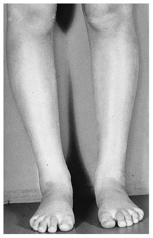
You are looking at a 4 year year with a condition called genu (and tibial) recurvatum. Genu recurvatum is operationally defined as knee hyperextension greater than 5 degrees. The knee is hyperextended, and in this case, the tibia is literally “bent backward”. Look at the 2 pictures of her tibia.

Generally speaking, the tibial plateau usually has a slight posterior inclination (as it does in this case; look carefully at the 1st picture) causing the knee to flex slightly when standing. Sometimes, if it is parallel with the ground and the center of gravity is forward of the knees, the knee will hyperextend (or in this case, the tibia will bend) to compensate.

In this particular case, the tibia has compensated more, rather than the knee itself. The knee joint is stable and there is no ligamentous laxity as of yet. She does not have a neurological disorder, neuromuscular disease or connective tissue disorder. She has congenitally tight calves.

As you can imagine, her step length is abbreviated and ankle rocker is impaired.

So what did we tell her Mom?

  • keep her barefoot as much as possible (incidentally, she loves to be barefoot most of the time, gee, go figure!)
  • have her walk on her heels (she’s a kid, make a game of it)
  • showed her how to do calf stretches
  • balance on 1 leg with her eyes open and closed
  • keep her out of backless shoes (like the clogs she came in with)
  • keep her out of flip flops and sandals where she would have to “scrunch” her toes to keep them on.
  • follow back in 3 months to reassess

There you have it. Next time you don’t think Wolff’s (or Davis’s) law* is real, think about this case. Want to know more? Consider taking our National Shoe Fit Program, available by clicking here.

The Gait Guys. Making you gait IQ higher with each post.

*Wolff’s law: Bone will be deposited in areas of stress and removed in areas of strain. or put another way: bone in a healthy person or animal will adapt to the loads under which it is placed

Davis’s law: soft tissue will adapt to the loads that are placed on it

tumblr_mrrca4X47t1qhko2so1_1280.jpg
tumblr_mrrca4X47t1qhko2so2_1280.jpg
tumblr_mrrca4X47t1qhko2so3_1280.jpg

Holy Late Cretaceous Therapods. Those Veliciraptors were twisted!

The dinosaur made famous by Jurassic Park (We never understood why they put this dinosaur in the movie, the Jurassic period was many millions of years earlier, but that’s another story).

Dr Ivo was able to take some pictures of a rare, preserved skeleton from Mongolia at the dinosaur museum in Fruita, CO, while visiting with his family.

These bad boys (and girls) were fast predators, and one of the things that made them that way, was the fact that they were built for speed!

Take a look at theses hips! Note the extreme retro torsioned angle of the femur heads. We remember that femoral retro torsion limits internal rotation of the hips (OK, so you don’t remember? click here for a review).

Now lets think about this. Externally rotate your thigh and lower leg. What do you notice? Hopefully you notice it puts your foot in more supination. This makes it into a more rigid lever, better for pushing off and better for sprinting!

Have you ever seen a sprinter? do they run on their toes? Is their foot more supinated? Ever see a velociraptor run? Check out this sequence from the “Dinosaur Planet” series. Remember, only their toes are on the ground and the thing that looks like a backwards knee is actually their ankle. 

Since their legs are so close to the body, there is little need for internal rotation, so why not maximize the effect and assist in supination?

Wow! Are you finally convinced that torsions are cool? After all, they appear to have been around for at least the last 75 million years and probably longer. 

The Gait Guys. Quarternary Geeks of the Cenozoic Era. Yes, we study dinosaur gait too…

Standing Propriosensory Exercise Progression

If you have been with us for any length of time, you know how we feel about proprioception. Here are a few samples from some of our posts on this subject. 

Here is an exercise progression we like to use, and we use it often. You are free to use it in your clinic, but please do not “rip it off” or print this copyrighted material without our permission. 

Enjoy, 

Ivo and Shawn

Standing Propriosensory Exercises

©1997 Ivo F. Waerlop DC, DABCN

patient positioning: patient stands in a door way with arms outstretched near door jam (for support if needed).

progression of exercise: when the patient can perform exercise pain free and with appropriate proficiency for 60 seconds, the next exercise is introduced. That exercise receives periodic review by the examiner, and is discontinued in a supervised setting. The patient may continue that exercise at home for 1-3 minutes, 3 times per day.

duration of exercise: each exercise is performed one time for up to 60 seconds or to patient pain threshold, whichever comes first. As the patient improves, time and may be increased up to 3 minutes.

frequency of exercise: exercises are performed daily under supervision until the patient demonstrates proficiency in that exercise. The patient then continues that exercise at home for at least 1 minute, at least 3 times per day.

standing with eyes open:  the patient stands in the door way with both feet together, eyes open, and arms outstretched. The patient should feel stable and confident before progressing.

standing with eyes closed: the patient stands in the door way with both feet together, eyes closed, and arms outstretched.

exercise #1 is repeated while standing on one leg: the non weight bearing leg and thigh are brought to 90E flexion.

exercise #2 is repeated while standing on one leg: the non weight bearing leg and thigh are brought to 90E flexion.

one legged dips/ eyes open: The patient assumes a one legged standing posture (as previously described with arms outstretched  for stability if needed). The patient then lowers their torso as low as possible without losing their balance.

exercise #5 is repeated with the eyes closed

the entire sequence is repeated with the head extended 60 degrees

More proof for the Cross Over Gait for the non-believers and debaters.

For those of you who have been with us for a few years, you are no stranger to our articles and videos on the web for piecing together many aspects of the CROSS OVER GAIT in a manner more comprehensive and more clear.  If you are not familiar with our work on this, please click here.

Today we add a little more “proof to our pudding”.

“Changing step width alters lower extremity biomechanics during running.” Brindle et al.
http://www.gaitposture.com/article/S0966-6362(13)00291-9/abstract

  • Step width influences frontal plane biomechanics of all body parts
  • Changes in step width affects arm swing symmetry and often creates arm abduction
  • Hip and knee biomechanics change from their normal predicted path and mechanics
  • Hip adduction, rearfoot eversion and internal tibial spin decrease as step width increases
  • Knee adduction/valgus stress decreases as step width increased.
  • Increased step width improves cephalad stacking of all lower extremity joints
  • The swing limb is a hinging pendulum. Striving for a level pelvis and normal step width promotes a normal sagittal pendulum path and improves the likelihood of a recurring sagittal pendulum swing for the opposite leg. 

As Brinkle et al. say in their paper, “step width is a spatiotemporal parameter that may influence lower extremity biomechanics at the hip and knee joint.”  We would argue that it is even more far reaching than the hip and knee. You have likely learned here at the Gait Guys that arm swing is heavily predicated on the dynamics of contralateral leg function and positioning.

The above video shows a classic cross over gait. The limbs can be seen crossing over the midline thus guaranteeing that the pendulum is moving through an arc and not along a straighter progression. This adduction of the limb virtually guarantees that the foot is striking greater on the lateral heel and forefoot than it should, that the rear foot is going to move through eversion with greater speed and force and internal tibial spin and arch control will need to be controlled better.  And if they are not controlled better, pathology may eventually occur.  Do you want any of this to occur at an accelerated rate as occurs in running ? One doesn’t need to just heel strike to suffer these problems, midfoot strike will still see them if the cross over occurs.

Shawn and Ivo, the Cross Over Guys.

Sometimes, you just need to add a little pressure….

Cyclists are no different than runners; often when the effort is increased (or the conditions reproduced), the compensation (or problem) comes out.

Take a good look at this video of a cyclist that presented with right sided knee pain (patello femoral) that begins at about mile 20, especially after a strong climb (approx 1000 feet of vertical over 6 miles through winding terrain).

The first 7 seconds of him are in the middle chain ring, basically “spinning” ; the last portion of the video are of him in a smaller (harder) gear with much greater effort.

Keep in mind, he has a bilateral forefoot varus, internal tibial torsion, L > R and a right anatomically short leg of approximately 5mm. His left cycling insole is posted with a 3mm forefoot valgus post and he has a 3mm sole lift in the right shoe.

Can you see as his effort is increased how he leans to the right at the top of his pedal stroke of the right foot and his right knee moves toward the center bar more on the downstroke? Go ahead, stop it a few time and step through it frame by frame.  The left knee moves inward toward the center bar during the power stroke from the forefoot valgus post.

So what did we do?

·      Worked on pedal stroke. We gave him drills for gluteal (max and medius) engagement on the down stroke (12 o’clock to 6 o’clock) to assist in controlling the excessive internal spin of the right leg. Simple palpation of the muscle that is supposed to be acting is a great start.

·      Did manual facilitation of the glutes and showed him how to do the same

·      Worked on abdominal engagement during the upstroke (the abs should initiate the movement from 6 o’clock to 12 o’clock)

·      Manually stimulated the external oblique’s

·      Placed a (temporary, hopefully) 5mm varus wedge in his right shoe to slow the internal spin of the right lower extremity

·      Taught him about the foot tripod and appropriate engagement of the long extensors; gave him the standing tripod and lift/spread/reach exercise (again to tame internal spin and maintain arch integrity)

Much of what you have been learning (for as long as you have been following us) can be applied not only to gait, but to whenever the foot contacts anything else.

The Gait Guys. Experts in human movement analysis and providing insight into biomechanical faults and their remediation.

All material copyright 2013 The Gait Guys/The Homunculus Group. Please use your integrity filter and ask before using our stuff. 

tumblr_mqf5re5X3Q1qhko2so1_1280.jpg
tumblr_mqf5re5X3Q1qhko2so2_1280.jpg
tumblr_mqf5re5X3Q1qhko2so3_1280.jpg

Trojan horses for knee menisci.

 Orthotics and internal tibial torsion. Good? Bad? or Ugly? It depends…

Hopefully you remember about torsions, especially internal tibial torsion (see above). Tibial torsions are deviations (in this case, in the transverse plane) of the long axis of the bone. The bone is basically twisted along its long axis, like wringing out a wet towel. They are measured by drawing an imaginary line through the medial and lateral malleoli, as well as through the two halves of the tibial plateau, and measuring the angle between them (see 2nd picture above). For a more complete review of torsions, click here.

 At birth there should be little to no angular difference between the proximal and distal tibia, and this changes to about 19-22 degrees in the adult; the shaft of the tibia rotates outward (externally) with growth resulting in a normal tibial external version (see 3rd picture above).  Sometimes, the angular difference is less than zero at birth and the tibia does not rotate outward (externally) resulting in internal tibial torsion.

Internal tibial torsion usually results in a decreased progression angle (more on those here). This often causes a “toed in gait” and the foot remains in supination for a longer period of time (supination is adduction, inversion and plantar flexion), making the foot a rigid lever. When we examine the person in a standing position with the knees in the coronal plane, the feet point inward. When we move the feet to a more normal posture, the knees rotate outward from the coronal plane.

Folks with internal tibial torsion often have a forefoot varus (the forefoot is inverted with respect to the rear foot) because of the amount of supination they are in, which we talked about in the previous paragraph, (see also here). When folks have a forefoot varus, they have a tendency to pronate more through the forefoot, and when people pronate more other folks like to typically put them in orthotics to “get rid of that pronation”(because we all know that pronation is the scourge of humanity, and if there were less pronation in the world, there would probably be fewer wars, famine and poverty : )

 So what happens to the knee when we place an orthotic in the shoe? Most orthotics are designed to slow pronation of the midfoot, so they basically supinate the foot, causing the talus to dorsiflex, abduct and invert. This rotates the leg (and thus the knee) externally. With internal tibial torsion, often the knee is already externally rotated because your brain will not allow you to progress forward with your toes in too far, you would trip. So, the orthotic rotates the knee out further, bringing it outside the sagittal plane. This does not bode well long term, as it creates a rotational and friction conflict at the knee (remember the knee is basically a hinge between two ball and socket joints). Guess where the conflict manifests itself? At the meniscus. This, over time, is a great way to macerate a meniscus and create a problem.

Does this mean an orthotic is never indicated? No it does not. It means that if you use one, you should probably make sure the part of the orthotic anterior to the styloid of the 5th metatarsal has a valgus post built into it. This valgus moment will help to bring the knee back to the midline during the propulsive phase of gait. See our recent post here about forefoot valgus posting. Do you think this is ever considered in stores when dispensing foot beds for shoes ?  Not all foot beds are evil or a problem mind you, but we have seen some in stores that are real risky business if you ask us.

The bottom line? Know how to use the tools you have available, or someone is going to get hurt. When in doubt, exercise is usually a safer alternative and often has less likelihood of creating a Trojan Horse.  

Want to learn more about these kinds of things, foot beds, foot types etc ?  Our National Shoe Fit program will help you get smarter about this stuff. email us at : thegaitguys@gmail.com 

Gait Guys online /download store:http://store.payloadz.com/results/results.aspx?m=80204

The Gait Guys. Raising questions and providing answers and guidance, with each and every post.


all material copyright 2013 The Gait Guys/ The Homunculus Group. All rights reserved. Please ask before lifting our wares. 

tumblr_mo8dnmvLAB1qhko2so1_1280.jpg
tumblr_mo8dnmvLAB1qhko2so2_1280.jpg

So, what kind of shoes do I put this guy in?


The answer is, well…it depends.

This gentleman has a large Q angle (need to know more about Q angles? click here). The second photo is taken from above looking down at his knee.


If he has medial (inside) knee pain (possibly from shear forces), you would want to unload the medial knee, so a more flexible shoe that would allow more pronation of the foot and INCREASE the amount of valgus would open the medial joint space and probably be more appropriate.

If he had lateral (outside) knee pain (possibly from compressive forces), then a shoe with more support (like a motion control shoe) would help to unload the lateral knee and create more space may be appropriate. And that just covers the local knee issue. What if he has a pes planus and needs more than a “more stable” shoe ? And, what if that pes planus is rigid and won’t accept a more rigid arch supporting device ? What are you gonna do then ?

The caveat?

There are no hard and fast rules AND there is no substitute for examining the person and asking LOTS of questions BEFORE putting them in a shoe. You must approach each case on a case-by-case basis with all factors brought into the fold to make the best clinical decision.  Simply watching them walk, as you have heard it over and over again here on The Gait Guys, will lead you into wrong assumptions much of the time. Sometimes the obvious fix is not possible or won’t be tolerated by the person’s foot, knee, hip or body.  So, sometimes you have to settle with something in-between. 

Need to, or dying to, know more? Take our 3 part National Shoe Fit Program and be a shoe guru!

Email us at thegaitguys@gmail.com for details.

tumblr_mo22vpR5zv1qhko2so1_1280.jpg
tumblr_mo22vpR5zv1qhko2so2_1280.jpg
tumblr_mo22vpR5zv1qhko2so3_1280.jpg

So, what’s in a test? The standing tripod test

Many of you probably recognize this as the standing tripod test (see here for video of standing tripod exercise). You have the individual stand on both foot tripods (center of calcaneus, head of 1st metatarsal and head of 5th metatarsal). Then have the person lift one leg and remain on the other tripod. This individual was featured in last thurdays post.

watch for:
collapse of arch
body lean
hip sway
falling to either side
spontaneous combustion (OK, this is a RARE complication).

What do we see here?

top picture, L leg

  • collapse of arch
  • forefoot eversion
  • valgus angulation of knee
  • pelvic shift to L
  • arm moves to compensate on right

middle picture, R leg

  • mild collapse of arch
  • pronounced pelvic shift to left
  • body lean to R
  • compensatory arm movement on L

Bottom picture

  • note the pronounced appearance of the head of the 1st met on the L foot
  • bilateral hallux abducto valgus most likely means bilateral uncompensated forefoot varus
  • more hammering (flexion) of digits on the R foot
  • note the prominence of the tail or tubercle of the 5th metatarsal on the L foot

Some questions for you:

Q: why does he have a pelvic shift to the left in both r and L leg standing?

A: look at the feet. He is able t make a better tripod on the L foot, probably because of the prominence of the head of the 1st metatarsal. also note the valgus angulation of the knee, which helps to shift the center of mass to the midline. this is most likely a long term compensation

Q: Why does he have more body lean to the R during r leg standing?

A: see previous question AND he probably has weaker hip abductor muscles on the right

Q: did you notice that the hand and forearm were more supinated in the top (L standing) picture than the middle (r standing) picture (where he is more pronated)? What gives?

A: Wow, this is some subtle stuff, eh? Look to the brain. remember coordinate arm swing? (if not, look here and here) Supination accesses more of the extensors of the arm and pronation more of the flexors. When we have less extensor activity (remember flexor dominance? if not, click here) you have a tendency to use your flexors more to compensate (you use what you have available to you). It appears that he has a much tougher time standing on his r leg (judging from the increased compensation)

Q: Wow,  nice floors! Are they hardwood?

A: No, laminate

The Gait Guys. Helping you help others each and every post. Keep your eyes and your mind open : )

Lateral Forefoot loading. Why do we see so many runners laterally strike on the forefoot ?

This was from a reader on our Facebook PAGE. It was a great observation and a great topic to continue on our dialogue here on the blog and on our last 2 podcasts. We discussed this on the last podcast but we feel that there needs to be further clarification. (FB link) and (Pod link)

I think Runblogger or someone like that showed video clips of footstrike at an elite (or pro) level race…virtually all the elites (or pros) were first contacting the ground on the outside of their forefoot and rolling to the inside.

The Gait Guys response:

For some people, their anatomy “works” or can tolerate the forefoot contact better than others. Remember, the natural walking gait foot progression is heel, lateral forefoot, medial forefoot. The natural running foot strike is under greater debate as you all know if you have been following the materials here on our blog, facebook, twitter and podcasts. Our last two podcasts (#19 and #20) have gone into this in greater depth.

What you likely are seeing (the more lateral forefoot loading pattern) for these elite pro runners in the video you spoke of is normal clean biomechanics (for them), but for many people, you are not seeing that (by the way, we saw plenty of nice squared off forefoot loading responses as well in other pics and videos); rather you are seeing a coping compensation or just simply poor biomechanics that will lead them to injury. The question is when does it become excessive for a person via poor running form choice, forefoot varus foot type or internal tibial torsion etc ? Perhaps a more important question is whether the person has a flexible mid foot and fore foot that will allow the drop of the first metatarsal (medial tripod) to the ground to complete the foot tripod without having to over pronate through the midfoot or forefoot ? That is the key ! 

And these are valid concerns. Many of people have this, the elites you saw obviously have tolerant anatomy and tolerant biomechanics, for them. For them, they orchestrate all of the parts, perfect or imperfect, into a symphony. This is not as common as many of us would wish. Sure a more (not 100%) squared off forefoot strike is more perfect but not many people have perfect anatomy, in fact we are taught in med school that anatomic variance is the norm. And besides, what is perfect for any given person ?  Perfect and clean biomechanics for a given person could arguably be debated as that which enables them to be most efficient without injury long term. Meaning that which may not look pristine but that which acts as such over the long term.

Classically, a brief, controlled, and non-excessive lateral strike may be  normal, and with a normal and progressive transition to the medial side of the foot however, many people have a rigidity-flexibility issue between the forefoot and rearfoot (ie. rigid or uncompensated forefoot varus for example) and these people often become patients as runners.  This was what we were referring to in podcast #20 which spurred the readers inquiry.  These folks cannot adequately, safely and efficiently drop the medial tripod down (1st metatarsal head) without having to so much of the movement more grossly through the midfoot and excessive pronation.  Many people try to fix this with shoes or orthotics but it is a bit more complicated than that, although on the surface it seems logical and simple.

Obviously those pros that were viewed do not have these issues, hence why they are pros, meaning optimal mechanics, rarely injured for long combined and with gifted cardio fitness. To be a pro you need all of the pieces, just wanting to run fast or simply training hard is often just not enough to become elite. The pros are a small percent of the population. Many others are not in that category and thus remain at risk injury or become statistics. We have had plenty of elite runners in our offices who had the cardio and the will but not the anatomy and biomechanics to stay out of our offices long term (injury free) to compound the necessary training.  Many of these folks were converted to triathletes and have been able to compete at world class levels because we found a way for them to dampen the impact miles on tortured running anatomy. 

Sometimes a person’s will is not enough, sometimes you have to have the complete package. And that means competent anatomy and a tolerance system to aberrant biomechanics.  In our opinion our dialogue here is critical in runners, unfortunately there are some big gaps from the medical and biomechanics side in  many of the dialogues on the internet.  But that is were we find our niche, and it is where we are best positioned to help the masses. 

Join us weekly on our podcasts,  here on our blog, or our other social media sites. Join the Gait Guys brethren !
Shawn and Ivo

The Gait Guys

all material copyright 2013 The Gait Guys/ The Homunculus Group. All rights reserved. Please ask before lifting our stuff!

YET MORE compensations for short legs…

We remember from from the last few weeks, there at least SIX common compensations for a short leg. Last week we looked at hip hiking. Here is the list, in case you needed a reminder:

  • hip hike on long leg side (seen as contraction of hip abductors, obliques and quadratus  lumborum on short leg side)
  • excessive knee bend on the long leg side
  •  pronation of the longer side, supination of the shorter
  • leaning to he shorter leg side
  • circumduction of the longer leg around the shorter
  • excessive ankle plantar flexion on short side

This time we will look at excessive knee bend on the long leg side. Normally the knee bends about 20 degrees at loading response/ midstance, and about 50 degrees during swing phase to create “clearance”.

Watch this gals R knee during swing. Yes, she has an abnormality of the R great toe extensor (torn extensor hallicus longus and brevis), but also a L short leg. It makes no difference if the leg is functionally or structurally short, the body still needs a strategy to move around the longer leg.

Remember here is that what you are seeing is the compensation, not necessarily the problem. When one leg is shorter, something must be done to get the longer leg through swing phase.

Excessive knee flexion. Yet another compensation to look out for. 

Ivo and Shawn. …still bald…still good looking…still geeky…… The Gait Guys

Circumducting gait.

This is a great video. This is a video we found on the internet. There is another one showing her with a much more pathologic steppage gait, we may bring this one to your attention another time.  She may have had a cerebral event or mild stroke from what the other video showed.  But we like this one, because it is subtle and much more likely what you are going to see day to day in your world.

We see here on the video a left sided circumducting gait.  What this means is that the left limb is not swinging purely through the sagittal plane from heel lift-toe off phase to repeat heel strike again.  Instead we see the foot sweep laterally. The foot moves immediately into an increased progression angle (turned out), and it sweeps around a half circular motion to repeat the contact phase again at heel strike.

In this case here it is clearly evident, especially on the lateral side view, that she is blocked out of left ankle rocker (dorsiflexion) for some reason. She does not even get to 90 degrees dorsiflexion range at the ankle mortise.  She can no longer allow the treadmill to draw her left foot backwards because she hits this limitation.  As the limitation is met, the foot must come forward now (because it cannot go anywhere else, certainly not backwards). But, she does not display enough right gluteus medius use to create hiking of the left hip to create the clearance necessary for this premature forward left leg swing.  Thus, she has to spill the left leg out to the side in a circular-circumducting fashion to clear the limb from hitting the ground.  She is also likely doing it because if you are not able to dorsiflex the ankle, you will be relatively plantarflexed at the ankle.  And if you are plantarflexed, you are much more likely to catch your toe on the treadmill-ground during the swing through phase. This case is a problem of not creating sufficient clearance for the left foot. 

Also take note of the subtle foot drop on the left. The foot cannot maintain adequate dorsiflexion, due to weakness of the anterior compartment (possibly from a cerebral event?). Did you see the lack of inversion of the left foot as well. This is usually accomplished by the tibialis anterior and long extensors, especially EHL, neither of which is seen (take a look at the amount of hallux extension on the R on the lateral view and compare it with the left). How about that subtle midfoot collapse as well?

There are other reasons for a circumducting gait.  This is just one.  Some are functional limitations, some are ablative orthopedic type limitations such as post surgical, some are neurological, and some are a combination.  Taking a good history, knowing your gait parameters and assimilating the information will bring you to an answer.

Then you have to figure out what to do.  In this case, we do not know, because we do not know what parameter is causing the compensation you see here.  This is a good case of “what you see is not what is wrong, you are just seeing what they are doing while working around limitations and finding parts to use and cheat with” to still be able to more forward.

We are The Gait Guys, —- Ivo and Shawn

Forefoot Rocker and Premature Heel Rise:


Remember the rockers? We did a series on this a few weeks ago. Remember there are three: heel, ankle and forefoot. We are going to concentrate on the forefoot today. As a reminder, forefoot rocker occurs at the 1st metatarsal phalangeal joint (big toe knuckle) as the tibia progresses over the forefoot during forward movement. There are 4 things that SHOULD happen at this point to ensure the heel comes up :


1. continued forward momentum  of the body

2. the posterior compartment (primarily the gastroc/soleus group and tibilais posterior) contract to accelerate the rate of forward limb movement.

3. passive tension in the posterior compartment muscles

4  the windlass effect of the plantar fascia (see diagram)

Watch this slow motion video and what do you see? You should see some midfoot collapse and premature heel rise, especially on the right foot. Did you notice the little “bounce” in his step? How about the subtle adduction of his heel, L > R?  Watch it again until you see it. (The bounce is generated by the premature heel lift and premature firing of the calf compartment muscles.  Normally the body mass is further forward of the heel rise event, and thus contraction of the calf generates a more forward directed vector, however, when the heel rise is premature the body mass is still somewhat over the foot.  Thus, if the calf were to fire at this moment, it would cause a vertical body mass movement vector.  When this occurs bilaterally these clients will have a very “bouncy” vertically oriented gait strategy.  This is very inefficient gait when it occurs. Plus there is a dramatic reduction in the pronation phase of gait, so shock absorption is severely reduced.)

Does he have forward progression of the body mass? Yes

Do you think the posterior compartment is actively contracting? Probably too much. Remember the medial gastrocnemius adducts the heel at the end of terminal stance to assist in supination.

Does there appear to be increased passive tension in the posterior compartment? Yes, it appears to be the case !

How is his windlass mechanism? Good but not good enough. (see our next blog post regarding the Windlass)

Premature heel rise… Coming to a midfoot overpronator and people with loss of hip extension near you.

Telling it like it is. We are the Gait guys…..

tumblr_loa5ivO33C1qhko2so1_400.jpg
tumblr_loa5ivO33C1qhko2so2_400.jpg

Photographs of a 6 yr. old.

What do you see ? How does the alignment look ? Are they developing normally ?
At what point should you intervene to ensure proper alignment and
development occurs in this child ? Can we standardize our visual
screen to fit all cases in all children ?  So many questions !

* this case goes very nicely with a prior case on the blog published on July 15th.

Click back and forth between the two photos above on the blog.  Youcan see that in the first photo that when the feet are parallel, the patellae point inward (normal compensation).

In photo 2, we can see that when we put the patellae in the saggittal plane (pointing
forward) the foot progression angle is severely positive (externally postured or out-toed).  

This is a severe case of FEMORAL ANTETORSION
with compensatory EXTERNAL TIBIAL TORSION.  The external tibial
torsion is an external spin along the long axis of the shaft in a
response to try to correct alignment.  You can see that in this case
there is no happy medium.  The torsion in the long bones is so extreme
that either the knees are positioned inwards when the feet are
normally aligned or the feet are spun outwards when the knees are
properly aligned.  Regardless, there is much abnormal stress on the
hip and knee joints and the labrum of the hip and menisci of the knees
not to mention the challenges into the foot mechanics and gait.

______________________________

Taking this into a gait perspective:

Patients with antetorsion are forced to externally rotate the limbs to bring the knee forward to a normal alignment from an internally rotated position.
This takes up some, occasionally all, of the lateral (external) rotation needed for the hips to function normally during gait causing compensatory rotation of the spine during single leg stance; the spine being really the only place the rotation can be achieved.

Normally, during gait the hip rotates laterally from toe off through to heel strike and is present during the initial loading response.
When this rotation does not occur in the hip, it must occur as a compensatory motion somewhere else in the kinetic chain and this is usually the pelvis or lumbar spinal joints.
Furthermore, the abnormal alignment of the femoral head in the acetabulum can repetitively irritate the hip joint and labrum let alone place abnormal torque on the tib-femoral joint (knee).
Antetorsion patients who compensate and realign the feet to a normal progression angle via tibial external torsion will force the femoral head anteriorly into the anterior hip capsule and this is a common source of pain.  It is a “hammocking effect” into the anterior capsule and it can leave the femoral head and it’s cartilage uncovered and uncompressed thus advancing arthritis degeneration. 
This is a similar symptom phenomenon as in “anterior femoral glide syndrome” as described by Shirley Sahrmann.  It is not uncommon to see a sway back lumbar posture in these clients.
These patients may have anterior contractile tissue deficits, namely stretch-weak iliopsoas as described by Kendall.
Remember to check for limb length symmetry in these clients who’s anteversion is not symmetrical. 
Watch for a future series on torsions and versions

We may be twisted, but the last time we looked, we are still …… The Gait Guys

The Dumpling Walk !

Its time for Gait Guys Gait Gaffs again ! 

This is one of our all time favorite gait styles. Aside from the atonic arm swing, what we love about this one is the vertical nature of this gait style.  If you look around you will see this one often. These are the bouncy folks.  There is a bucket load of vertical body movement when in fact the head should stay static in gait and in running.

This gait style is caused by a premature heel rise from premature engagement of the gastrosoleus (and sometimes even the long toe flexors, you will see them hammering and curled). The person will never get to full late-midstance of gait and thus never achieve full hip extension nor adequate ankle dorsiflexion / ankle rocker. These are timely events and specific things are supposed to happen during these phases of gait that are omitted and thus passed into other areas for compensation. This gait style is very inefficient in that the gluteals cannot power into hip extension into a forward progression drive, because the calf is prematurely generating vertical movement through ankle plantarflexion.  This strategy is sometimes deployed because the person actually is significantly ankle dorsiflexion (ankle rocker) deficient.  Meaning, they hit the limitations of dorisflexion and in order to progress forward they first have to go vertical. Obviously, the remedy is to find the functional deficit, remove it and retrain the pattern.  We will be going over this pattern quite a bit in our new DVD line when we can get to it, because there are other deficits that could drive this one such as short hip flexors and quads to name one. 

See you again on Gait Guys Gait Gaffs !…….Shawn and Ivo, we are…….The Gait Guys