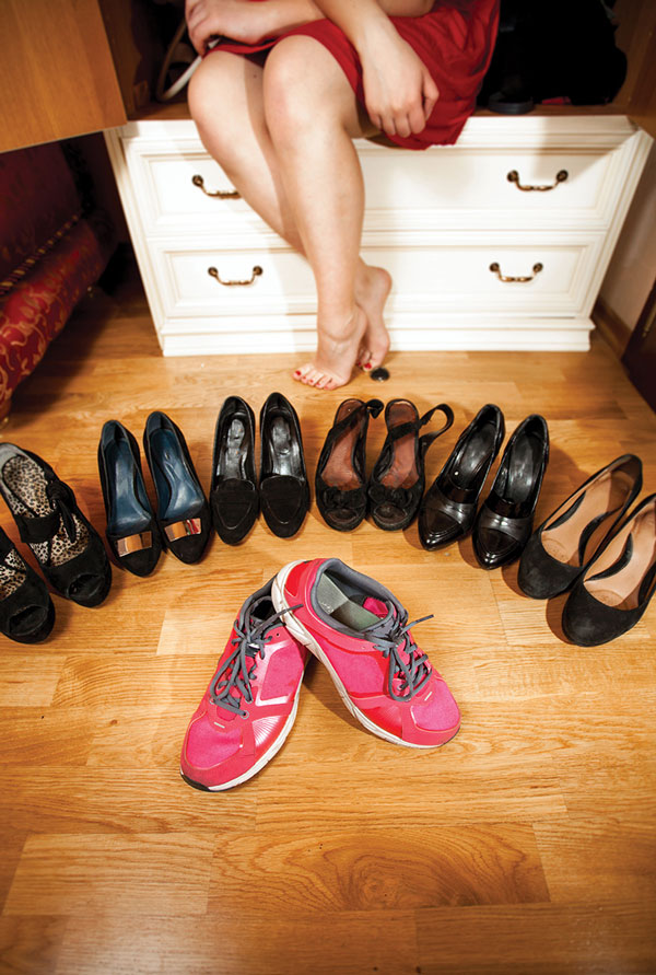
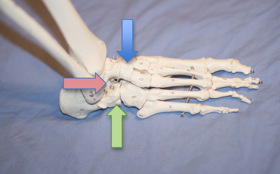
Do you know SQUAT? Have you seen SQUAT? Have patients/clients that LIKE to squat? Seen a foot that looks like this? Can you say REARFOOT VALGUS?

 "Significant changes in lower limb kinematics may be observed during bilateral squatting when rearfoot alignment is altered. Shoe pitch alone may significantly reduce peak pronation during squatting in this population, but additional reductions were not observed in the subtalar neutral position. Further research investigating the effects of footwear and the subtalar neutral position in populations with lower limb pathology is required."
 
So, what does this study tell us?

when rearfoot aliment changes, so do the kinematics (duh)
the surface (tilted into varus or inversion) or shoes (which are medially posted) can make or break the man (or women) when it comes to "peak" pronation (we knew that already; confirmation is always nice)
inverting the rearfoot can change ankle dorsiflexion (read "ankle rocker"); inverting the rearfoot seems to reduce it
inverting the rearfoot can change knee flexion; inverting the rearfoot seems to increase knee flexion
inverting the rearfoot can change hip abduction (and thus knee valgus); reducing it

Learn about the gait kinematics and clinical findings associated with this foot type, along with video clip examples and always entertaining discussion with us tomorrow night on onlinece.com: Biomechanics 308: Focus on the Rear Foot.  5PST, 6MST, 7 CST, 8EST


Power V, Clifford AM. The Effects of Rearfoot Position on Lower Limb Kinematics during Bilateral Squatting in Asymptomatic Individuals with a Pronated Foot Type. J Hum Kinet. 2012 Mar;31:5-15. doi: 10.2478/v10078-012-0001-0. Epub 2012 Apr 3.

#rearfootvalgus #squat #foottype

2012 Mar;31:5-15. doi: 10.2478/v10078-012-0001-0. Epub 2012 Apr 3.

The Effects of Rearfoot Position on Lower Limb Kinematics during Bilateral Squatting in Asymptomatic Individuals with a Pronated Foot Type.

Power V1, Clifford AM.

Author information

Abstract

Clinicians frequently assess movement performance during a bilateral squat to observe the biomechanical effects of foot orthotic prescription. However, the effects of rearfoot position on bilateral squat kinematics have not been established objectively to date. This study aims to investigate these effects in a population of healthy adults with a pronated foot type. Ten healthy participants with a pronated foot type bilaterally (defined as a navicular drop >9mm) performed three squats in each of three conditions: barefoot, standing on 10mm shoe pitch platforms and standing on the platforms with foam wedges supporting the rearfoot in subtalar neutral. Kinematic data was recorded using a 3D motion analysis system. Between-conditions changes in peak joint angles attained were analysed. Peak ankle dorsiflexion (p=0.0005) and hip abduction (p=0.024) were significantly reduced, while peak knee varus (p=0.028) and flexion (p=0.0005) were significantly increased during squatting in the subtalar neutral position compared to barefoot. Peak subtalar pronation decreased by 5.33° (SD 4.52°) when squatting on the platforms compared to barefoot (p=0.006), but no additional significant effects were noted in subtalar neutral. Significant changes in lower limb kinematics may be observed during bilateral squatting when rearfoot alignment is altered. Shoe pitch alone may significantly reduce peak pronation during squatting in this population, but additional reductions were not observed in the subtalar neutral position. Further research investigating the effects of footwear and the subtalar neutral position in populations with lower limb pathology is required.

Things often work better in pairs… Especially with Exercise

You have heard us always talk about how the lower kinetic chain is connected, how ankle rocker effects hip extension and how important hallux (great toe) extension is.

What can we conclude form this study?

  • toe spreading exercises are important for reducing navicular drop (and thus mid foot pronation, at least statically)
  • In addition to increased abductor hallucis recruitment in ascending and descending stairs, when hip external rotation exercises were added along with toe spreading exercises folks had more recruitment of the vastus medialis (a closed chain external rotator of the leg and thigh)

Keep in mind:

  • the exercises given were all non weight bearing and open chain for the external rotators. Imagine what might have happened if they were both closed chain AND weight bearing!
  • They concentrated on the effects of toe spreading (AKAlift/spread/reach) on the abductor hallucis. It also has far reaching effects on the dorsal interossei, long and short extensors of the toes.


Goo YM, Kim DY, Kim TH. The effects of hip external rotator exercises and toe-spread exercises on lower extremity muscle activities during stair-walking in subjects with pronated foot. J Phys Ther Sci. 2016 Mar;28(3):816-9. doi: 10.1589/jpts.28.816. Epub 2016 Mar 31.

link toFREE FUL TEXT: https://www.ncbi.nlm.nih.gov/pmc/articles/PMC4842445/


Podcast 120: Runner's Brains & Glute Rabbit holes


Show links:
http://traffic.libsyn.com/thegaitguys/pod_120_real_final.mp3

http://thegaitguys.libsyn.com/podcast-120-runners-brains-glute-rabbit-holes

Key tag words:
running, running form, running tricks, gait, gait analysis, the gait guys, brain, statins, glutes, runner's brain, 
 
Show sponsors:
newbalancechicago.com
 
www.thegaitguys.com  is all you need to remember. Everything you want, need and wish for is right there on the site.
Interested in our stuff ? Want to buy some of our lectures or our National Shoe Fit program? Click here (thegaitguys.com or thegaitguys.tumblr.com) and you will come to our websites. In the tabs, you will find tabs for STORE, SEMINARS, BOOK etc. We also lecture every 3rd Wednesday of the month on onlineCE.com. We have an extensive catalogued library of our courses there, you can take them any time for a nominal fee (~$20).
 
Our podcast is on iTunes, Soundcloud, and just about every other podcast harbor site, just google "the gait guys podcast", you will find us.
 
Show Notes:
 
Neuroscience:
 
Statins and exercise
http://www.nytimes.com/2017/01/04/well/move/a-fitness-downside-to-statin-drugs.html?utm_medium=email&utm_source=flipboard&_r=0
 
Cholesterol reference:
http://circ.ahajournals.org/content/early/2016/11/21/CIR.0000000000000461
 
Joe Rogan Experience Podcast: #842, Dr. Chris Kresser
 
Follow Chris Beardsley at "Strength and Conditioning Research" and Bret Contreras, "the glute guy". They always have great research based stuff.
 
Young runners have stronger brain connections
http://www.futurity.org/running-functional-connectivity-1317802-2/
 
Runners and connected brains
http://www.sciencealert.com/runners-brains-are-more-connected-than-most-study-says
 
Runners brains:
http://journal.frontiersin.org/article/10.3389/fnhum.2016.00610/full
 
Early sport diversification vs. late specialization
http://www.humankinetics.com/excerpts/excerpts/late-specialization-is-recommended-for-most-sports

Ankle stiffness, foot collapse, achilles tendonitis and its blood flow.

Yesterday, we posted on some important concepts on ankle stiffness and arch collapse, discussing some correlations and conflicts.  We believe this is an important concept to never ignore. 

Today we find this article on achilles tendonopathy, blood flow to the tendon and pronation. These concepts are deeply intermingled with yesterday's writings

These authors investigated whether there was an underlying association between foot pronation and blood flow.  The took twenty-five experienced runners, aged 34.5±10.2 years, and put them through barefoot and shod running evaluating their frontal and sagittal planemechanics. Blood flow of the Achilles tendon was measured before and after barefoot and shod running, using an oxygen-to-see device. The results of their study showed a "significant effect of eversion excursion on the increase in Achilles tendon blood flow after shod running. More specifically, the more the eversion excursion observed, the lower the increase in blood flow (P=.013)." 

This article is very germane to what we wrote about yesterday, which can be reviewed from the links above.

Is Achilles tendon blood flow related to foot pronation?
 E. Wezenbeek,T. M. Willems,N. Mahieu,I. Van Caekenberghe,E. Witvrouw,D. De Clercq

http://onlinelibrary.wiley.com/doi/10.1111/sms.12834/full

Ankle stiffness and foot collapse, correlation ?

A client who comes in with calf tightness and ankle stiffness can't be clumped into the catch all group that they need more ankle rocker or to just stretch out the posterior mechanism.

Screen Shot 2017-03-02 at 7.57.05 AM.png

In all likelihood they probably don't have a stable enough foot/arch and are passing their body mass over that unstable structure, collapse ensues before ankle rocker is completed during stance phase of gait. Thus, the body goes into a strategy the next joint complex up the chain and attempts to gain stability at the ankle complex and the most available tools, the posterior mechanism. The foot should be stable and the ankle should be mobile through sagittal ankle rocker. When the foot is unstable, things often switch; the once mobile ankle rocker shifts towards stability attempts. Not everyone needs ankle rocker work ! Don't force it, make them earn it once you find the root of the problem. In a huge chunk of the population, that stiffness and loss of ankle rocker is there as a coping mechanism to find stability. Don't take it away from them ! 
PS: raising someones arch with an orthotic doesn't earn any stability, it is borrowed, it is false, so keep that in mind. Not that it doesn't have value or a purpose, but nothing has been intrinsically fixed, only extrinsically and that cannot be forgotten. Someone has to pay for these loads coming into the system.
-Dr. Allen's rant of the day

Varus Thrust Gait, Trampoline ankle Part 2: When ankle rocker is lost.

In several previous case videos we have shown a case of traumatic ankle injury causing ankle rocker loss and subsequent knee hyperextension during sagittal gait progression, and we have shown a case of a classic Varus Thrust gait (search our site).

Today, I will shows you a case where the 2 phenomenon are connected. If you know your normal anatomy, you should be able to put this together.

Case background, video #1:  *Impaired ankle rocker (severe) in action. This was a case of ankle talus dislocation while trampoline'ing :) No surgery, but ankle was bagged up for 6 weeks. This is a TIGHT and blocked ankle rocker now, better for it to be more stable than unstable since every ligament was torn completely. These are his first steps in 6 weeks. 90 ankle dorsiflexion on the table, which is insufficient for anyone to have normal gait. Here is a great view of what happens when there is insufficient ankle rocker, one scenario at least (there are several ways around an insufficient ankle rocker). Here you can see the knee hyperextension strategy at the moment the body mass attempts to pass over the ankle, the ankle says "Nope, not today bud, try throwing the knee into extension to get over me.". And so, that is what happens here. Imagine what message the hip and glutes get from that strategy ! So, you won't see this every day, but imagine all the cases of minor ankle rocker impairment you do get in a few of your clients, and the micro knee extension strategies you can't see, that are fiddling with optimal mechanics. If you do not look, you will not find. It is why I mentioned the case last week of the ankle ROM looking normal on the exam table, but it not being used during gait. Again, not everyone needs more ankle rocker, often they need more S.E.S. (skill, endurance, strength). Skill includes, proprio, balance, coordination, motor patterning, etc. Make no mistake, this fella needs more ankle rocker !

in the sagittal video below, and more obviously in a separate video further below to more clearly demonstrate a more classic Varus Thrust gait, one should be able to see the knee undergoing a sudden abrupt varus (lateral) shift during the gait loading response.  The tib-femoral joint is a sagittal hinge, not a frontal-lateral plane hinge, so this is clearly pathomechanical movement. This knee will likely undergo premature knee cartilage and meniscal degeneration if the phenomenon is not resolved.
The cause of this issue is likely more simple than complicated however there may also be multiple factors coming together in a perfect storm. However, make no mistake, in order to understand a varus thrust gait, one has to understand the why and how of the gait presentation. Additionally, one must have a clinical knowledge of the restraining systems of the knee, both active and passive, and have a high degree of clinical suspicion and working knowledge of how to assess for these types of problems. It this immediate case below, with the severe ankle rocker loss (see in the first video) the client hits the loss of ankle rocker/dorsiflexion and must attempt to move forward. In video #1 we see knee hyperextension, but what you need to see on the video below is knee varus thrust. This is a soft case, it is not a TRUE varus thrust, but the mechanism is there. It is there on that left leg/knee if you know what to look for, and is in part because he is supinating the foot excessively, while moving through neutral knee and into terminal knee extension, to try and find some kind of lateral frontal plane strategy to get around the blocked ankle rocker. Remember, there is lots of medial and lateral joint play at neutral zero degree extension, and very little if any in terminal knee extension lock out. So the shift occurs mostly around the zero degree range and then is thrusted into terminal extension giving it that "sudden abrupt" appearance. Remember the knee is not a frontal plane hinge, but it does have some frontal plane wiggle room at zero degrees, test it out for yourself !  Why does this phenomenon occur in this client with zero posterolateral corner knee injury ? Well, it is simple anatomy. The medial condyle is longer and deeper than the lateral (see xray photo below showing this relationship) and with such far lateral foot supination combined with terminal knee extension, he is likely only bearing weight on the medial condyle and the joint pivots and shifts in this zero degree extension through to hyperextension lock out (not a true instability pivot-shift but the mechanism remains present) until the LCL (lateral collateral ligament) complex and iliotibial band and other lateral structures engage. Because there is no true lateral laxity, there is only a subtle lateral shift,  unlike the 3rd video below of the lady walking on tiles. So, this is a case of knee hyperextension and mild varus thrust gait from a blocked ankle rocker motion joint. 


Below are some thoughts from a prior video on Varus Thrust gait (see video to the right). You must understand all of these components to help these clients fully. 

Things to consider:  
- old ACL/PCL and posterolateral corner damage (search our site for articles we have composed)
When the posterolateral corner complex of the knee is torn up from a blow to the knee or a torsional loading failure, the 3 components of the posterolateral corner (the lateral collateral ligament (LCL), the popliteal tendon, and the popliteo-fibular ligament complex). This complex attaches just in front of the origin of the lateral gastrocnemius tendon off the lateral femoral epicondyle. This complex can be blown out from either a PCL or ACL injury mechanism, these big player ligaments are rarely torn in isolation.
- is there a Pivot Shift phenomenon, likely.  A positive Pivot Shift test will be present. One must know how to perform this test to confirm its presence, it can be a tricky test if one does not know the load vectors to apply and what the shift feels like and where it occurs during the test. This can be a very subtle positive test, again, first hand experience is everything. 
- one must find this before surgery occurs for the ACL or PCL. Failure to find and address this damaged complex will likely result in rotational stability problems once return to play occurs. IT will not likely be noted in the initial post-operative months as the aggressive loading response will not be performed early on. Failure to address this problem will likely put ACL-PCL reconstruction success at a high risk.


Other critical factors to consider in the Varus Thrust Gait:
- is there medial knee osteoarthritis ?
- what is the foot type and what are the mechanics ?  ie. Forefoot varus, Forefoot supinatus, rearfoot variances
- does the patient have excessive pronation challenges that create massive internal spin into the tibia ?
- is the hip frontal and rotation plane stable?  Can the patient adequately control rotation at the hip level ?
- is there a Cross Over gait phenomenon with narrow based step width ? (search our blog and youtube for  "gait guys crossover gait").  A narrow step width will create an "unstacked" limb and promote more rotational risk into the limb, often playing out at the least tolerable joint to rotation . . . the knee.
- Does the client have Tibial Varum ? Genu Varum, Genu Valgum ? These can promote and complicate the Varus Thrust gait.
- Does the client have Tibial torsion or Femoral Torsion variants ? These can promote and complicate the Varus Thrust gait.

- is there weakness of the lateral gastrocnemius or biceps femoris (to name just two the directly cross over this posterolateral interval and can offer joint compression/stability ? What about weaknesses in the medial leg ? Not that these are anywhere sufficient to offset a PLRI (posterolateral rotatory instability), but, they are secondary helpers/restraints.

One should clearly see now that the Varus Thrust gait is potentially complicated and multifactorial. One MUST understand:
1. many components of normal gait and normal anatomy from foot to pelvis, at least.
2. be able to assess for aberrant mechanics and pathologies within all joints of the lower limb
3. be able to assess for post operative rotational stability and laxity (*even a healed, yet partially attenuated, Posterolateral corner complex that was not noted or addressed in the ACL-PCL reconstruction can come back to haunt even the best reconstruction. Those little rotational instabiliites will build over the years and render attenuation of the other secondary posterior restraints in the knee. Like a Lisfranc injury, sometimes things take a few years to brew and blossom before the "career ender" instability shows up. Trust us, we have seen it enough times.  

Rule: if one does not know it exists, one will miss it. If one does not know how to assess it, one will miss it. If one does not know normal anatomy, torsional variants, foot types and gait types, one is likely to be lost and left fumbling.  Our clients deserve more. 

Clinical pearl: if you are radiographically sharp, you should have noted the Pellegrini-Stieda lesion at the medial tibial epicondyle (this is not a radiograph for this case, it was used to show the longer medial condyle reach). These are ossified post-traumatic lesions near the medial femoral collateral ligament attachment. This avulsion injury of the medial collateral ligament can calcificy after a few post-trauma weeks. 

- Dr. Shawn Allen, the other gait guy

Cerebellar impairment = Gait Changes = Happy Patient

This is a fairly info dense post with many links. please take the time to explore each one to get the most out of it. 

If you have been with us here on TGG long enough, you know the importance of the cerebellum and gait. Mechanoreceptor information travels north to the cortex via the dorsal (and ventral) spinocerebellar pathways to be interpreted (and interpolated, in the case of the ventral pathway), with the information relaying back to the motor cortex and vestibular nucleii and eventually back down to the alpha (and gamma) motor neurons that proved the thing you call movement and thus gait. (Cool video on spinocerebellar pathways here and here).

This FREE FULL TEXT paper has some cool charts, like this one, that show the parameters of gait that change with cerebellar dysfunction (in this case, disease, although idiopathic means they really don't know. Anatomical or physiological lesions will behave the same, no? Doesn't the end result of a functional short leg look the same as an anatomical one?)

Looking tat this chart, what do we really see? People with cerebellar dysfunction:

  • a shorter step length
  • a wider base of gait
  • decreased velocity
  • increased lateral sway
  • slower overall gait cycle

Hmmmm...Beginning to sound like a move toward more primitive gait. Just like we talked about in this post on the 5 factors and proprioception here several years ago. We like to call this decomposition of gait. 

They go on to talk about specific anatomic regions of the cerebellum and potential correlation to specific gait abnormalities, like the intermediate zone and interposed nucleii controlling limb dynamics and rhythmic coordination like hypermetria (overshooting a target), especially when walking in uneven surfaces or when gait is perturbed, like walking into something or changes in surface topography, or the lateral zone of the cerebellum, for voluntary limb control, such as where you place your foot. Definitely gait nerd material.

There aren't any direct tips on rehab, but it would stand to reason that activities that activate the cerebellum and collateral pathways would give you the most clinical gains. Lots of propriosensory exercises like here, here, here and here for a start.

Happy cerebellum = Happy patient

The Gait Guys


Winfried Ilg, Heidrun Golla, Peter Thier, Martin A. Giese; Specific influences of cerebellar dysfunctions on gait. Brain 2007; 130 (3): 786-798. doi: 10.1093/brain/awl376  FREE FULL TEXT

More hip and foot clues: connections we all know (or should).


You've heard it  here many times before on TGG, the whole organism must be considered when it comes to movement. Humans will find a way to move, the question is, are we moving right, with compensations that will eventually plague us, or are we moving mostly cleanly ?  We like to say, "the load is going to go somewhere", the body will adapt.  The problem is, we believe the body often looks for the easiest opportunity, not often the best one. And, like a teenager, we believe that the body does not have the foresight to anticipate the consequences of its choices in the future. As long as the adaptation is not immediately painful, the choice often seems reasonable and thus is implemented. 
Today's article suggests something we have said over and over, when things are working wekk,  the hip and foot are a team, they discuss the motor plan to a degree and attack the loading and unloading together.  That is when it works well.
The purpose of the referenced study was to examine the effects of abductor hallucis and gluteus maximus strengthening exercises on pronated feet. Yes, this study has limitations, most do. But we like to take in the information because it forces us to deepen our well of knowledge and facts. As Neil Degrasse Tyson recently laid out the continuum, things move from facts, to knowledge, to wisdom, to insight. It takes decades to get to the wisdom and insight, it is a process. There are many out there teaching without these last two, and that is scary. There is an abundence of "repackaging" of information and turning it into something apparently new and unique. Spend your time deepening your well.  Articles like this make Ivo and sit back and have long discussions that go down tangents and rabbit holes (like on our Podcast), that is where the good stuff is. Anyone who takes this article as, "strengthen the glutes and the feet and perhaps consider orthotics" are in the discovery phase, no doubt, but far from wisdom and insight.  In our silly stupid opinion, for what little it might be worth to some.  

*photo: one thing we would be adding pattern correction to is the inward drift of the right knee during this high knee posturing. This is a real problem in runners and sprinters, it is a speed suck/power leak.

The effects of gluteus maximus and abductor hallucis strengthening exercises for four weeks on navicular drop and lower extremity muscle activity during gait with flatfoot

Young-Mi Goo, MS, PT,1 Tae-Ho Kim, PhD, PT,1,* and Jin-Yong Lim, MS, PT1  J Phys Ther Sci. 2016 Mar; 28(3): 911–915.

Published online 2016 Mar 31. doi:  10.1589/jpts.28.911

https://www.ncbi.nlm.nih.gov/pmc/articles/PMC4842464/

High Heels...A requirement?

More on high heels.

We think high heel wear is largely geographical. Dr Ivo lives in the mountains of Colorado and rarely sees ANYONE in high heels, even at social events. Dr Allen, in Chicago, sees plenty of them.

Their popularity may be waning but the problems continue (see yesterdays post here). Required to wear them? Really?

"At the 2015 Cannes Film Festival, multiple women—some with medical conditions—were famously turned away from screening events for wearing rhinestone flats rather than heels. Cocktail waitresses at Foxwoods Resort Casino in Mashantucket, CT, complied with their employer’s 2-inch heel requirement for decades until their union successfully quashed it in 2013. A similar 2015 case in East London, involving a receptionist sent home for failing to wear shoes with a 2- to 4-inch heel, led to union involvement as well as scrutiny of prime minister Theresa May’s well-known fondness for kitten heels. And in 2013, the US Defense Intelligence Agency—helmed at the time by Lt. Gen. Mike Flynn, president-elect Donald Trump’s choice for national security advisor—included a “no flats” policy among its dress-code requirements for women."


http://lerfoothealth.com/archives/2016/popularity-of-high-heels-wanes-but-issues-remain/

Something a little different for a change: Case Management of a Post Surgical Foot

In this series, we will follow the progress of a post surgical, post rehab foot. These are the actual case notes and you can follow our thought process as we move along. 

History:

JM presented with left-sided content foot pain. On July 24 she broke her left navicular and cuboid (pretty unusual, as these fractures are rare. Navicular fractures are usually stress fractures (1), occurring in about .6% of fractures in one study (2).  Cuboid fractures are also rare and occur in less than 1.8 per 100,000 (3) ) She also tore the reticular ligaments. this happened when she fell down the stairs, inverting and plantar flexing the foot.

She has had extensive physical therapy as well as plate fixation of the navicular but is still having constant discomfort; she feels a pinching and shock like sensation in the right arch, particularly when loading the foot (whenever you hear about a "shock like pain, begin thinking about nerve related pain). She has been on gabapentin in the past which helped but she stopped it 3 weeks ago with no regression of her symptoms. She feels frustrated.

She was in physical therapy until the end of December. She has continued with exercises consisting of plantar flexion/toe raises, mobilization and inversion/eversion, squats/lunges as well as massage. She has improved but not completely better. She is able to hike 4-5 miles with little pain (boy, those Colorado women are tough!). The foot generally feels better with non weight bearing and rest as well as avoiding impact. The foot feels stiff in the morning and  she limps for the first 10 minutes after getting out of bed. Most recently she has had x-rays at VSO with Dr. X.

What did we find?

There was swelling noted over the extensor digitorum brevis with significant weakness of it as well as the extensor longus. No sensory deficits, reflexes intact. She had an anatomically short left short leg which appeared to be functional. There was a scar visible over the dorsum of the foot approximately 2 inches in length (see photos) and some discoloration lateral just anterior to the lateral malleolus. palpation along the medial plantar nerve revealed increased sensitivity below the navicular and into the medial heel. 

She has external tibial torsion bilaterally and limited eversion of the forefoot on the right. Her cuboid was moving appropriately but talonavicular articulation was not. She has adequate hip extension, 15+ degrees and ankle dorsiflexion bilaterally in excess of 15 degrees.

One leg standing with eyes open was less than 10 seconds. Loss of flexion and extension about the L SI joint. 

no x rays available for this visit

What we think was wrong:

Left foot pathomechanics (talonavicular) secondary to surgery and fracture. She has significant weakness of the short and long extensors as well as limited eversion and proprioceptive difficulties.

Discussion:

The talonavicular articulation is one of the key joints in mid foot pronation. Pathomechanics appear to be compromising the medial plantar nerve. This is exacerbated by her inability to fully evert the forefoot and pronate through the mid and forefoot (pronation is dorsiflexion, eversion and abduction). 

What We did:

We manipulated the sacroiliac joint and metatarsophalangeal articulations. We held off on any mobilization of the foot until we see her x-rays, hopefully available next visit.

We treated with neuromuscular acupuncture at the origin/insertion of the long extensors as well about the short extensor mass where the swelling was located. She was given the tripod standing, lift spread reach, toes up walking, and tiptoe waiting exercises to perform 2-3 times daily. She is scheduled for followup next week with x-ray review.


1. http://emedicine.medscape.com/article/85973-overview

2. http://journals.sagepub.com/doi/pdf/10.1177/2473011416S00299

3. http://www.uptodate.com/contents/cuboid-and-cuneiform-fractures

Podcast 119: Prof. Carlos Lemos Jr. & Dr. Shawn Allen

Key Words: BJJ, jiu jitsu, carlos lemos, carlos lemos jr, gracie barra, gbdownersgrove, shawn allen, movement, 


Website link:   www.thegaitguys.com
Podcast links:

http://traffic.libsyn.com/thegaitguys/calos_pod_1f.mp3

http://thegaitguys.libsyn.com/podcast-119-prof-carlos-lemos-jr-dr-shawn-allen
 
www.thegaitguys.com
That is our website, it is all you need to remember. Everything you want, need and wish for is right there on the site.
Interested in our stuff ? Want to buy some of our lectures or our National Shoe Fit program? Click here (thegaitguys.com or thegaitguys.tumblr.com) and you will come to our websites. In the tabs, you will find tabs for STORE, SEMINARS, BOOK etc. We also lecture every 3rd Wednesday of the month on onlineCE.com. We have an extensive catalogued library of our courses there, you can take them any time for a nominal fee (~$20).
 
Our podcast is on iTunes, Soundcloud, and just about every other podcast harbor site, just google "the gait guys podcast", you will find us.

Show Notes:

Special guest today, Professor Carlos Lemos Jr, a 4th degree black belt Brazilian Jiu Jitsu professor at Gracie Barra Downers Grove.

Join us for a great 90 minute podcast with my good friend, mentor, teacher and all around great man.

We talk about many interesting things, including movement, learning, history and so much more. IF you thought you new all about jiu jitsu, you are in for a lesson or two today. This certainly opens up things for Part 2 in the near future.

gbdownersgrove.com
Downers Plaza Shopping Center, 130 Ogden Ave, Downers Grove, IL 60515
Phone: (630) 964-1414

One of life's great mysteries....Some folks will do what they want anyway....

The origins of the species, gravity and women...Just a few of life mysteries. Reading this article (1) made us sad in many ways. It's like smoking. You know it's bad for you but you keep doing it. Why? The mystery remains to us.

Vanity seems to often trump biomechanics, as we see in pencil skirts (see our post here), droopy pants (see here)  and high heels (here).

Yet, here is yet another study about women, heels and bunion surgery. 

"Almost two thirds (31) of the 50 patients who said they wanted to go back to wearing heels after surgery did so, and 24 of these women said their postoperative use equaled or exceeded the frequency of their preoperative wear. There were no differences between pre- and postoperative heel heights.

In the study, women older than 65 years were more likely than younger women to report high-heel use prior to hallux valgus surgery.

However, 58.5% of study participants reported difficulty with heel wear, and 13.9% said they had significant restriction, and couldn’t wear anything without pain but custom orthopedic shoes or slippers. Most women (86%) were able to return to comfortable shoes after surgery with minimal or no discomfort; 27.7% said their footwear choice was unrestricted, meaning they could wear both comfortable shoes and heels with minimal discomfort. The 23 women older than 65 years were twice as likely to report significant restriction as those in the younger cohort; compared by operative type, patients who had the most extensive procedures had the highest rates of restriction. The findings were published in June by the World Journal of Methodology. (2)"

Bunions are believed to be caused by an inability to anchor the 1st ray and the untoward action of the adductor hallucis, acting from the transverse and oblique insertions more proximally on the foot, make the hallux head west. This is under the purview of the peroneus longus, extensor hallucis brevis as well as the short flexors of the lesser toes (see here).

The components of supination are plantar flexion, inversion and adduction. Why would you continue to wear a shoe with a narrow toe box that forces the big toe medially and that puts you in plantar flexion? We won't even begin to talk about the loss of ankle rocker.....

We guess folks will continue to do what they will do....


1. Robinson C, Bhosale A, Pillai A. Footwear modification following hallux valgus surgery: The all-or-none phenomenon. World J Methodol 2016;6(2):171-180.

2. http://lerfoothealth.com/archives/2016/most-women-who-want-to-wear-heels-after-bunion-surgery-do-so/

Flexible hammer toes

IF you've been with us all along you know this one by heart. All you have to do is test it to confirm (never assume, ever !). 
And remember, sometimes people present with their problem, and sometimes they present with a layered compensation to strategize and cope with the underlying problem. It is your job to determine that.
You must recognize here:
flexible hammer toe early development. It is a compensation for weakness somewhere in the chain. It is a gripping strategy to make up for something somewhere. It is not normal, it is a clue.

1- over activity of the short extensors & and long flexors
2- under activity of the long extensors & and short flexors
3- thusly, distal displacement of the Metatarsal fat pad complex
4- and lotsa other things (but we will not detract from the major cursory observation point here today)

Loading this forefoot is troubled, in many ways...... so many that we will not go it here. We have written about these things in long form so many times, just head over to the blog and search. This client did not have any foot or ankle issues, they had impaired hip rotation and extension and were complaining of low back pain. They have obviously been coping through the entire chain for awhile. Be Sherlock Holmes in your practice today, look for the clues.

-Dr. Allen

Trampoline ankle case: Part 2

Trampoline ankle: case progression:

*We have not uploaded this video to youtube yet. It is on our Facebook page on Feb 11th. Go watch it there. We will compose this case as it progresses and put together a complete video then. But you can see what is discussed below, in the FB video,February 11th.

Impaired ankle rocker (severe) in action. We showed you this case last week, the ankle talus dislocation while trampoline'ing :) No surgery, but ankle was bagged up for 6 weeks. This is a TIGHT and blocked ankle rocker now, better for it to be more stable than unstable since every ligament was torn completely. These are his first steps in 6 weeks. 90 ankle dorsiflexion on the table, which is insufficient for anyone to have normal gait. Here is a great view of what happens when there is insufficient ankle rocker, one scenario at least (there are several ways around an insufficient ankle rocker). Here you can see the knee hyperextension strategy at the moment the body mass attempts to pass over the ankle, the ankle says "Nope, not today bud, try throwing the knee into extension to get over me.". And so, that is what happens here. Imagine what message the hip and glutes get from that strategy ! So, you won't see this every day, but imagine all the cases of minor ankle rocker impairment you do get in a few of your clients, and the micro knee extension strategies you can't see, that are fiddling with optimal mechanics. If you do not look, you will not find. It is why I mentioned the case last week of the ankle ROM looking normal on the exam table, but it not being used during gait. Again, not everyone needs more ankle rocker, often they need more S.E.S. (skill, endurance, strength). Skill includes, proprio, balance, coordination, motor patterning, etc. Make no mistake, this fella needs more ankle rocker !

-Dr. Allen

* again, this video does not play, read above

Talus dislocation: Trampoline ankle

Trampoline gyms are fun but risky places. It is fun leaping like spiderman from one trampoline to the next. 
But, Foot placement from such a height onto a pliable sloped surface (ie. if you do not hit the next trapoline smack dab in the center) is just a bad place to load a foot. Hence the foot that i saw today in the office, 4 weeks post talus and forefoot dislocation, complete, as you can see here. Oy, no trampolines for my peeps. Nope. Never.

When the ankle lies to you

When the ankle lies to you.
Yesterday I saw something I see quite often. It was a client with dorsal foot pain, nothing shocking. But, this client had plentiful ankle dorsiflexion on the table during examination but when they walked, there was barely any use of ankle dorsiflexion-ankle rocker. Heel rise was premature.

It once again proves that just because you have it, doesn't mean it is available to be used. There was adequate hip extension and glute strength so it wasn't coming from there, though that is a frequent source. The examination was detailed, but to keep it brief here today, this client, had decent strength about the ankle from what could be determined, but they failed the hop test, control was terrible, and they could tell. Once again, if you cannot control the joint under load, the body will often not give you the full range, merely out of self preservation mode to protect the joint. This client was attempting to get more ankle rocker motion via arch collapse and over pronation to get the tibia to progress forward enough for normal gait. The collapse was causing a dorsal impingement on the foot. Lots more to come on these ideas in future posts. 
None the less, it is a good lesson to all those people out there that think that everyone just needs more ankle rocker strength and range of motion. The truth is, not everyone does, and forcing it in some will cause them pain or problems or compensations . . . . and that is your fault if it is the case.

-Dr. Allen

The buck DOES NOT stop here...

The buck doesn't stop here..

image from: http://www.aofas.org/footcaremd/treatments/pages/triple-arthrodesis.aspx

image from: http://www.aofas.org/footcaremd/treatments/pages/triple-arthrodesis.aspx

One of the most pervasive problems following a ankle arthrodesis, particularly a triple arthrodesis which involves fusing the subtalar (talocalcaneal), calcaneocuboid , and talonavicular joints results in a loss of ankle rocker. The "buck" needs to be passed somewhere and this usually will mean proximally in the lower kinetic chain.

Seeing adjacent joints with osteoarthritic changes following hypomobility have another joint is nothing new; you probably see it all the time in practice. Remember that it is not always have to be a "fusion". Simple longstanding pathomechanics or longstanding hypomobility will often cause the same problems.

A nice, full text referenced review 1 of her favorite journals. Some nice side discussions as well. Enjoy : )

"Altered biomechanics after ankle arthro­desis often increase stress on the adjacent joints in the foot, which can cause or exacerbate osteoarthritic degeneration in those joints. Clinicians and researchers are working to better understand this process and how to minimize patients’ risk."

http://lermagazine.com/…/adjacent-joint-arthritis-after-ank…
#anklemobility #anklerocker #triplearthrodesis #hypomobility


Dry Needling and Muscle Activation Patterns

A nice study looking at how sequential muscle activation patterns can change with dry needling. Think about the applications for gait?

"Removing LTrPs changes the order of muscle recruitment to a more sequential, stable pattern that is not significantly different to that displayed by the control group prior to fatiguing exercise. This suggests that removing LTrPs may allow subjects to better cope with the effects of fatigue, as evidenced by the reduced variability in activation times and the reduced co-activation of the muscles investigated. "

FREE FULL TEXT here: https://isbweb.org/images/conf/2003/longAbstracts/LUCAS_198-208_SB_LONGE.pdf

Dry Needling and Myofascial Pain

Regardless of the mechanism, dry needling and ischemic compression both seem to reduce myofascial pain. How about some more studies looking at muscle function and activation patterns?

"This study compared these treatment techniques to one another using the Neck Disability Index (NDI), a numeric rating scale (NRS), pressure pain threshold and muscle characteristics. 42 female patients with myofascial neck pain were randomly assigned to a treatment group and the 4 most painful MTrPs were treated using DN or MPT. No difference was found between the two techniques on the short and long term. Both techniques showed an improvement in NDI on the short and long term. "

Dry needling or manual pressure in myofascial pain? - Anatomy & Physiotherapy

The aim of this study was to compare dry needling to manual pressure in patients with myofascial pain.

ANATOMY-PHYSIOTHERAPY.COM|BY <A HREF="/AUTHORLIST/3:JOANNA1988" TITLE="VIEW ALL ARTICLES FROM JOANNA TUYNMAN">JOANNA TUYNMAN</A>