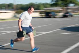
You need toe extension, more than you might think.

Screen Shot 2018-07-20 at 9.24.08 AM.png

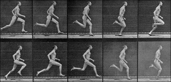
There is a major difference in these 2 photos.One foot is ready for foot loading, the other has one foot over the starting line, and is going to possibly have the risks related to inappropriate loading.

In clients with one of several possible issues related to poor control of the arch during weight bearing loading, it is not all too uncommon for us to bring to their attention that not only do they NOT utilize toe extension appropriately, and at the right time, they just simply have poor strength and endurance of the toe extensors (we will not be bringing up the complicated orchestration of the long and short toe extensors today, lets just keep it loosely as looking at them as a whole for today).

We know we say it an awful lot, that clients need more toe extension endurance and strength. But more often than not, they need more awareness of how little they are actually using their toe extensors during foot loading. This is why we despise flip flops and foot wear without a back strap on them, the flexors have to dominate to keep the footwear on the foot.  And, if you are into your toe flexors, you are definitely not into your toe extensors.

Screen Shot 2018-07-20 at 9.23.54 AM.png

You can easily see in this photo that there is a major difference in the integrity and preparation of the foot arch prior to foot loading in these 2 sample photos. One the toes are up in extension, the other the toes are lazy and neutral.  The toe up photo demonstrates well that when the toes are extended, the Windlass mechanism draws the forefoot and rearfoot together and raises the arch. Go ahead, lift your toes, it will happen on you as well (unless your arch is so collapsed that the first metatarsal actually dorsiflexes during toe extension, in this case, you are a whole different management tier). From this arch raised position, the first metatarsal is adequately plantarflexed, this means the joint complexes proximal and distal to the metatarsals are all in the right position to load and cope with loads. In the toe neutral picture, these components are not prepared, the arch is already getting ready to weight bear load from a half-baked position. One cannot expect the foot complex to load well when it is starting from a position of "half way there". One should start the loading of the foot from the starting line, not 3 steps over the line and not 3 steps before the starting line.  There is no athletic or mechanical endeavor that does well when we start the challenge too soon or too late, timing is everything.

How you choose to prep your foot for contact loading, and yes, there is some conscious choice  here, one is lazy the other is optimal, can determine to a large degree if you or your client is about to fall into the long list of problems related to poorly controlled pronation (too much, too soon, too often, too fast). Any of those bracketed problems lead to improper loading and strains during time under tension.

We will almost always start our clients on our progressing protocol of arch awareness and we will loosely say arch restoration, and attempts at better optimizing the anatomy they have, with toe up awareness.  Many clients will have poor awareness of this component issue, on top of poor endurance and frank weakness. The arch is to a great degree build from a lifting mechanical windlass effect, from the extensors and foot dorslflexors, not from the foot flexors. This is one of our primary beefs with the short foot exercise of Janda, there needs to be a toe extensor component in that exercise (search our blog for why the short foot exercise is dead). The short foot exercise is not actually dead, all exercises have some value when placed and performed properly, but the short foot exercise is based off of the toes being down and utilizing the plantar intrinsics to push the arch up and shorten the foot, this is a retrograde motion and it is not how we load the foot, but, it does have value if you understand this and place it into your clients repertoire appropriately.  This is also why we have some conceptual problems in stuffing an orthotic under someones arch to "lift it up", ie. slow its fall/pronation.  There are times for this, but why not rebuild the proper pathways, patterns and mechanics ?

Teach your clients about toe extension awareness. TEach them that they need to relearn the skill that when the toes drop down to the ground that the arch does NOT have to follow them down, that the client can relearn, "toe up, arch up . . . . . then toes down, but keep the arch up".  IT is a mantra in our office, "don't let your arch play follow the leader".  Reteach the proper neurologic disassociation between the toes and arch.

Perhaps the first place you should be starting your clients with foot and ankle issues, is regaining awareness of proper toe extension from the moment of toe off, maintaining it through swing, and then keeping it until the forefoot has purchase on the ground again, and not any time sooner than that ! If their toes are coming down prior to foot contact, it is quite likely their arch is following the leader.

So, if your client comes in with any of the following, to name just a few:  tibialis posterior tendonitis, plantar fascitis, heel pain, forefoot pain, painful bunions, arch pain, hallux limitus, turf toe, . . . . and the list goes on. Perhaps this will help you get your client to the starting line.

Shawn & Ivo, thegaitguys.com


Step width: Head over foot ?

Step width, "head over foot"?

There has been some decent debate on "head over foot" running biomechanics. We postulate from our years of reading research and studying people's gait (coupled with physical examination, a neuromuscular assessment, not just a visual assumption) that the head should remain within the limits of the step width. This theory falls apart if someone is a crossover gait runner or walker (search our blog for this "cross over gait" idea). IF one is a narrow step width (cross over gait, not a literal cross over of course) then the head must basically be over the foot on each step. But this is a gait with severe limitations and lots of risks and biomechanical problems as we have written about many times, though one can say is has some economical aspects which we have proposed many times.
But, if the head is outside the step width, one is leaning and this resembles a pathologic Trendelenburg gait. Can we definitively then say that when the head is outside the foot contact (beyond the limits of a person's step width) that it is problematic? No, but it is likely pathologic and clearly uneconomical.

So the fence seems to be the head over the foot.
If you are outside that fence even a little, you may be (we strongly believe) on the wrong side of the fence. Look at a CP gait (photo below) for example, point made. So, would you rather be on the other side of the fence? We would, we want to be inside the step width and we are fine going right up against the fence (the head foot) but not over top of it. One cannot just say that the head over the foot is better. What about hip and pelvis stability ? If the hips-pelvis are drifting into the frontal plane, this will put the head over the foot as a default. So does this validate the head over foot theory as good in this client ? No, we see this as a problematic gait all the time, lots of hip and spinal stability issues in these clients. One cannot stand and preach on head over foot alone. We just made the case that in a frontal plane drift pelvis client, this is a compensatory default, but it doesn't make it a good thing, far from it.

For now, we will stay put that, with all other faulty mechanics not present, a more sound head position is to be found between the limits of the step width. Yes, right up to the fence of "head over the foot", but not over top as a sound pattern to play with. Why risk falling over the fence on some steps, Try this, stand on one foot, put your head over the foot. In this position, you had to drift the pelvis laterally into the frontal plane. Now try to effectively engage your glute. Enough said, for now. So why would we promote this as an effective running form? More to come we are certain, but we are open to debate, and to being schooled wrong. If you wish, go into our blog (link below) and read up on the effects of step width on gait, and all of the risks/problems that a narrow step width promotes (ie. head over foot).

https://www.google.com/search?q=the+gait+guys+step+width&ie=utf-8&oe=utf-8&client=firefox-b-1-ab

This simple screening test becomes a form of exercise.

Today we look at a simple CNS screen for your “central pattern generators” or “CPG’s”. If you do not pass, then the exercise becomes the rehab exercise. If you (or your client) does not have good coordination between the upper and lower extremity, then they will not be that efficient, physiologically or metabolically. 

The “cross crawl” or “step test” looks at upper and lower extremity coordination, rather than muscular strength. If performed for a few minutes, it becomes a test that can look at endurance as well. 

It is based on the “crossed extensor” response, we looked at last week. That is, when one lower limb flexes, the other extends; the contralateral upper limb also flexes and the ipsilateral upper limb extends. It mimics the way things should move when walking or running. 
 

  • Stand (or have your client stand) in a place where you will not run into anything.
  • Begin marching in place.
  • Observe for a few seconds. When you (or your client) are flexing the right thigh, the left arm should flex as well; then the left thigh and right arm. Are your (their) arms moving? Are they coordinated with the lower extremity?
  • What happens after a few minutes? Is motion good at 1st and then breaks down?
  • Now speed up. What happens? Is the movement smooth and coordinated? Choppy? Discoordinated?
  • now slow back down and try it with your (their) eyes closed


If  movement is smooth and coordinated, you (they) pass

If movement is choppy or discoordinated, there can be many causes, from simple (muscle not firing, injury) to complex (physical or physiological lesion in the CNS).

  • If movement is not smooth and coordinated, try doing the exercise for a few minutes a day. You can even start sitting down, if you (they) cannot perform it standing. If it improves, great; you were able to help “reprogram” the system. If not, then you (they) should seek out a qualified individual for some assistance and to get to the root of the problem.

Physiological Mechanisms of PRP

image source: https://commons.wikimedia.org/wiki/File:Blausen_0740_Platelets.png

image source: https://commons.wikimedia.org/wiki/File:Blausen_0740_Platelets.png

We were discussing the efficacy of PRP as well as some of the pros/cons this weekend while recording an upcoming podcast.

This paper did a nice job explaining what we know versus what we do not know at this point in time, regarding the basics science behind it. A referenced, full text article for you:

Yiqin Zhou and James H-C. Wang, “PRP Treatment Efficacy for Tendinopathy: A Review of Basic Science Studies,” BioMed Research International, vol. 2016, Article ID 9103792, 8 pages, 2016. https://doi.org/10.1155/2016/9103792.

link to free full text: https://www.hindawi.com/journals/bmri/2016/9103792/

Running with deep core weakness.

You see something in your client's movement that your eyes (and your present knowledge base) do not like. You decide to implement come well intended corrective work to change these issues you do not like. The risk is, without a prudent clinical examination, which could quite possibly dismiss what you are seeing, or discover that the source is from another cause, that you could be adding a compensation to a client's compensation to an underlying problem.

We discuss this all the time. It is nice to find new paper that supports our suspicion that when we do not fix a problem, merely because we are changing something we see which we do not like, "Muscular compensations may increase risk of muscular fatigue or injury and increased spinal loading over numerous gait cycles and may result in damage to spinal structures. Therefore, insufficient strength of the deep core musculature may increase a runner's risk of developing LBP."

Biomechanical consequences of running with deep core muscle weakness. Raabe ME, Chaudhari AMW
J Biomech. 2017 Dec 6. pii: S0021-9290(17)30692-9. doi: 10.1016/j.jbiomech.2017.11.037. [Epub ahead of print]

Controlling, or at least reducing spinal shear forces is always a desirable outcome. We should all have sufficient strength through the thoraco-lumbo-pelvic interval to protect undesirable forces and loads through the spine. We have discussed this many times on The Gait Guys site/blog during our "antiphasic" biomechanical discussions, ones that have incorporated arm swing, spinal twist/torsional loading and leg swing and whatnot. McGill has discussed this previously as well, mentioning that the degree of antiphasic counter-rotation between the thorax and pelvis reduces as spinal pain is present to reduce compression and shear components.

This paper suggests that improper function of the deep core musculature, defined by this paper as the multifidus, quadratus lumborum, psoas, and deep fascicles of the erector spinae, may lead to abnormal spinal loading, muscle strain, or injury to spinal structures, all of which have been associated with increased low back pain (LBP) risk. "The purpose of this study was to identify potential strategies used to compensate for weakness of the deep core musculature during running and to identify accompanying changes in compressive and shear spinal loads."-Raabe et al.

In this paper, the deep core muscles were weakened individually and together.

"The superficial longissimus thoracis was a significant compensator for 4 out of 5 weakness conditions (p < 0.05). The deep erector spinae required the largest compensations when weakened individually (up to a 45 ± 10% increase in compensating muscle force production, p = 0.004), revealing it may contribute most to controlling running kinematics. With complete deep core muscle weakness, peak anterior shear loading increased on all lumbar vertebrae (up to 19%, p = 0.001). Additionally, compressive spinal loading increased on the upper lumbar vertebrae (up to 15%, p = 0.007) and decreased on the lower lumbar vertebrae (up to 8%, p = 0.008). Muscular compensations may increase risk of muscular fatigue or injury and increased spinal loading over numerous gait cycles may result in damage to spinal structures. Therefore, insufficient strength of the deep core musculature may increase a runner's risk of developing LBP." -Raabe et al.

https://www.ncbi.nlm.nih.gov/pubmed/29249454

J Biomech. 2018 Jan 23;67:98-105. doi: 10.1016/j.jbiomech.2017.11.037. Epub 2017 Dec 6.Biomechanical consequences of running with deep core muscle weakness. Raabe ME1, Chaudhari AMW2


So, you do weighted carries?

METHODS:

Participants were instructed to ascend and descend a three-step staircase at preferred pace using a right leg lead and a left leg lead for each load condition: no load, 20% body weight (BW) bilateral load, and 20% BW unilateral load. L5/S1 contralateral bending, hip abduction, external knee varus, and ankle inversion moments were calculated using inverse dynamics.


Nothing earthshaking here (1) , but a few takeaways:

  • Asymmetric loading of L5-S1 will most likely become more significant if the individual has a L5-S1 facet tropism, where one (or both) of the facets is (are) facing saggitally, as loading will be be even greater.  This has been associated with disc derangement (2) and degeneration (3).


  • The body does seem to adjust for the load, but it takes at least to the second step. We need to make sure the proprioceptive feedback loops (joint and muscle mechanoreceptors and their associated pathways) are functioning well. Manipulate, mobilize, facilitate, inhibit as appropriate.


  • The increased varus moment and hip abduction on the unweighted side are most likely to move the center of gravity more to the midline, which makes sense. This may become problematic with folks with increased internal tibial torsion, especially with femoral retroversion/torsion as they already have limited internal rotation available to them at the hip


1. Wang J, Gillette JC. Carrying asymmetric loads during stair negotiation: Loaded limb stance vs. unloaded limb stance. Gait Posture. 2018 Jun 19;64:213-219. doi: 10.1016/j.gaitpost.2018.06.113. [Epub ahead of print]
2. Chadha M, Sharma G, Arora SS, Kochar V. Association of facet tropism with lumbar disc herniation. European Spine Journal. 2013;22(5):1045-1052. doi:10.1007/s00586-012-2612-5.
3. Berlemann U, Jeszenszky DJ, Buhler DW, Harms J (1998) Facet joint remodeling in degenerative spondylolisthesis: an investigation of joint orientation and tropism. Eur Spine J 7: 376-380.


Abstract

BACKGROUND:

Individuals often carry items in one hand instead of both hands during activities of daily living. Research Question The purpose of this study was to investigate low back and lower extremity frontal plane moments for loaded limb stance and unloaded limb stance when carrying symmetric and asymmetric loads during stair negotiation.

METHODS:

Participants were instructed to ascend and descend a three-step staircase at preferred pace using a right leg lead and a left leg lead for each load condition: no load, 20% body weight (BW) bilateral load, and 20% BW unilateral load. L5/S1 contralateral bending, hip abduction, external knee varus, and ankle inversion moments were calculated using inverse dynamics.

RESULTS:

Peak L5/S1 contralateral bending moments were significantly higher when carrying a 20% BW unilateral load as compared to a 20% BW bilateral load for both stair ascent and stair descent. In addition, peak L5/S1 contralateral bending moments were significantly higher during step one than for step two. Peak external knee varus and hip abduction moments were significantly higher in unloaded limb stance as compared to loaded limb stance when carrying a 20% BW unilateral load.

SIGNIFICANCE:

General load carriage recommendations include carrying less than 20% BW loads and splitting loads bilaterally when feasible. Assessment recommendations include analyzing the first stair step and analyzing both the loaded and unloaded limbs.

What were you thinking?

We were just talking about cognitive tasking and arm swing, and have talked about cognition altering gait. Here is a another paper on cognition (or inattention in this case) boding not so well


ABSTRACT: We examined factors related to conditions of life function and falls, including eye movements and gait variability, in community-dwelling older adults in Japan.

METHODS: Participants were 82 older adults (21 men, 61 women, mean age 76.1 years). We measured eye movements and gait variability during walking, and cognitive, attentional and life function. We compared two groups according to their fall history, and used a multiple logistic regression analysis to determine its relevance.

RESULTS: Fixation time, which was estimated from eye movements during obstacle crossing, and gait variability (vertical) were significantly associated with falls. There was also a significant correlation between fixation time and gait variability during obstacle crossing. In other words, the higher the gait variability in older adults, the higher the risk of falls, which was due to reduced attention estimated from eye movements during obstacle crossing that required obstacle avoidance.

CONCLUSIONS: These results show that poor attention during gait is a critical risk factor for falls in community-dwelling older adults. For fall prevention, it is necessary to promote exercises for attention, and to maintain an older adult-friendly pedestrian environment.


Inoue T, Kamijo K, Haraguchi K, Suzuki A, Noto M, Yamashita Y, Nakamura T. Risk factors for falls in terms of attention during gait in community-dwelling older adults. Geriatr Gerontol Int. 2018 Jun 27. doi: 10.1111/ggi.13462. [Epub ahead of print]


Gait help: How and where to carry a cane, and why.

Screen Shot 2018-02-04 at 2.45.32 PM.png

Test Question from this photo:

This lady on the right is using her cane and purse correctly IF, she has a degenerative hip on the RIGHT or LEFT ?

Answer: LEFT hip

Why, because the cane in the right hand pressing down creates a ground reactive force back up through the cane, helping to tip her torso to the left, the passive cane-generated lean in effect reduces the left gluteus medius compressive load across a painful degenerative hip. Result, less painful gait.

But, she is also brilliant to use the purse in the left hand, to effectively PULL her torso over the left hip (again, limiting g.medius joint compressive forces through more passive means).
End result, less compressive pain loading across a degenerative joint.
*IF we were her daughter we would help by putting a 10 pound brick in the purse, just to help of course.

IF she however has a painful degenerative right hip, school her.

Gait changes with your weight change?

Does a sudden gain in one's weight change their gait? Well, in this study, another form of weight change sure seemed to change the gait of these ladies.

The primary aim of this study was investigate the changes in gait and postural control as factors of stability during walking. Gait and posture of thirty-five (35) pregnant women and the results indicated that there were significant associations noted between:
- step width,
- lateral trunk lean, and
- medio-lateral deviations in center of gravity and center of pressure.

They found among other things, that the lateral trunk lean is the primary factor women use in pregnancy to keep the center of gravity closer to the base of support. "Postural changes and those in gait kinematics were largely affected by the relative mass gain, rather than the absolute mass. Considering the importance of relative mass gain, more attention during healthy pregnancy should be given to monitoring the timing of onset of musculoskeletal changes, and design of antenatal exercise programs targeting core strength and pelvic stability."

We concur, we can see this lateral trunk lean in clients all the time who have challenges in hip-pelvis-core stability in the frontal plane. Stability work, primarily of the hips is critical for these clients. This work often serves pregnancy clients well, it helps to fend off some of the late term pelvis and low back problems as the frontal mass develops.

If this is new information for you, you should head over to our website/blog and start your learning process with the fundamentals of the "cross over gait" because that in essence is what we are talking about here.

https://www.ncbi.nlm.nih.gov/pubmed/29254847

Hum Mov Sci. 2017 Dec 15. pii: S0167-9457(17)30750-9. doi: 10.1016/j.humov.2017.12.011. [Epub ahead of print]  Changes in gait and posture as factors of dynamic stability during walking in pregnancy.  Krkeljas Z1.

The Power of Facilitation: How to supercharge your run.

While running intervalsone morning, something dawned on me. My left knee was hurting from some patellar tracking issues, but only on initial contact and toe off. I generally run with a midfoot strike. I began concentrating on my feet, lifted and spread my toes and voila! my knee pain instantly improved. Very cool, and that is why I am writing this today. 

Without getting bogged down in the mire of quad/hamstring facilitation patterns, lets look at what happened.

I contracted the long extensors of the toes: the extensor digitorum longus and the extensor hallicus longus; the short extensors of my toes: the extensor digitorum brevis, the extensor hallucis brevis: as well as the dorsal interossei.the peroneus longus, brevis and tertius were probably involved as well.

Do you note a central theme here? They are all extensors. So what, you say. Hmmm… 

Lets think about this from a neurological perspective:

In the nervous system, we have 2 principles called convergence and divergence. Convergence is when many neurons synapse on one (or a group of fewer) neuron(s). It takes information and “simplifies” it, making information processing easier or more streamlined. Divergence is the opposite, where one(or a few) neurons synapse on a larger group. It takes information and makes it more complicated, or offers it more options.

In the spinal cord, motor neurons are arranged in sections or “pools” as we like to call them in the gray matter of the cord. These pools receive afferent information  and perform segmental processing (all the info coming in at that spinal cord segment) before the information travels up to higher centers (like the cerebellum and cortex). One of these pools fires the extensor muscles and another fires the flexor muscles.. 

If someone in the movie theater keeps kicking the back of our seat, after a while, you will say (or do) something to try and get them to stop. You have reached the threshold of your patience. Neurons also have a threshold for firing.  If they don’t reach threshold, they don’t fire; to them it is black and white. Stimuli applied to the neuron either takes them closer to or farther from threshold.  When a stimulus takes them closer to firing, we say they are “facilitating” the neuron. If it affects a “pool” of neurons, then that neuronal pool is facilitated. If that pool of neurons happens to fire extensor muscles, then that “extensor pool” is facilitated.

When I consciously fired my extensor muscles, two things happened: 1. Through divergence, I sent information from my brain (fewer neurons in the cortico spinal pathway) to the motor neuron pools of my extensor muscles (larger groups of motor neurons) facilitating them and bringing them closer to threshold for firing and 2. When my extensor muscles fired, they sent that information (via muscle spindles, golgi tendon organs, joint mechnoreceptors, etc) back to my cerebellum, brain stem and cortex (convergence) to monitor and modulate the response.

When I fired my extensor muscles, I facilitated ALL the neuronal pools of ALL the extensors of the foot and lower kinetic chain. This was enough to create balance between my flexors and extensors and normalize my knee mechanics.

If you have followed us for any amount of time, you know that it is often “all about the extensors” and this post exemplifies that fact.

 Next time you are running, have a consciousness of your extensors. Think about lifting and spreading our toes, or consciously not clenching them. Attempt to dorsiflex your ankles and engage your glutes. It just may make your knees feel better!

When the going gets rough, we have a tendency to look down...

While working with a patient with runners dystonia the other day, I had one of those epiphanies. I thought I would share it with you here. Here is some food for thought. 

We remember that we have 3 systems that keep us upright in the gravitational plane: The visual system, the vestibular system and the proprioceptive system. As we age, we seem to become more dependent on the visual system, but that is a story we have told before here, and could certainly been expanded on in another post or three... 

tumblr_mvtqrj0cqk1qhko2so1_500.jpg

The long story today involves the vestibular system. It is a part of the nervous system that lives between your ears (literally) and monitors position, velocity and angular acceleration of the head. There are three hula hoop type structures called “semicircular canals” (see picture above) that monitor rotational, tilt position and angular acceleration, as well as two other structures, the utricle and saccule, which monitor tilt and linear acceleration. 

The vestibular apparatus (the canals and the utricle and saccule) feed into a part of the brain called the floccular nodular lobe of the cerebellum, which as we are sure you can imagine, have something to do with balance and coordination. This area of the cerebellum feeds back to the vestibular system (actually the vestibular nucleii, all 4 of them! superior, inferior, medial and lateral); which then feed back up to the brain (medial, inferior and superior nuclear pathway) as well as down the spinal cord (the lateral pathway) to predominantly fire the extensor muscles.

So, what do you think happens if we facilitate (or defaciltate) a neuronal pool? We alter outcomes and don’t see a clear picture. Most actions in the nervous system are a system of checks and balances, or positives and negatives, and the one the one that predominates, is the one that wins : )

Look at the picture above. Notice the lateral semicicular canals are 30 degrees to the horizontal? If you are standing up and extending your head , that lateral canal becomes vertical and the fluid inside (emdolymph) cannot flow, making it much less useful to the nervous system. Thats why it is hard to stand with your head extended and eyes closed and maintain balance (go ahead and try it, feeling is believing). Conversely, when we flex our head forward(like looking down to see what our footing looks like), we move this lateral canal onto a more physiologically advantageous position, enhancingour balance.  If you are on uneven ground, have an injury or are having issues with proprioception (like many folks do), this actually helps the vestibular system (as well as the proprioceptive and visual systems) to work more efficiently. 

OK, have that? Now one more concept..

So if we look down, we put a slow stretch into our neck extensor muscles, which just happen to have some great postural receptors in them, called muscle spindles, along with mechanoreceptors in the capsules of the joints. So, facilitating (ie. exciting) these receptors, fires more information into our cerebellum, the queen of balance in the nervous system. What do you think happens? Even better balance and coordination! The 2 systems work together, summate to improve movement and balance!

Wow. All this from head position…The key here is to realize what and why you are doing what you are doing....

Dominance of the lumbosacral girdle over the cervicothoracic is probably preserved in humans

. . . dominance of the lumbosacral girdle over the cervicothoracic is probably preserved in humans
This suggests that arm swing is, to a notable degree, subservient to leg swing.

Research thus far has strongly suggested two pieces to arm swing, a passive and an active swing component. Without muscle activity, passive swing amplitude and relative phase decrease significantly. As phase decreases, it is referred to as in-phase swing pattern of the arms. The Goudriaan et al paper referenced below concluded that "muscle activity is needed to increase arm swing amplitude and modify relative phase during human walking to obtain an out-phase movement relative to the legs."
But it is more complicated that this . . . .

Research continues to suggest that interlimb coordination is achieved at the brainstem and cortical level, which this study suggests as to why we can dual task and walk with something in our hands, carry objects and even walk and run with said objects and changes in our gait . . . . because, the program is running off a top down neurologic mediated process with predictable, economically CPGs(central pattern generator) in place.
"The coordination of arm and leg movements takes the form of an in-phase relationship between diagonal limbs [64]. The dominance of the lumbosacral girdle over the cervicothoracic is probably preserved in humans as well. For example, Sakamoto et al. [65] showed that during combined arm and leg cycling, the cadence of the arms was significantly altered when leg cycling cadence was changed. The opposite, however, was not true, i.e. the arms did not affect the leg cadence."-Preece et al.

Human Movement Science 45 (2016) 110–118
The coordinated movement of the spine and pelvis during
running
Stephen J. Preece, Duncan Mason, Christopher Bramah
School of Health Sciences, University of Salford, Salford, Manchester M6 6PU, United Kingdom

Gait Posture. 2014 Jun;40(2):321-6. doi: 10.1016/j.gaitpost.2014.04.204. Epub 2014 May 6.
Arm swing in human walking: what is their drive? Goudriaan M1, Jonkers I2, van Dieen JH3, Bruijn SM4.

You cannot make gait corrections based on "visual assessment and oral instructions"

our Christmas #facepalm of the day

These clients changed their gait habits with visual and verbal cues. We can only hope that for the rest of their lives they have this software and someone walking beside them to give them the visual and verbal cues for the rest of their lives so that they can continue to walk "normally" again, which is likely a compensation to their compensatory deficits (instead of earning the changes through championing their way through their deficits.)

Uggg. We have said this over and over again, and will say it again here.
You cannot make gait corrections based on "visual assessment and oral instructions" (as this paper mentions). This is borderline foolish. A person's gait has changed for a reason, they did not do it consciously. Thus, they should not lean towards a simple conscious correction. Their body made the adaptive changes one can see on gait evaluation because of an adaptive deficit, weakness, pain, compensatory motor strategy etc. There is a reason their gait has changed. Thus, the fix must come from addressing these causes, not merely from a visual cue or a verbal instruction. This is foolish. This is what is wrong with the gait assessment world. This is why you cannot and should not give corrective exercises from a gait analysis, not until you examine your client clinically for deficits, weakness, faulty motor patterns, sensory deficits, etc. This is just not prudent work without the clinical evaluation, hands on stuff, smart stuff.
These clients changed their gait habits with visual and verbal cues. We can only hope that for the rest of their lives they have this software and someone near by to give them the visual and verbal cues for the rest of their lives so that they can walk normally again (instead of earning the changes through championing their way through their deficits.)
#facepalm of the day

http://www.jbiomech.com/article/S0021-9290(17)30570-5/abstract

A gait retraining system using augmented-reality to modify footprint parameters: Effects on lower-limb sagittal-plane kinematics . Sami Bennour, Baptiste Ulrich, Thomas Legrand, Brigitte M. Jolles, Julien Favre

The "interference effect", between strength and endurance. Some Alex Hutchinson thoughts.

"I asked the coaches to name the single biggest change in elite triathlon training in 2017 compared with a decade earlier. The answers had nothing to do with wearable tech or secret workouts. Instead both gave the same answer: strength training." -Alex Hutchinson

This article by Alex discusses the "interference effect", between endurance and strength training.
According to Hutchinson, the most interesting point, in his view, is "Baar’s suggestion that you should design your strength workout to use heavy weights so that you reach failure after relatively few reps. This will maximize the metabolic signals for muscle growth, while minimizing the calories burned and metabolic stress."
And, "As you get stronger and hit the upper end of these ranges before hitting failure, increase the weight for the next workout."

As Hutchinson suggests at the end of this nice article, is that you shouldn't freak out unless you are in beast mode on a regular basis. He says that unless you are not undergoing endurance training 4+ times a week, or pushing beast mode 80%VO2 max sessions, you are unlikely to impact your strength gains with 4+ endurance sessions.

Find the article here:

https://www.outsideonline.com/2270846/how-build-strength-and-endurance-simultaneously

We hope you are standing up while you read this….

21273746_1703488743024127_8985666039537126258_o.png

A newborn’s brain is only about one-quarter the size of an adult’s. It grows to about 80 percent of adult size by three years of age and 90 percent by age five (see above). This growth is largely due to changes in individual neurons and their connections, or synapses.

The truth is, most of our brain cells are formed at birth, In fact, we actually have MORE neurons BEFORE we are born. It is the formation of synapses, or connections between neurons, that actually accounts for the size change (see 1st picture above). This is largely shaped by experience and interaction with the environment.

Do you think children’s brains are less active than adults? Think again, your 3 year old’s brain is twice as active as yours! It isn’t until later in life that you actually start dialing back on some of those connections and those pathways degenerate or fade away…a process scientists call “pruning”.

How does this apply to gait? Gait depends on proprioception, or body position awareness. Your brain needs to know where your foot is, what it is standing on and so on. Proprioception, as we have discussed in other posts, is subserved by muscle and joint receptors called mechanoreceptors (muscle spindles, golgi tendon organs and type 1-4 joint mechanoreceptors to be exact). This information is fed to 2 main areas of the brain: the cerebral cortex and the cerebellum. These 2 parts of the central nervous system are interconnected on many levels.

The cerebellum is intimately associated with learning. Try this experiment. you will need a tape recorder (guess we are showing our ages, digital recorder), a timer and a moderately difficult book.

Sit down and pick a section of the book to read. start the recorder and timer and read aloud for 2 minutes. Stop reading, stop the recorder and stop the timer.

Stand up, somewhere you won’t get hurt if you fall. Stand on 1 leg (or if available, stand on a BOSU or rocker board). Open the book to a different spot. Start the timer, the recorder and start reading again for 2 minutes.

Sit back down and grab a snack. Listen to the 2 recordings and pay attention to the way you sound when you were reading, the speed, fluency and flow of words. Now think about recall. Which passage do you remember better?

The brain works best at multitasking and balance and coordination activities intimately affect learning. Having children sit in a class room and remain stationary and listen to a lecture is not the best way to learn. We always tel our students to get up and move around…

This article looks at this relationship in a slightly different way.

The Gait Guys….Sorting it out so you don’t have to.

We hope you are still standing : )

Scand J Med Sci Sports. 2011 Oct;21(5):663-9. doi: 10.1111/j.1600-0838.2009.01027.x. Epub 2010 Mar 11

Motor coordination as predictor of physical activity in childhood.

Lopes VP, Rodrigues LP, Maia JA, Malina RM.

Source

Department of Sports Science, Research Center in Sports Sciences, Health Sciences and Human Development (CIDESD), Bragança, Portugal. vplopes@ipb.pt

Abstract

This study considers relationships among motor coordination (MC), physical fitness (PF) and physical activity (PA) in children followed longitudinally from 6 to 10 years. It is hypothesized that MC is a significant and primary predictor of PA in children. Subjects were 142 girls and 143 boys. Height, weight and skinfolds; PA (Godin-Shephard questionnaire); MC (Körperkoordination Test für Kinder); and PF (five fitness items) were measured. Hierarchical linear modeling with MC and PF as predictors of PA was used. The retained model indicated that PA at baseline differed significantly between boys (48.3 MET/week) and girls (40.0 MET/week). The interaction of MC and 1 mile run/walk had a positive influence on level of PA. The general trend for a decrease in PA level across years was attenuated or amplified depending on initial level of MC. The estimated rate of decline in PA was negligible for children with higher levels of MC at 6 years, but was augmented by 2.58 and 2.47 units each year, respectively, for children with low and average levels of initial MC. In conclusion MC is an important predictor of PA in children 6-10 years of age.

© 2009 John Wiley & Sons A/S.

Iliocapsularis Update

We have written before about one of our favorite muscles for dysfunctional hips, the iliocapsularis. See here and here to catch up.

Screen Shot 2018-06-05 at 11.03.04 AM.png

As you are probably (hopefully?) aware it has its proximal attachment at the anterior-inferior iliac spine and the anterior hip capsule (1), though it does not attach to the labrum (2). Its inserts distally just south of the lesser trochanter, sometimes inseting into the iliofemoral ligament and/or the trochanteric line of the femur (3,4). It is innervated by a branch of the femoral nerve (L2-4) (4) and is believed to raise the capsule of the hip with hip flexion and be an accessory stabilizer of the hip (1, 2-4, 5)

Since our last article, there has been a few papers published, so here's the update. There has been only one EMG study to date of the iliocapsularis. It found the greatest muscle activity occurred during resisted hip flexion at 90° and lowest activity during hip extension (6). This fits well with its believed function.

We had discussed previously how it becomes hypertrophied with dysplastic hips (1). A newer study (7) looked at comparing its size in developmentally dysplastic hips vs hips with pincer/CAM type lesions, looking at the iliocapsularis/rectus femoris ratio with it being increased (again) in dysplastic hips.

Finally, 2 new(er) anatomical study (8, 9) confirmed its extensive attachment to the hip capsule, along with the gluteus minimus, reflected head of the rectus femoris, obturator externus and conjoined tendon of the iliopsoas. A bonus of the study revealed the hip capsule was thickest posterosuperior and superolateral on the acetabular side and anterior on the femoral side.


And here is a video on how to needle it...


1. Babst D, Steppacher SD, Ganz R, Siebenrock KA, Tannast M. The iliocapsularis muscle: an important stabilizer in the dysplastic hip. Clin Orthop Relat Res. 2011 Jun;469(6):1728-34. doi: 10.1007/s11999-010-1705-x. Epub 2010 Dec 3.link to full text: http://www.ncbi.nlm.nih.gov/pmc/articles/PMC3094621/

2. Anatomy of the iliocapsularis muscle. Relevance to surgery of the hip.Ward WT, Fleisch ID, Ganz R Clin Orthop Relat Res. 2000 May; (374):278-85.

3. HOLLINSHEAD W. H. (ed), Anatomy for surgeons: the back and limbs, vol. 3, Harper & Row, New York, 1969, 707.

4. SUJATHA D’COSTA, LAKSHMI A. RAMANATHAN, SAMPATH MADHYASTHA, S. R. NAYAK, LATHA V. PRABHU, RAJALAKSHMI RAI, VASUDHA V. SARALAYA, PRAKASH An accessory iliacus muscle: a case report Romanian Journal of Morphology and Embryology 2008, 49(3):407–409 link to full text : http://www.rjme.ro/RJME/resources/files/490308407409.pdf

5. Retchford TH1, Crossley KM, Grimaldi A, Kemp JL, Cowan SM Can local muscles augment stability in the hip? A narrative literature review. J Musculoskelet Neuronal Interact. 2013 Mar;13(1):1-12. link to full text: http://www.ismni.org/jmni/pdf/51/01RETCHFORD.pdf

6. Lawrenson P, Grimaldi A, Crossley K, Hodges P, Vicenzino B, Semciw AI. Iliocapsularis: Technical application of fine-wire electromyography, and direction specific action during maximum voluntary isometric contractions. Clin Orthop Relat Res. 2015 Dec;473(12):3725-34. doi: 10.1007/s11999-015-4382-y.

7. Haefeli PC, Steppacher SD, Babst D, Siebenrock KA, Tannast M. An increased iliocapsularis-to-rectus-femoris ratio is suggestive for instability in borderline hips. Clin Anat. 2015 Jul;28(5):665-71. doi: 10.1002/ca.22539. Epub 2015 Apr 14.

8. Walters BL, Cooper JH, Rodriguez JA. New findings in hip capsular anatomy: dimensions of capsular thickness and pericapsular contributions. J Musculoskelet Neuronal Interact. 2013 Mar;13(1):1-12.

9. Cooper HJ, Walters BL, Rodriguez JA1.Anatomy of the hip capsule and pericapsular structures: A cadaveric study. Arthroscopy. 2014 Oct;30(10):1235-45. doi: 10.1016/j.arthro.2014.05.012. Epub 2014 Jul 23.

image credit: Haefeli, Pascal Cyrill et al. “An increased iliocapsularis-to-rectus-femoris ratio is suggestive for instability in borderline hips.” Clinical orthopaedics and related research 473 12 (2015): 3725-34.

Early motor experiences.

The early locomotor experience , a free play spontaneous study
Once again, we learn from our mistakes, or we should at least.
This natural locomotion study suggests that better walkers spontaneously walk more and fall less.

"Twelve- to 19-month-olds averaged 2,368 steps and 17 falls per hour. Novice walkers traveled farther faster than expert crawlers, but had comparable fall rates, which suggests that increased efficiency without increased cost motivates expert crawlers to transition to walking. After walking onset, natural locomotion improved dramatically: Infants took more steps, traveled farther distances, and fell less. Walking was distributed in short bouts with variable paths--frequently too short or irregular to qualify as periodic gait. Nonetheless, measures of periodic gait and of natural locomotion were correlated, which indicates that better walkers spontaneously walk more and fall less. Immense amounts of time-distributed, variable practice constitute the natural practice regimen for learning to walk."

Psychol Sci. 2012;23(11):1387-94. doi: 10.1177/0956797612446346. Epub 2012 Oct 19.
How do you learn to walk? Thousands of steps and dozens of falls per day.
Adolph KE1, Cole WG, Komati M, Garciaguirre JS, Badaly D, Lingeman JM, Chan GL, Sotsky RB.

The Adductor Magnus; Not just for adduction anymore...

add mag.png
brunkine6_ch12_f0018-2.png


Yet another paper (1) to support the notion that the adductors (particularly the adductor magnus, in this paper), act as external rotators (especially eccentrically), not internal rotators as is commonly purported in many anatomy texts (2) . Remember that the lower extremity is internally rotating (as a whole) from initial contact to midstance and externally rotating from midstance to pre swing. SOMETHING needs to help attenuate some of that internal rotation (and pronation) that occurs during the 1st part of stance phase and assist in external rotation (and supination); now you can add the adductor magnus to the popliteus, deep six external rotators, anterior and posterior compartments of the lower leg to the hamstrings and quads.

"This study suggests that adductor magnus has at least two functionally unique regions. Differences were most evident during rotation. The different direction-specific actions may imply that each segment performs separate roles in hip stability and movement. These findings may have implications on injury prevention and rehabilitation for adductor-related groin injuries, hamstring strain injury and hip pathology."


1. Benn ML, Pizzari T, Rath L, Tucker K, Semciw AI1 . Adductor magnus: An emg investigation into proximal and distal portions and direction specific action. Clin Anat. 2018 Mar 9. doi: 10.1002/ca.23068. [Epub ahead of print]

2. Leighton RD. A functional model to describe the action of the adductor muscles at the hip in the transverse plane.Physiother Theory Pract. 2006 Nov;22(5):251-62.


add mag.png

Gait: How we stay upright in the gravitational plane.

We remember that we have 3 systems that keep us upright in the gravitational plane: The visual system, The vestibular system and the proprioceptive system. As we age, we seem to become more dependent upon the visual system to maintain stability of the head (which is largely under the purview of the vestibular system). Older folks seem to be less stable than younger ones....At least when it comes to head stability.

This study looked at the neuromechanical mechanisms of head stability in younger and older women during gait initiation, a time when we employ the visual system for things like balance and foot placement. The visual system in this case is king and trumps the other systems in that it will do whatever it need to to keep the eyes level with the horizon.

They used a stereophotogrammetric system to measure angular displacement and acceleration and surface emg (no, not the best) to look at activation latencies of neck (particularly the SCM), trunk and pelvic muscles. Older women had higher variability in angular displacement of the head (possibly age related breakdown of the vestibular system?), decreased ability to attenuate accelerations in the sagittal (forward) plane, and increased SCM activation latencies.

The bottom line?

Make sure the older women you are working with have:

- better functioning joints
- better functioning muscles (appropriate skill, endurance and strength)
- better proprioception

You could help these by:

- manipulating and mobilizing joints that have pathomechanics
- improving muscular function through endurance and strength based exercises of the neck as well as core
-improving muscular function through modalities you use
-give them more proprioceptive based exercises, especially ones which incorporate the head, like head repositioning exercises,
-encourage them to engage in proprioceptive heavy activities, like rock climbing (which also works the axial extensors), cross country skiing, bike riding, etc

Maslivec A, Bampouras T, Dewhurst S, Vannozzi G, Macaluso A, Laudani L. Mechanisms of head stability during gait initiation in young and older women: A neuro-mechanical analysis.
J Electromyogr Kinesiol. 2017 Nov 23;38:103-110. doi: 10.1016/j.jelekin.2017.11.010. [Epub ahead of print]

link to free full text: https://www.sciencedirect.com/science/article/pii/S1050641116302036?via%3Dihub

Gait and spinal cord reflexes

Gait and the spinal cord reflexes and inhibitory processes.

It is important that we all understand this, especially those that think they can retrain or correct people's gait and running form just by adding mere corrective suggestions from data and video.

Research by the lab of Martyn Goulding reveals that specific neurons called RORbeta (RORβ) interneurons inhibit transmission of potentially disruptive sensory information during walking in order to promote a fluid gait.

This new research shows us that so much more is going on in our spinal cord that we previously knew. The cord seems to know when to process information and when to ignore it so as to not be distracted. This is good, because the brain cannot process all of this information all the time, some of it has to be spinal cord reflexively dealt with. We have discussed presynaptic inhibitory interneuron work a few times on our The Gait Guys podcast over the years and this new research is confirming this process even further. In many respects, this new research is nothing new, but it seems to go deeper, which is very exciting.

"The work, appearing in the journal Neuron on December 7, 2017, reveals that specific neurons called RORbeta (RORβ) interneurons inhibit transmission of potentially disruptive sensory information during walking in order to promote a fluid gait. The research illustrates a high level of sophistication in spinal cord information processing.

"When we are moving, motor circuits in the spinal cord are constantly being barraged by information from sensory receptors in the skin and muscles, telling these circuits what our limbs are doing or what the ground underfoot feels like. This information is critical for actions like walking or standing still. Often these actions are at odds with each other, so a big question in neuroscience has been how our spinal cord “gates” or traffics different kinds of sensory information that might cause conflicting actions, to ensure that each movement is performed properly."

" . . . RORβ interneurons are gating–inhibiting–irrelevant sensory information that would interfere with the normal stepping pattern. When RORβ is present, each step is a smooth fluid motion, but when absent, the legs become excessively flexed (bent) and each step is awkward. In humans this would be akin to your knee continuing to stay bent for too long with each step."

http://neurosciencenews.com/rorbeta-spinal-cord-8147/