Pain on the outside of one leg, inside of the other. 

Whenever you see this pattern of discomfort, compensation is almost always at play and it is your job to sort it out. 

This patient presents with with right sided discomfort lateral aspect of the right fibula and in the left calf medially. Pain does not interfere with sleep.  He is a side sleeper 6 to 8 hours. His shoulders can become numb; left shoulder bothers him more than right.

PAST HISTORY: L shoulder surgery, rotator cuff with residual adhesive capsulitis. 

GAIT AND CLINICAL EVALUATION: see video. reveals an increased foot progression angle on the right side. Diminished arm swing from the right side. A definite body lean to the right upon weight bearing at midstance on that side.

He has external tibial torsion bi-lat., right greater than left with a right short leg which appears to be at least partially femoral. Bi-lat. femoral retrotorsion is present. Internal rotation approx. 4 to 6 degrees on each side. He has an uncompensated forefoot varus on the right hand side, partially compensated on the left. In standing, he pronates more on the left side through the midfoot. Ankle dorsiflexion is 5 degrees on each side. 

trigger points in the peroneus longus, gastroc (medial) and soles. 

Weak long toe extensors and short toe flexors; weak toe abductors. 

pathomechanics in the talk crural articulation b/l, superior tip/fib articulation on the right, SI joints b/l

WHAT WE THINK:  

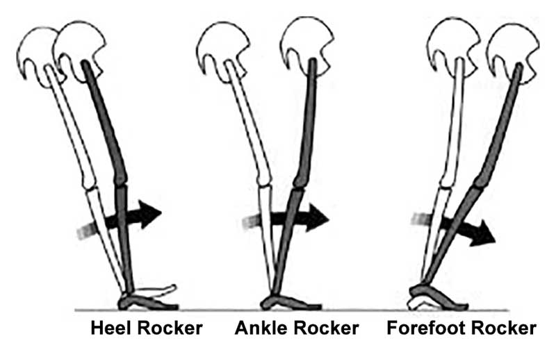
1.    This patient has a leg length discrepancy right sided which is affecting his walking mechanics. He supinates this extremity as can be seen on video, especially at terminal stance/pre swing (ie toe off),  in an attempt to lengthen it; as a result, he has peroneal tendonitis on the right (peroneus is a plantar flexor supinator and dorsiflexor/supinator; see post here). The left medial gastroc is tender most likely due to trying to attenuate the midfoot pronation on the left (as it fires in an attempt to invert the calcaneus and create more supination). see here for gastroc info

2.    Left shoulder:  Frozen shoulder/injury may be playing into this as well as it is altering arm swing.

WHAT WE DID INITIALLY (key in mind, there is ALWAYS MORE we can do):    

  •  build intrinsic strength in his foot in attempt to work on getting the first ray down to the ground; EHB, the lift/spread/reach exercises to perform.
  • address the leg length discrepancy with a 3 mm sole lift
  • address pathomechanics with mobilization and manipulation. 
  • improve proprioception: one leg balancing work
  • needled the peroneus longus brevis as well as medial gastroc and soles. 
  • follow up in 1 week to 10 days.

Pretty straight forward, eh? Look for this pattern in your clients and patients

Ankle sprains and the reorganization of the sensorimotor system

“Our subjects with unilateral chronic ankle sprains had weaker hip abduction strength and less plantar-flexion range of motion on the involved sides. Clinicians should consider exercises to increase hip abduction strength when developing rehabilitation programs for patients with ankle sprains.”-Friel et al

Awhile back we wrote about the principle that if the hip abductors are weak, the leg will posture more adducted (ie, cross over type pattern) and this places the foot more directly below the body midline plumb, this will posture the foot in inversion and thus at greater risk for future inversion sprains.  This sets up the vicious cycle of hip abductor weakness, frontal plane drift of pelvis, inversion of the foot and more ankle sprain risks/events.  The cycle must be broken. The hip must be addressed. That lateral chain must be restored all the way up from the foot.  

Another newer study by Bowker discusses the somatosensory feedback necessary for postural adjustments, walking, and running stating that they may be hampered by a decrease in soleus spinal reflex excitability.  The study adds more validity to what we are all growing to know more clearly, that the central nervous system via supraspinal circuitry plays deeply into chronic ankle instability (CAI). The studies suggest that CAI may be more about coordination and control of dynamic stabilizers and changes in the motor neuron excitability rather than the function of static stabilizers.

“A successful reorganization of the sensorimotor system after an initial ankle sprain is the critical point when individuals suffer chronic ankle instability or become copers [individuals who do not develop chronic instability after an ankle sprain] who break the cycle of recurrent injuries and disabilities seen in CAI,” Masafumi Terada, PhD

According to LER and the Terada work, 

The slow-twitch fibers in the soleus muscle are mostly innervated by small alpha motoneurons, Terada explained, so the study findings suggest that some people may restore their ability to reflexively recruit alpha motoneurons after ankle injury, and some may not.

“Therapeutic interventions that can increase the H-reflex in the soleus may help to break the cycle of recurrent injuries and disabilities seen in CAI,” he said. “Lower-intensity transcutaneous electrical stimulation, joint manipulations, and reflex conditioning protocols may be effective in increasing the soleus spinal excitability.”

The Gait Guys


Reference:

CAI and the CNS: Excitability may influence instability. Larry Hand

http://lermagazine.com/news/in-the-moment-sports-medicine/cai-and-the-cns-excitability-may-influence-instability

Taken from original source:

Bowker S, Terada, M, Thomas AC, et al. Neural excitability and joint laxity in chronic ankle instability, coper, and control groups. J Athl Train 2016 Apr 11. [Epub ahead of print]

J Athl Train. 2006; 41(1): 74–78.PMCID: PMC1421486Ipsilateral Hip Abductor Weakness After Inversion Ankle SprainKaren Friel,Nancy McLean,Christine Myers, and Maria Caceres
http://www.ncbi.nlm.nih.gov/pmc/articles/PMC1421486/

Can you guess why this person has left-sided plantar fasciitis?This question probably seem somewhat rhetorical. Take a good look at these pedographs which provide us some excellent clues.First of all,  note how much pressure there is over the metata…

Can you guess why this person has left-sided plantar fasciitis?

This question probably seem somewhat rhetorical. Take a good look at these pedographs which provide us some excellent clues.

First of all,  note how much pressure there is over the metatarsal heads. This is usually a clue that people are lacking ankle rocker and pressuring these heads as the leg cantilevers forward.  This person definitely have a difficult time getting the first metatarsal head down to the ground.

Notice the overall size of the left foot compared to the right (right one is splayed or longer). This is due to keeping the foot and somewhat of a supinated posture to prevent excessive tension on the plantar fascia.

The increase splay of the right foot indicates more mid foot pronation and if you look carefully there is slightly more printing at the medial longitudinal arch. This is contributing to the clawing of the second third and fourth toes on the right. Stand up, overpronate your right foot and notice how your center of gravity (and me) move medially.The toes will often clench in an attempt to create stability.

The patient’s pain is mostly at the medial and lateral calcaneal facets, and within the substance of the quadratus plantae with weakness of that muscle and the extensor digitorum longus. She has 5° ankle dorsiflexion left and 10 degrees on the right and hip extension which is similar.

The lack of ankle rocker and hip extension or causing her to pronate through her midfoot, Tensioning are plantar fascia at the insertion. The problem is worse on the left and therefore that is where the symptoms are.

Pedographs can be useful tool in the diagnostic process and provide clues as to biomechanical faults in the gait cycle.

tumblr_o84av4iR3i1qhko2so2_250.jpg
tumblr_o84av4iR3i1qhko2so1_1280.png

Shoe Drop and its effects on the shoe itself

An interesting, free, full text article we ran across entitled: “THE EFFECT OF WALKING IN FOOTWEAR WITH VARYING HEELSOLE DIFFERENTIALS ON SHANK & FOOT SEGMENT KINEMATICS”

Not exactly a page turner but some important pearls to glean here.

CONCLUSION: During 0-50% gait cycle stance phase shank kinematics do not change with changes in Heel Sole Differential. Actual foot angles do change, increasing with increasing Heel Sole Differential of footwear and by the angle of pitch of the footwear.

In other words, the kinematics (read: flex and physical characteristics) of the shank (the platform that the shoe is built on) do not change with increased ramp delta (ie: “drop”, from heel to toe), but foot kinematics (ie: how the foot moves) DOES change.

http://www.oandp.org/publications/jop/2016/2016-020.pdf

tumblr_o864jbYF2R1qhko2so1_1280.jpg
tumblr_o864jbYF2R1qhko2so2_1280.jpg
tumblr_o864jbYF2R1qhko2so3_1280.jpg
tumblr_o864jbYF2R1qhko2so4_1280.jpg

Short leg and mottling of the skin

Have you ever heard of Klippel-Trenaunay Syndrome? I hadn’t either, until I had a patient come in with low back pain and a gait issue and said she had it.

Evidently, in 1900, noted French physicians Klippel and Trenaunay first described a syndrome in 2 patients presenting with a port-wine stain and varicosities of an extremity associated with hypertrophy of the affected limb’s bony and soft tissue. Klippel-Trenaunay-Weber syndrome (KTWS) is characterized by a triad of port-wine stain, varicose veins, and bony and soft tissue hypertrophy involving an extremity (1).

Most cases KTWS are sporadic, although a few cases in the literature report an autosomal dominant pattern of inheritance (2). There is no racial predilection, even distribution between males and females and presents at birth or during early childhood (3). It generally affects a single extremity, although cases of multiple affected limbs have been reported. The leg is the most common site followed by the arms, the trunk, and rarely the head and the neck(4).

This patient had a history of low back pain with a recent epidural steroid injection. Exam highlights included a R sided leg length discrepancy approximately 5mm (tibial and femoral). Pelvic tilt to the right (for LLD) with anterior rotation of that side of the pelvis, posterior on the opposite side (counter clockwise pelvic distortion pattern). Lumbar flexion off 60/90 with all motion occurring in the lumbar spine (ie: no hip hinge), extension 20/30, lateral bending 30/45 BL with pain ipsilateral. Decreased low back endurance of <50 seconds in extension.

Right lower extremity was smaller (appeared hypoplastic) than left and had multiple discolorations in the skin (see pictures). L sided Q angle > R (12 vs 8 degrees). Less internal rotation of the right lower extremity compared to left, but with normal limits. Gait revealed a shift and hike to the right during stance phase with an increased arm swing on the right. Foot intrinsics were weak (lumbricals, EDL, FDB, dorsal intrerossei)

She walked in a pair of Chaco sandals with allowed much greater calcaneal eversion bilaterally R > L.

MRI revealed paraspinal marbling at the lower part of the lumbar spine, improving as you move rostrally. Small disc herniations at L3/4, 4/5, 5/S1, which did not effect the exiting nerve roots. Degenerative changes in the lumbar facet joints. There was no radiographic evidence of instability.

Impression:
It seems that she did not have enough intrinsic for the strength to stop calcaneal eversion in her Chaco’s and therefore this was causing increased foot pronation. This, combined with her leg length discrepancy, was contributing to increasing the lordosis in her lumbar spine, causing facet joint irritation. This was compounded by weakness and lack of endurance of the lumbar paraspinal musculature. The effects of the Klippel-Trenaunay Syndrome are evident with the IPO plasticity of the right lower extremity and accompanying musculoskeletal abnormalities.

What did we do?

  • Gave her endurance exercises for the lumbar spine.
  • Gave her propriosensorv exercises for the lumbar spine
  • Recommended she continue with the 5 mm sole lift.
  • Advised getting rid of the Chaco sandals as they allow too much calcaneal eversion and sticking to a shoe that has a stronger/larger heel counter.
  • acupuncture to improve circulation and proprioception as well as muscular function
  • we will monitor weekly for the next 4 to 6 weeks.

All in all, and interesting use with a little twist (not a torsion, of course!) : )


1. http://reference.medscape.com/article/1084257-overview
2. Ceballos-Quintal JM, Pinto-Escalante D, Castillo-Zapata I. A new case of Klippel-Trenaunay-Weber (KTW) syndrome: evidence of autosomal dominant inheritance. Am J Med Genet. 1996 Jun 14. 63(3):426-7.
3. Sung HM, Chung HY, Lee SJ, Lee JM, Huh S, Lee JW, et al. Clinical Experience of the Klippel-Trenaunay Syndrome. Arch Plast Surg. 2015 Sep. 42 (5):552-8.
4. http://reference.medscape.com/article/1084257-clinical

tumblr_ni6lv4HU1v1qhko2so2_r1_250.png
tumblr_ni6lv4HU1v1qhko2so1_500.jpg

Riding the inside edge of the sandal. Mystery hunting with Dr. Allen.

You can see it in the photo above, the heel is a third of the way off the sandal. (there are 2 photos provided today, find the arrow and tab to see both)

You either have it or have seen it. It is frustrating as hell if you have it. Your heel rides on only half of your flip flop or summer sandals. You do not notice it in shoes, only in sandals, typically ones without a back or back strap.  This is because the heel has no controlling factors to keep it confined on the rear of the  sandal sole. But there is a reason this happens to some, but not everyone. It is best you read on, this isn’t as simple as it might seem. 

These clients have restricted ankle rocker (dorsiflexion), restricted hip extension and/or adductor twist (if your reference is the direction the heel is moving towards). I could even make a biomechanical case that a hallux limitus could result in the same scenario. So what happens is that as the heel lifts and adducts it does not rise directly vertically off the sandal, it spins off medially from the “adductor twist” event. This event is largely from a torque effect on the limb from the impaired sagittal mechanics as described above, manifesting  at the moment of premature heel rise resulting in an slightly externally rotating limb (adducting heel). The sandal eventually departs the ground after the heel has risen, but the sandal will rise posturing slightly more laterally ( you can clearly see this on the swing leg foot in the air, the sandal remains laterally postured). Thus, on the very next step, the sandal is not entirely reoriented with its rear foot under the heel, and the event repeats itself. The sandal is slightly more lateral at the rear foot, but to the wearer, we believe it is our heel that is more medial because that is the way it appears on the rear of the sandal or flip flop.  Optical illusion, kind of… . . a resultant biomechanical illusion is more like it.

You will also see this one all over the map during the winter months in teenagers who swear by their Uggs and other similar footwear, as you can see in the 2nd photo above. This is not an Ugg or flip flop problem though, this is often a biomechanical foot challenge that is not met by a supportive heel counter and may be a product of excessive rear foot eversion as well.  This does not translate to a “stable” enough shoe or boot, that is not what this is about. This is about a rearfoot that moves to its biomechanical happy place as a result of poor or unclean limb and foot biomechanics and because the foot wear does not have a firm stable and controlling heel counter.  The heel counter has several functions, it grabs the heel during heel rise so that the shoe goes with the foot, it give the everting rearfoot/heel something to press against, and as we have suggested today, it helps to keep the rearfoot centered over the shoe platform.  To be clear however, the necessary overuse and gripping of the long toe flexors to keep flip flops and backless sandals on our feet during the late stance and swing phases of gait, clearly magnifies these biomechanical aberrations that bring on the “half heel on, half heel off” syndrome.

There you have it. Another solution to a mystery in life that plagues millions of folks. 

Dr. Shawn Allen, mystery hunter, and one of the gait guys.

Treadmills, motorized or nonmotorized can have some pitfalls. Here are seven of our biggest concerns.

More on non motorized treads from Mike Reinold which came to my attention via Scott Tesoro (thanks!).

1. Watch out for how much ankle dorsiflexion(and great toe extension) your client has to be able to take advantage of the “curve”

2. The treadmill, whether motorized or not, is constantly moving, opposite the direction of travel. With the foot on the ground, this provides a constant rate of change of length of the gastroc/soleus (ie, it is putting it through a slow stretch); so, once the muscle is activated, it contracts for a longer period of time because of the treadmill putting a slow stretch on the gastroc and soleus.

3. The moving deck also has a tendency to put the ankle in dorsiflexion ( see point number one) initiating a stretch reflex in the tricep surae (gastroc/soleus) facilitating toe off through here and pushing you through the gait cycle, rather than pulling you through (with your hip extensors).

4. Likewise, the treadmill pulls the hip into extension and places a pull on the anterior hip musculature, especially the hip flexors including the rectus femoris, iliopsoas and iliacus. This causes a slow stretch of the muscle, activating the muscle spindles (Ia afferents) and causing a mm contraction (ie the stretch reflex). This acts to inhibit the posterior compartment of hip  extensors through reciprocal inhibition, especially the glute max, making it difficult to fire them.

5. Because the deck is moving, the knee is brought into extension, with stretch of the hamstrings, the quads become reciprocally inhibited.

6.  the moving deck forces you to flex the thigh forward for the next footstrike (ie footstance), firing the RF, IP and Iliacus, and reciprocally inhibit the g max

7. If your core isn’t engaged, the pull of the rectus femoris and iliopsoas/iliacus pulls the ilia and pelvis into extension (ie increases the lordosis) and you reciprocally inhibit the erectors and increase reliance on the multifidus and rotatores, which have short lever arms and are supposed to be more proprioceptive in function.

We are not saying they are bad and in fact, we tend to like self-propelled models more than motorized ones  and agree with many of the points made. We are just saying that treadmills are not the same as walking on a flat surface and approximate but do not simulate actual gait.

Podcast 107: Unilateral Training: Warping the Nervous System

Plus: Changing an existing orthotic to make it work, Meniscal tear truths, Shoe Insole truths, Plantar Pressures

Show Sponsors:

softscience.com
Altrarunning.com

Other Gait Guys stuff

A. Podcast links:

http://traffic.libsyn.com/thegaitguys/pod_107f.mp3

http://thegaitguys.libsyn.com/episode-107-0

B. iTunes link:
https://itunes.apple.com/us/podcast/the-gait-guys-podcast/id559864138

C. Gait Guys online /download store (National Shoe Fit Certification & more !)
http://store.payloadz.com/results/results.aspx?m=80204

D. other web based Gait Guys lectures:
Monthly lectures at : www.onlinece.com type in Dr. Waerlop or Dr. Allen, ”Biomechanics”

-Our Book: Pedographs and Gait Analysis and Clinical Case Studies
Electronic copies available here:

-Amazon/Kindle:
http://www.amazon.com/Pedographs-Gait-Analysis-Clinical-Studies-ebook/dp/B00AC18M3E

-Barnes and Noble / Nook Reader:
http://www.barnesandnoble.com/w/pedographs-and-gait-analysis-ivo-waerlop-and-shawn-allen/1112754833?ean=9781466953895

https://itunes.apple.com/us/book/pedographs-and-gait-analysis/id554516085?mt=11

-Hardcopy available from our publisher:
http://bookstore.trafford.com/Products/SKU-000155825/Pedographs-and-Gait-Analysis.aspx

________________________

Show Notes:

Running helps mice slow cancer growth
https://www.sciencedaily.com/releases/2016/02/160216142825.htm

The future of Wearables
http://readwrite.com/2016/02/19/future-of-wearables

mensicus surgery is dead ?
http://www.regenexx.com/should-i-have-meniscus-surgery/#

Why you should be training your CNS
http://www.outsideonline.com/2055066/cross-educate-your-body#article-2055066

The business of insoles
http://www.outsideonline.com/2057156/business-insoles-support-system-or-super-rip

Altered plantar pressures
http://link.springer.com/article/10.1007%2Fs00167-016-4015-3

The diaphragm and chronic ankle instability.

I have been treating the global manifestations of unaddressed chronic ankle sprains for decades now. I am never unsurprised to find frontal plane hip weakness and dysfunction of the same side obliques , shoulder and spinal stabilizers. Here is one more piece of proof that unaddressed ankles are monster problems, slowly eroding the stability of the system.
But, shame on those who attempt to simplify this, just correcting the breathing and throwing some corrective spinal stability work at this problem. This approach will fail, repeatedly. At some point the ankle has to be addressed and the impaired supra spinal programming. Gait will have to be retrained as well, forget to do this and your efforts will be muted.
-Dr. Allen

“Previous investigations have identified impaired trunk and postural stability in individuals with chronic ankle instability (CAI). The diaphragm muscle contributes to trunk and postural stability by modulating the intra-abdominal pressure. A potential mechanism that could help to explain trunk and postural stability deficits may be related to altered diaphragm function due to supraspinal sensorimotor changes with CAI.”

Reference:

Diaphragm Contractility in Individuals with Chronic Ankle Instability.

Terada, Masafumi; Kosik, Kyle B.; McCann, Ryan S.; Gribble, Phillip A.  Medicine & Science in Sports & Exercise:

http://journals.lww.com/acsm-msse/Abstract/publishahead/Diaphragm_Contractility_in_Individuals_with.97497.aspx

tumblr_o80wf8tU9u1qhko2so1_400.jpg
tumblr_o80wf8tU9u1qhko2so2_1280.jpg

Do you know where your rocker is?

At 1st pass, some articles may seem like a sleeper, but there can be some great clinical pearls to be had. I recently ran across one of these. It was a presentation from the  42nd annual American Academy of Orthotists and Prosthetists meeting in Orlando, March 2016 entitled “ Shifting Position of Shoe Heel Rocker Affects Ankle Mechanics During Gait”. The title caught my eye.

They looked at ankle kinematics while keeping the toe portion of rocker constant at 63% of foot length, angled at 25 degrees and shifting the base of a rockered shoe from 1cm behind the medial malleolus, directly under it and 1cm anterior to it. Knee and hip kinematics did not differ significantly, however ankle range of motion did.

The more forward the ankle rocker, the less plantarflexion but more ankle dorsiflexion at midstance. So, the question begs, why do we care? Lets explore that further…

  • Think about the “average” heel rocker in a shoe. It largely has to do with the length of the heel and heel flare (base) of the shoe. The further back this is (ie; the more “flare”) the more plantar flexion at heel strike and less ankle dorsiflexion (and thus ankle rocker, as described HERE) you will see. Since loss of ankle dorsiflexion (ie: rocker) usually means a loss of hip extension (since these 2 things should be relatively equal during gait (see here), and that combination can be responsible for a whole host of problems that we talk about here on the blog all the time. Picking a shoe with a heel rocker based further forward (having less of a flare) would stand to promote more ankle dorsiflexion.
  • Having a shoe with a greater amount of “drop” from heel to toe (ie: ramp delta) is going to have the same effect. It will move the calcaneus forward with respect to the heel of the shoe and effectively move the rocker posteriorly.
  • Lastly, look a the shape of the outsole of the shoe. The toe drop is usually clear to see, but does it have a heel rocker (see the picture above)?

These are  a few examples of what to look for in a clients shoe when examining theirs or making a recommendation, depending on whether you are trying to improve or decrease ankle rocker. We can’t think of why you would want to decrease ankle rocker, but with conditions like rigid hallux limitus, where the person has limited or no dorsiflexion of the great toe, you may want to employ a rockered sole shoe. We would recommend one with the rocker set more forward.

Walking in sync makes enemies seem less scary

Walking in sync makes enemies seem less scary.
“When men match each others’ steps, purported criminals seem less physically formidable, a new study shows. The results, published August 27 in Biology Letters, suggest that matched movements in men may foster fighting alliances, a behavior seen in apes and some dolphins and whales.”
What this does not discuss, but that which we do all over our blog, is whether they are talking about in-phase or out-of-phase synchrony. This is an important distinction, though not likely when it comes to the topic at hand here. We discussed this and many other things on podcast 74.

http://thegaitguys.tumblr.com/post/96787952409/podcast-74-cross-fit-more-on-squatting-and-hip

https://www.sciencenews.org/blog/science-ticker/walking-sync-makes-enemies-seem-less-scary

tumblr_o7p469lND91qhko2so1_1280.jpg
tumblr_o7p469lND91qhko2so2_1280.jpg
tumblr_o7p469lND91qhko2so3_1280.jpg
tumblr_o7p469lND91qhko2so4_1280.jpg
tumblr_o7p469lND91qhko2so6_1280.jpg
tumblr_o7p469lND91qhko2so8_1280.jpg
tumblr_o7p469lND91qhko2so9_1280.jpg
tumblr_o7p469lND91qhko2so5_r1_1280.png

Pain on the outside of the leg? Could it be your orthotic? What you wear on your feet amplifies the effect of the orthotic.

This woman presented with right-sided pain on the outside of her leg after hiking approximately an hour. She noticed a prominence of the arch in her right orthotic. She hikes in a rigid Asolo boot ( see below). Remember that footwear amplifies the effect of an orthotic!

In the pictures below you can see the prominent arch. The orthotic has her “over corrected” so that she toes off in varus on that side. The rigid footwear makes the problem worse. The peroneus group is working hard (Especially the peroneus longus)  to try and get the first Ray down to the ground.

The “fix” was to soften the arch of the orthotic and grind some material out. Look at the pictures where the pen is pointing to see how some of the midsole material was taken out. Notice how I ground it somewhat medial to further soften the arch.

She felt better much better after this change and is now a “happy hiker” :-)

Looking for the subtle clues will help you. You should have hypotheses and work to prove or disprove them. “Remember, this client is displaying these weight bearing differences side to side for a reason, this is their adaptive strategy. It is your j…

Looking for the subtle clues will help you. You should have hypotheses and work to prove or disprove them. 

“Remember, this client is displaying these weight bearing differences side to side for a reason, this is their adaptive strategy. It is your job to prove that this is the cause of their pain, their adaptive strategy to get out of pain, or this is now a failed adaptive strategy causing pain, yet still not the root of the problem.”

We used to call this a “windswept” presentation. It is not that it is incorrect, but it is so vague.  

Look at these fippy floppers. Look closely at the dark areas, where foot oils and whatnot have played their changes in the leather upper of the flops. The right f.flop displays more lateral heel loading, rear foot inversion if you will. You can even see that there is less big toe pressure on this right side and even some increased lateral forefoot loading. This client appears to be more supinated clearly. You can even see there is more lightness to the arch leather on the right, again, more supination is suggested.

The left f.flop suggests the opposite. More medial heel pressures and more over the medial forefoot and arch. 

Now this clients f.flops tell a story.  So, this client is being windswept to the right we used to say, appearing to pronate more on the left and supinating more on the right.  Why are they doing this? Is the left leg functionally longer and by pronating they reduce the functional length of the leg (yet, increase internal spin of the limb and the host of naughty things that come with that). Is the right leg shorter, and by supinating they are raising the ankle mortise and arch which helps reduce the length differential ?  MAybe a bit of both, finding common ground for a more symmetrical pelvis ?  Who knows. This is where you need your physical exam, but, now you have some hypotheses to prove or disprove. 

“Remember, this client is displaying these weight bearing differences side to side for a reason, this is their adaptive strategy. It is your job to prove that this is the cause of their pain, their adaptive strategy to get out of pain, or this is now a failed adaptive strategy causing pain, yet still not the root of the problem.”

Is there some right hip pain from the right frontal pelvis drift creating some aberrant loading on the greater trochanter from ITB tension ? Perhaps a painful right hallux big toe, and they are unloading it to avoid pain? Maybe some knee pain or low back pain ? Who knows? Take your history and start putting the pieces together, it is your job. Just don’t screen them and throw corrective exercises at them, you owe it to them to examine them, take their history, watch them walk, teach them about what you see, and then sit down, spread the puzzle pieces out, look for the straight edges and corner pieces, and begin to build their puzzle. 

Clues, they are everywhere, if you look for them.

Dr. Shawn Allen, one of the gait guys

Landing strategies focusing on the control of tibial rotation in the initial contact period of one-leg forward hops - Chen - 2016 - Scandinavian Journal of Medicine & Science in Sports - Wiley Online Library

“If the knee is whining and doing things it should not be doing, the wise clinician first looks at the foot-ankle and the hip-pelvis complexes, where the blood has dried. Don’t look for the fresh blood at the knee” - Dr. Allen

If you cannot control pelvis position on the femoral head, or hip rotation or initial foot arch mechanics, the knee is going to give in to the directional loading response and that typically means medial valgus movement. This is internal tibial rotation or spin.  

Here is an analogy i use with all my patients. The knee is like the middle child. In the simplest terms, you have 3 lower limb joint complexes. The foot/ankle, the knee and the hip. The knee is the middle of these 3 joint complexes.  

Similarly if you put 3 children in the back of the car, the one sitting in the middle is the one directly impacted by the child on the right and the left.  When you hear the middle child screaming and whining, the smart parent first looks at the two apparently “innocent” children looking out the windows (with blood dripping off their elbows). 

Similarly, the knee takes this same seat. IF the knee is whining and doing things it should not be doing, the wise clinician first looks at the foot-ankle and the hip-pelvis complexes, where the blood has dried. Don’t look for the fresh blood at the knee

Changing landing strategies with the focus of control of tibial rotation, requires the astute clinician to look at all the children.

Dr. Shawn Allen, one of the gait guys.

Does hill running equate to biking when it comes to pathomechanics ?Think about it, when  you are hill running, one leg is in extension while the lead leg is in more extremes (compared to road running) of hip flexion reaching up the hill for the nex…

Does hill running equate to biking when it comes to pathomechanics ?Think about it, when  you are hill running, one leg is in extension while the lead leg is in more extremes (compared to road running) of hip flexion reaching up the hill for the next step. Isn’t this similar to biking ? On the bike one is bent over leaning forward, the lead leg is in extremes of flexion while the foot on the bottom crank has that same hip in extension.  So does hill running equate to biking ? Well, no. But then it comes to approximating anterior hip structures, there are some similarities. You cannot deny that there seems to be some similarities to pathomechanics.This was a post from a few weeks ago, but this week in our online teleseminar class we went over these principles.  We talked about some of the same biomechanical principles and vulnerabilities in hill running and when in biking.Might be a good time to revisit this brief blog post and see why we had hill running and biking in the same conversation.

Dr. Shawn Allen

Here is the hill running blog post where we mentioned a few things.
http://thegaitguys.tumblr.com/post/143841190479/when-you-run-up-a-hill-most-of-the-cross-over

Awkward photos&hellip;Date smarter but necessarily walk that way&hellip;Take a look at this photo from an airplane magazine I was leafing through on my way home from teaching a recent needling seminar.I am not really interested in matchmaking (which…

Awkward photos…Date smarter but necessarily walk that way…

Take a look at this photo from an airplane magazine I was leafing through on my way home from teaching a recent needling seminar.

I am not really interested in matchmaking (which this ad is for), but the “awkwardness” of the gait caught my eye (not much on the gait cam this time, sorry).

Yes, their feet and legs seem to be in synch (for the most part), but take a look at the arms and hands. First of all, he has his right hand in his pocket, which will restrict its motion during forward movement of his left leg. In the shot, his right leg is forward (as is hers), though they are slightly out of synch. His left arm and hand don’t move forward that much in that he adducts it across his body, so he seems to move his left shoulder up and forward to compensate. She appears to be resisting this motion somewhat with her right arm as her right leg comes forward and she needs to lean her body to the right. Also note the increased abduction of her left arm and forearm as it extends in tandem with her left leg and thigh.

Try walking with your right arm moving forward with your right leg. Notice how your right shoulder resists moving forward in tandem with the right hip? This is phasic, as Dr Allen likes to say, and because there is not an opposite force to counteract the forward movement of the hip in the saggital plane, you often lean to move the center of gravity to that side in the coronal plane.

Wouldn’t it make slightly more sense, when walking hand in hand to have the opposite legs in synch, rather the same ones? Hmm…Food for thought and fodder. All that from a  little picture : )

tumblr_o7941xdZHo1qhko2so7_1280.jpg
tumblr_o7941xdZHo1qhko2so2_1280.jpg
tumblr_o7941xdZHo1qhko2so1_1280.jpg
tumblr_o7941xdZHo1qhko2so6_1280.jpg
tumblr_o7941xdZHo1qhko2so5_1280.jpg
tumblr_o7941xdZHo1qhko2so4_1280.jpg

1st met pain in an orthotic?

This patient came in with pain at the base of the first metatarsal that she believed was related to her orthotic. The first picture shows the foots relationship to the orthotic. Notice how the sesamoid bones and distal aspect of the first metatarsal under lap the orthotic shell. In other words, the shell is longer than her foot. When she dorsiflexes her big toe, she’s hitting the distal of the orthotic.

The next view shows the orthotic with a typical first ray cutout. Notice how far forward the shell of the orthotic goes (next picture). I have placed a pen pointing to the area where the orthotic shell is too long.

In addition to reviewing her first ray descending exercises, a simple fix was to grind back the orthotic shell and be careful to bevel the edge so that it was not hitting the sesamoids and it did not impinge upon the descending first ray. I have placed a pen where the cut out now is (pre and post gluing in the pictures). The cork underlying the base of the first ray was also ground away (last picture)

A simple fix for a common problem. Make sure that your orthotic shell lengths fall just short of the 1st ray and not impinge on the sesamoids!