From our post earlier today:
Part 2:
Considered another way, from the top down this time, if at the moment of heel contact the gmedius is delayed (as suggested in the study below from achilles pain), the pelvis is likely to drift laterally and this could cause a reactive inversion strategy of the rearfoot, and maybe even forefoot as well, as an instinctive measure to try and draw support beneath the laterally drifting body mass center of gravity. (This in essence sets up the “cross over gait” deployment strategy we have talked about here for years now).This too could cause a change in load to the achilles mechanism, resulting in tendonopathy thus putting one into a vicious cycle of achilles causing glute and then glute perpetuating altered strike and thus abnormal achilles loads . This is also a major cause of ankle inversion sprains, so be extra aware of this pattern. We see this all the time in practice, we hope you do as well. IF you haven’t already, If you wish to dive deeper, goto our blogwww.thegaitguys.tumblr.com and type in Cross Over Gait and it will get you going.
* Remember this, if you are spending time moving sideways, you are taking from time moving forwards, in the allotted amount of time given for the stance phase of gait. Yet, you will still move forwards, so one way around this is a premature heel rise (ie. speed up certain mechanical events) via premature plantarflexion mechanism loading (calf-achilles complex). Remember to also look for all the other reasons for premature heel rise (ie. loss of ankle rocker etc).
Franettovich Smith MM1, Honeywill C, Wyndow N, Crossley KM, Creaby MW. : Neuromotor control of gluteal muscles in runners with achilles tendinopathy.
Med Sci Sports Exerc. 2014 Mar;46(3):594-9.
______________
Part 1
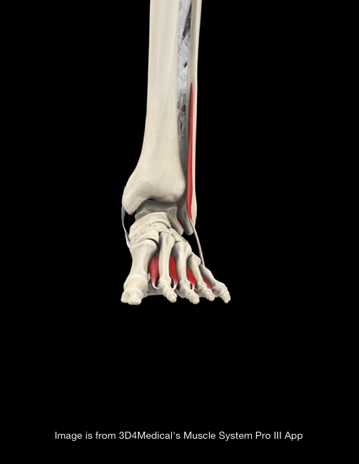
The mighty Gluteus Medius, in all its glory!
Perhaps the delayed action of the gluteus medius allows an adductory moment of the pelvis, moving the center of gravity medially. This could conceivably place additional stress on the achilles tendon (via the lateral gastroc) to create more eversion of the foot from midstance onward.
“The results of the study demonstrate altered neuromuscular control of the GMED and GMED in runners with Achilles Tendonitis. During running, GMED typically activates before heel strike so as to stabilize the hip and the pelvis. In runners with Achilles Tendonitis, GMED is activated with a delay, which consequently might affect the kinematics of knee and ankle resulting in rear foot inversion. Similarly, GMAX is activated with a delay and for a shorter duration in runners with Achilles Tendonitis. GMAX is the primary hip extensor and via a kinetic chain, a decreased hip extension moment might be compensated by an increased ankle plantarflexion moment which could potentially increase the load on the Achilles tendon.”
Franettovich Smith MM1, Honeywill C, Wyndow N, Crossley KM, Creaby MW. : Neuromotor control of gluteal muscles in runners with achilles tendinopathy.
Med Sci Sports Exerc. 2014 Mar;46(3):594-9.