


What do you see here? And how does it relate to reciprocal inhibition? Details to follow tomorrow!
Part 2 of a case study from Northern Ireland. This video discusses the dynamic findings and how they correlate clinically with the history. Treatment recommendations are discussed as well.
Follow up question from a doctor…..
Thanks for the post. Interesting case study. Are most hernias at this point a result of overactive hip flexors? What would be your exercise dosage/prescription of the exercises mentioned in part 2? The Gait Guys In our experience, most inguinal hernias are due to weakness of the lower abdominal wall, in this case, the external obliques, not being able to fire appropriately to guard against the load. Exercise would most likely progress along the lin…es of skill 1st (can he perform the exercise appropriately), endurance 2nd (increased reps to increase capillarization, myoglobin content, mitochondrial content; beginning with 8-12 reps and increasing to 5-10 sets daily) and strength last (low reps, high weight; dependent on progress)

When the Short Toe Extensors Try to Rule the World !
A case of a runner with forefoot pain.
This is a runner of ours, one of the fastest young men in the state of illinois, top 10 in the country in mid-distance, top 20 in the USA in cross country.
He came in with left forefoot plantar pain. He explained (in a matter of words) that he was having pain at full forefoot loading at heel rise /push off.
We watched him walk, saw this visual problem present itself in dynamic motion (yup, no stop frame video on this one, not when you see it about 10 times a month !) and noted a subtle left lateral hip/pelvis shift past what would be considered normal for frontal plane mechanics.
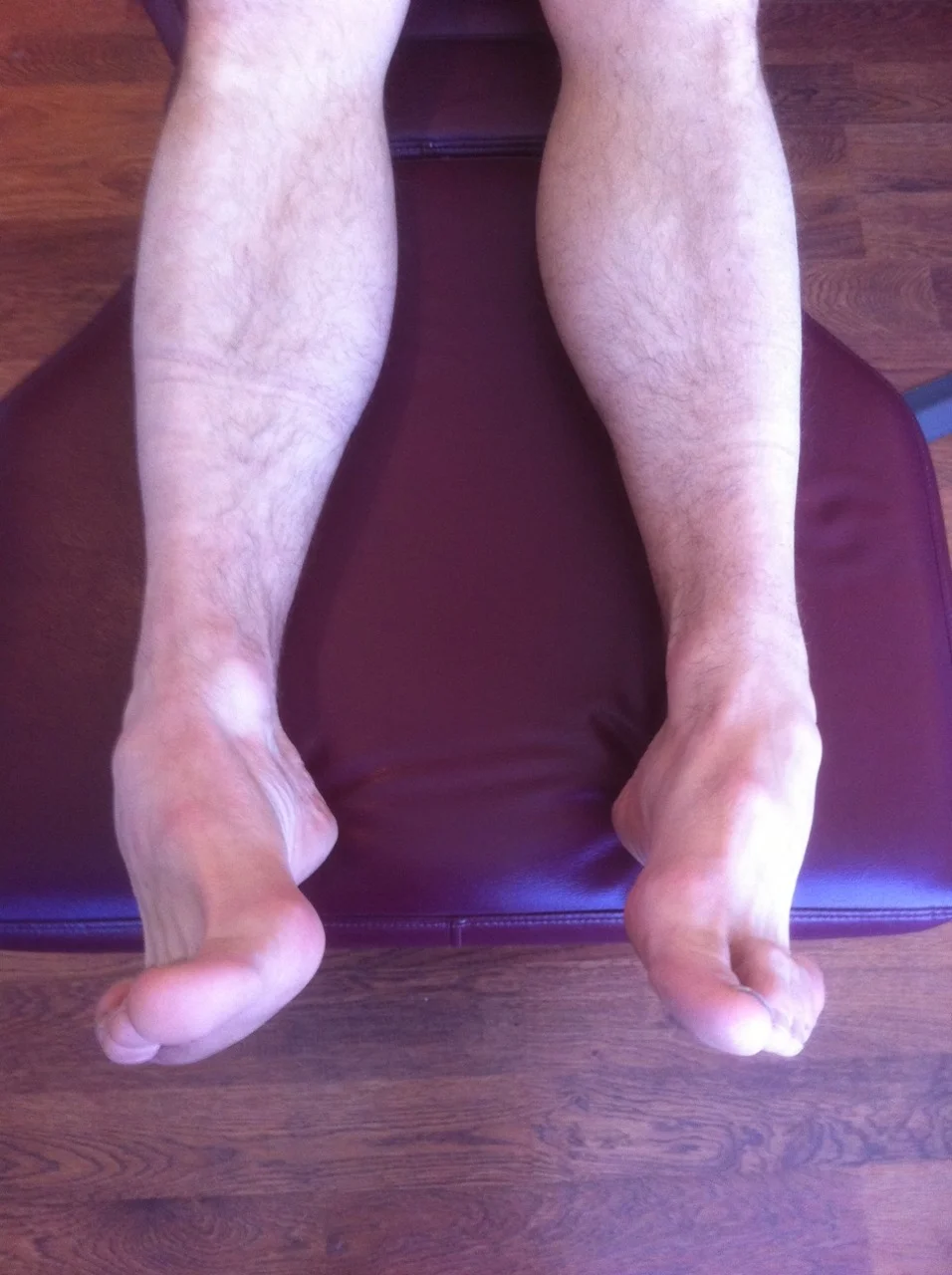
On the table this is a photo of his feet. What do you see ?
We see a suspected (which you will try to confirm on examination) increase in short extensor (EDB, extensor digitorum brevis) muscle tone. Increased long extensor (EDL, extensor dig. longus muscle) tone would have represented itself with the distal toes also extended but here we see a relative dominance of the long flexors (FDL, Flexor dig. longus) with the heightened short flexor increase.
We also see more confirmation of heightened long flexor tone (FDL) by the degree of heavy callus formation on the very tip of the 2nd toe (it was on all 4 lateral toes but the photo is not clear enough to demonstrate). You can also see supporting evidence of heightened long flexor dominance by the subungual hematoma (bleeding under the 2nd toe nail). (How does this correlate ? Well, in most runners with excessive long flexor tone/use not only do they flex and claw so much in the shoes that the callus is on the tip of the toes but the nail also begins to lift as the nail is caught on the sock liner of the shoe as the toe flexes, slowly, mile by mile pulling the toe nail from the nail bed thus bleeding underneath it). Yes, it is NOT from the toes hitting the front end of the shoe !
Our examination confirmed weakness of all lumbrical muscles and of the flexor digitorum brevis and lateral quadratus plantae. The patient could feel the strength/engagement difference as compared to testing on the right foot of the same muscle groups (we always compare side to side, for us and for the patient’s awareness). The extensor digitorum brevis muscle mass on the lateral dorsum of the foot was tender as were the tendons along their course. There was also weakness higher up in the kinetic chain at the lower division of the transversus abdominus and internal abdominal oblique, and frontal plane hip stabilizers (gluteus medius; anterior-middle-and posterior divisions).The 2nd and 3rd metatarsal heads were remarkably tender to palpation and it was obvious that the metatarsal fat pads had migrated distally from the lumbrical muscle weakness.
Sometimes a grasp response by the long flexors can represent a propioceptive /balance deficit during single leg stance phase so be sure to test those centers as well (cerebellar, vision, joint position sense, inner ear-vestibular apparatus).
So, what is the take away for the non-medical person, the runner next door if you will ? Lets just say, symmetry wins and when asymmetry is apparent, bring it up to the people that do your body work. Hopefully, what you and they see will be assessed in a clinical light, and as a team you can get to the bottom of what is not working…….and in this case…..what was causing not only the plantar foot pain, but the left lateral hip sway outside the frontal plane.
———we are, The Gait Guys……Shawn and Ivo


The Quadratus plantae (Flexor accessorius) muscle. Do you have foot pain ?
(*There are two pictures here on the blog. Move your cursor over to the side of the photo and you will see that you can toggle between the photo and anatomy pic).
This is a great, but highly overlooked, muscle. The QP acts to assist in flexing the 2nd to 5th toes. Equally important is its effect of offsetting the oblique pull of the long toe flexor group (flexor digitorum longus). It has two heads, medial and lateral. The medial head is attached to the calcaneus, while the lateral head originates from the lateral border of the calcaneus, in front of the lateral process of the calcaneal tuberosity and the long plantar ligament.
The fact that we just love, and one that we believe is often overlooked is the acute angle at which the muscle heads attach into the tendons of the flexor digitorum longus (see picture) and has a rather dramatic alignment effect on the lateral 3 digits (since the line of pull on the long flexor tendons to these 3 digits is most dramatically changed by the purely posterior pull of the Quadratus Plantae. As you can see in this stripped down anatomy picture, without the QP pulling on the tendons of the FDL to these 3 lateral toes, those toes will have to curl medially and gently flex (*see the photo, a classic presentation!) By having a competent and active QP that oblique line of pull of the FDL /long flexors is rearranged to be more of a pure posterior pull and you will not see this classic lateral 3 digit curl and medial drift. This action is accentuated in a cavus foot type, where the pull of the FDL will be accentuated, due to the mechanical advantage afforded it and relative adduction of the forefoot with respect to the rear foot.
In the photo you can see a classic representation of a deficient Quadratus Plantae, in this case the patients lateral head was dramatically weaker than the medial, but both were weak. So, summary time….if you know your anatomy, know your biomechanics, and if you can test the muscle bundles specifically……..then you can see why form follows function (and in this case, why form has followed dysfunction). As we always say, “ya gotta know your stuff”, and you have to test what you suspect……there are other things that could also do this……so, let your eyes gain info, let your brain process and prove or disprove the information.
we are…….the gait guys !
Audio Podcast: The Gait Guys, Barefoot Concepts
This is a blast from the past from our parent company, The Homunuculus Group ! Our podcast from 2008 ! Still solid info several years later. Just trying to get you all up to speed before we start up the podcasts here in a month or so.
Here we talk about the foot, intrinsic foot musculature, Nike Free, Vibram 5Fingers and some of Dr. Ivo’s always brilliant neuromechanical discussions.
Enjoy !
Research to support that we are on target !
CONCLUSIONS AND CLINICAL RELEVANCE:
The abductor hallucis muscle acts as a dynamic elevator of the arch. Understanding this mechanism may change the way we understand and treat pes planus, posterior tibial tendon dysfunction, hallux valgus, and Charcot neuroarthropathy. (see our video attached, it is much of what we talked about in this video just a few months ago).
*From the article: “Most studies of degenerative flatfoot have focused on the posterior tibial muscle, an extrinsic muscle of the foot. However, there is evidence that the intrinsic muscles, in particular the abductor hallucis (ABH), are active during late stance and toe-off phases of gait.
RESULTS:
All eight specimens showed an origin from the posteromedial calcaneus and an insertion at the tibial sesamoid. All specimens also demonstrated a fascial sling in the hindfoot, lifting the abductor hallucis muscle to give it an inverted ‘V’ shaped configuration. Simulated contraction of the abductor hallucis muscle caused flexion and supination of the first metatarsal, inversion of the calcaneus, and external rotation of the tibia, consistent with elevation of the arch.
http://www.ncbi.nlm.nih.gov/pubmed/17559771
Foot Ankle Int. 2007 May;28(5):617-20.
Influence of the abductor hallucis muscle on the medial arch of the foot: a kinematic and anatomical cadaver study.
Wong YS. Island Sports Medicine & Surgery, Island Orthopaedic Group, #02-16 Gleneagles Medical Centre, 6 Napier Road, Singapore, 258499, Singapore. yueshuen@yahoo.com
A question from a doctor on the topic of limb alignment development.
/The following question was forwarded to us from an internist on the USA east coast.
Question:
“I have a large number of female patients, many of them elderly. I have noted that women in our society tend to progress to valgus knee deformity with age, and that TKR (total knee replacement) doesn’t seem to correct that deformity. Men tend more to the varus in our society. I had formerly chalked that up to inherit gender difference.
3 or 4 years ago, I had occasion to spend a lot of time waiting outside the main Tokyo train station and observed a large number of people coming and going. I observed the following:
1. Young women had legs that were either straight or varus.
2. Young women tended toward toeing in.
3. They did all this in ridiculous high heels.
After some thought, I tend to attribute it to prolonged sitting in sesa (knees folded under), though being barefoot or in slippers while inside may also contribute. Women in our society sit with their legs crossed. Additionally, extensive shoe wearing leads to foot pronation.
So, could you direct me to someone who might have an interest in this observation and can refer me to any research that might have been done in this area? I’ve had the dickens of a time trying to find anything on it, or even a specialized area of study that cares about such things.”
The GAIT GUYS RESPONSE:
Thanks for your confidence in us. Here are some thoughts:
Frontal plane deformities (or development) is twofold: genetic (and X linked) and developmental. Children usually go through a varus to straight to valgus to straight development (Ron Valmassey talks about this in his text Clinical Biomechanics of the Lower Extremities). Women generally have larger Q angles (from birth) and this angulation often causes assymetrical epiphyseal development (increased pressure on the lateral malleolus/tibial plateau stunts growth) with overgrowth of the medial femoral condyle. Developmental changes are secondary to weight (obesity causes increase in valgus angle) and posture/muscular devlopment. The increased genu valgus places weight medial to the midline (2nd met) of the foot and the foot accomodates by pronating (often excessively, as noted by both of you). This causes medial rotation of the lower leg and thigh, resulting in lengthening of the glutes (esp G max) resulting in stretch weakness and subsequent over reliance on the gastroc/soleus group for propulsion (remember this group tries to invert the heel in an attempt to cause supination once you go past midstance. Weak intrinsics (as pointed out by Dr Mark) further fuels this cycle. “W” sitting (sometimes a cultural development, as pointed out by Dr Birgit) plays in as well.
As for “toeing in”; may women have the combination of genu valgus with internal tibial torsion (often with femoral retroversion) which makes the condition difficult to treat (the rearfoot needs to be supported, but the forefoot needs to be valgus posted) otherwise the knee is placed outside the saggital plane and the meniscus becomes macerated due to conflicting biomechanics at the knee (Thus the short term fix with orthotics with a return of the pain later).
Yes, high heels and open back shoes are evil as are open backed shoes (we spoke at a convention in Chicago a few years back on this, before some of the research was out).
Thanks for allowing us to participate. below are some references for you.
-The GAIT GUYS…….Ivo and Shawn
______________________________________________________________
J Orthop Sports Phys Ther. 2008 Mar;38(3):137-49.
Differences in lower extremity anatomical and postural characteristics in males and females between maturation groups.
Shultz SJ, Nguyen AD, Schmitz RJ.Source
Applied Neuromechanics Research Laboratory, Department of Exercise and Sport Science, University of North Carolina at Greensboro, 1408 Walker Ave., Greensboro, NC 27402, USA. sjshultz@uncg.edu
RESULTS:
When comparing maturation groups, limb length, pelvic angle, and tibial torsion increased with maturation, and anterior knee laxity, genu recurvatum, tibiofemoral angle, and foot pronation decreased with maturation. Females had greater general joint laxity, hip anteversion, and tibiofemoral angles, and shorter femur and tibial lengths than males, regardless of maturation group. Maturational changes in knee laxity and quadriceps angles were sex dependent.
CONCLUSIONS:
We observed a general change of posture with maturation that began with greater knee valgus, knee recurvatum, and foot pronation in MatGrp1, then moved toward a relative straightening and external rotation of the knee, and supination of the foot in later maturation groups. While the majority of the measures changed similarly in males and females across maturation groups, decreases in quadriceps angles and anterior knee laxity were greater in males compared to females, and females were observed to have a more inwardly rotated hip and valgus knee posture, compared to males, particularly in later maturation groups.
_______________________________________________________________________
J Bone Joint Surg Br. 1995 Sep;77(5):729-32.
Development of the clinical tibiofemoral angle in normal adolescents. A study of 427 normal subjects from 10 to 16 years of age.
Cahuzac JP, Vardon D, Sales de Gauzy J.
Source
Centre Hospitalier Universitaire de Toulouse-Purpan, France.
Abstract
We measured the clinical tibiofemoral (TF) angle and the intercondylar (IC) or intermalleolar (IM) distance in 427 normal European children (212 male and 215 female) aged from 10 to 16 years. In our study, girls had a constant valgus (5.5 degrees) and displayed an IM distance of < 8 cm or an IC distance of < 4 cm. By contrast, boys had a varus evolution (4.4 degrees) during the last two years of growth and displayed an IM distance of < 4 cm or an IC distance of < 5 cm. Values above these for genu varum or genu valgum may require careful follow-up and evaluation.
The Psoas Muscle in a Runner: An Endurance Savy Muscle ?
/We received a question yesterday from a doctor. We felt it was worthy of sharing. Here it is, followed by our response.
Doctor: I do have a question about one of my athletes in particular. He is a fairly good (All-State in IL) high school track distance runner that has some left sided femoral acetabular impingement. He gets some capsular hip pain that also will ‘tighten up’ his low back during speed endurance/threshold running only. Moderate and easy distance runs cause no problem and neither do track/speed workouts. Only during speed endurance does he have issues. Upon evaluation after these sessions he does seem to have some low back QL tightness, but joint mobility is fairly good in his lumbar spine. He does show marked hypertonicity through his left hip joint. I’m not quite sure the mechanism here- why he would only flare up with speed endurance running- any insights?
Thanks a bunch and I look forward to hearing from you!
The Gait Guys response:
You state “only during speed endurance” does he have issues. We will assume you mean a long, hard anaerobic workout, which would tax type II b fibers. You also mention he has hypertonicity through his hip joint. Since the psoas crosses this joint it should be considered in sprinting and long, hard endurance activities, especially if the patient is flexor dominant. The psoas major muscle is composed of type I, IIA and IIX muscle fibers. It has a predominance of type IIA muscle fibers. The fiber type composition of the psoas major muscle was different between levels of its origin starting from the first lumbar to the fourth lumbar vertebra. The psoas major muscle has dynamic and postural functions, which supports the fact that it is the main flexor of the hip joint (dynamic function) and stabilizer of the lumbar spine, sacroiliac and hip joints (postural function). The cranial part of the psoas major muscle has a primarily postural role, whereas the caudal part of the muscle has a dynamic role. This is all very much supported in this journal article here (link) (http://www.ncbi.nlm.nih.gov/pubmed/19930517) and making it work in an endurance capacity would certainly cause issues. Flexor dominance is a common scenario we see clinically, due to insufficient extensor activity (and decreased vestibulo and reticulo spinal drive to extensors) and increased cortico spinal drive (to the flexors, including the iliopsoas). This would fuel the “bail out” (lack of stability) of the lower abs. The anterior tippage of the pelvis would drive the femur posteriorly, binding the joint (the opposite of an anterior femoral glide).
Video footage and some pix of your athlete would provide more insight for us to help.
we are……The Gait Guys
Tissue vibration in prolonged running.→
/J Biomech. 2011 Jan 4;44(1):116-20. Epub 2010 Sep 16.Order this article from Elsevier Ltd !!!!
Tissue vibration in prolonged running.
Friesenbichler B, Stirling LM, Federolf P, Nigg BM.Source
Human Performance Laboratory, Faculty of Kinesiology, University of Calgary, 2500 University Dr NW, Calgary, Alberta, Canada. berndf@kin.ucalgary.ca
Gait Gaff Time.
(Gaff: verb tr. (to stand or take the gaff) To receive severe criticism; to endure hardship.
The Foot Slap Gait Style:
This is a funny little video that shows a few important points.
Remember, our purpose here is to help train your eyes to the important things. We used to use slow/stop frame digital gait software programs to slow down the person to look for particular components of failure in the gait or running cycle. After many years of doing this, we found more and more that even before we could fire up the video camera and software that we had trained our eye to see these deficits. This is because, there are multiple clues in every gait compensation. There is head movement (which we will discuss in this case), there is arm swing (is it equal and symmetrical, topics we have posted research articles on in the last 48 hours on this blog), torso rotation, hip lateral sway in the frontal plane, violations of sagittal knee progression, and then the always difficult multiplanar foot and ankle motions as well as so many other parameters we consider. So, when one component goes wrong, with enough experience and skill, one can make predictions as to what is wrong. And, the more flaws (correlative compensations) that are noted, the higher the predictive value of the assumption. Now, many will say to us that there is no way one can do this, and that is ok. To each his/her own. But, after decades of doing this, as with anything, a skill is developed and an art to doing it begins to take shape, as we will see here (without stop frame, without foot mapping devices etc). One begins to form a mental algorithm to the process. We always start with, “is the head silent in the vertical, frontal and sagital plane?”. When a person’s gait is off, the head is almost never silent in space. And arm swing also begins an assymetrical pendulum effect. This could be called an energy conservation mode (as talked about in the article on the blog entitled, Dynamic Arm Swing in Human Walking, (http://www.ncbi.nlm.nih.gov/pubmed/19640879) where it was determined that
THE SKUNK FU GAIT:
The first thing we see is, the Sagittal head bob.…..each step there is a propulsive head anterior oscillation and then dropping downwards at the end. This can mean there is an apropulsive problem in midstance such as loss of ankle rocker but that is not so in this case, the ankle rocker is great. The head drop in this case coincides with successive heel strikes each time. This in essence means that they are dropping from a height each time. How can this be ? The little fella is on flat ground ! (more on this in a minute). This could mean a lack of core maintenance in the late midstance phase of gait (heel rise-toe off) and subsequent movement onto the next heel strike. This can come from overstriding, as in this case, but it can also come from an aggressive forward lean in a person’s gait style (like walking into a strong head wind). In this case, we have a more reasonable ASSUMPTION, it comes back to the “falling from a height issue”. In this case, lack of adequate anterior compartment lower limb strength (tibialis anterior and the long and short toe extensors, EDL, EDB, EHL, EHB) allows PEPE to move from heel strike to foot flat in an uncontrolled and abrupt fashion. When this occurs, pronation (even the normal amount of pronation) occurs fast. And we know that when a person moves from supination to pronation there is a drop in height of the arch and thus a drop in the body (try this to prove the point, …..stand up straight, look in a mirror and begin raising up your toes and then dropping them. If you do it right, each rise of the toes should raise the arch (The Windlass Effect), and each fall should drop the arch demonstrated in the mirror by a rise and fall of the head vertically.) And so here you have the height differential in this case. So, in a nutshell, PEPE is over-striding (as evidenced by his also aggressive arm swing), and falling hard from heel strike abruptly into foot flat, a double whammy ! There is basically zero eccentric phase activity of the lower anterior compartment musculature and so the foot accelerated to the ground from its starting peak height at heel strike. The poor fella probably has a raging anterior shin splint condition because of this but you would be hard to tell from the smile on the little stinkers face. …………but remember, prove your facts on the table……who knows, maybe he has posterior column spinal cord disease, but an examination will have to be done to confirm your findings and suspicions. In this case, we highly recommend an upwind exam table and plenty of air fresheners.
we remain,… The Gait Guys
Hip internal rotation
/* key……the leg is straight at zero degress of leg raise……ie. the leg is flush on the table. if you lift the leg…..you have effectively put them more in early midstance…..ideally you would love to drop them a bit further into hip extension to see if you can get the internal rotation with hip extension which would represent the approach to toe off.
Arm Swing privides clues to gait pathology
/Arm swing provides clues to gait pathology. We always talk about arm swing as a compensation mechanism. Here arm swing is used to increase hip extension in individuals that want to limit rotation of the lumbar spine.
http://www.ncbi.nlm.nih.gov/pubmed/21181483
get the article !
Eur Spine J. 2011 Mar;20(3):491-9. Epub 2010 Dec 24.
Gait adaptations in low back pain patients with lumbar disc herniation: trunk coordination and arm swing.
Huang YP, Bruijn SM, Lin JH, Meijer OG, Wu WH, Abbasi-Bafghi H, Lin XC, van Dieën JH.
Source
Department of Orthopaedics, First Affiliated Hospital of Fujian Medical University, Fuzhou, Fujian 35005, People’s Republic of China.
Abstract
Patients with chronic non-specific low back pain (LBP) walk with more synchronous (in-phase) horizontal pelvis and thorax rotations than controls. Low thorax-pelvis relative phase in these patients appears to result from in-phase motion of the thorax with the legs, which was hypothesized to affect arm swing. In the present study, gait kinematics were compared between LBP patients with lumbar disc herniation and healthy controls during treadmill walking at different speeds and with different step lengths. Movements of legs, arms, and trunk were recorded. The patients walked with larger pelvis rotations than healthy controls, and with lower relative phase between pelvis and thorax horizontal rotations, specifically when taking large steps. They did so by rotating the thorax more in-phase with the pendular movements of the legs, thereby limiting the amplitudes of spine rotation. In the patients, arm swing was out-of phase with the leg, as in controls. Consequently, the phase relationship between thorax rotations and arm swing was altered in the patients.
Arm Swing Matters !
/Arm swing matters! “The data thus suggest that the motor cortex makes an active contribution, through the corticospinal tract, to the ongoing EMG activity in arm muscles during walking.” Appropriate afferent feedback loops (from the joints in the upper and lower extremities) are necessary for the brain to run this motor engram; so if gait is altered, so are those feedback loops. You are witnessing a CORTICAL phenomenon! It’s about a lot more than pronation!
http://www.ncbi.nlm.nih.gov/pubmed/20123782
get the article !!!
J Physiol. 2010 Mar 15;588(Pt 6):967-79. Epub 2010 Feb 1.
Corticospinal contribution to arm muscle activity during human walking.
Source
Department of Exercise and Sport Science, University of Copenhagen, Blegdamsvej 3, DK-2200 Copenhagen, Denmark.
Abstract
When we walk, our arm muscles show rhythmic activity suggesting that the central nervous system contributes to the swing of the arms. The purpose of the present study was to investigate whether corticospinal drive plays a role in the control of arm muscle activity during human walking. Motor evoked potentials (MEPs) elicited in the posterior deltoid muscle (PD) by transcranial magnetic stimulation (TMS) were modulated during the gait cycle in parallel with changes in the background EMG activity. There was no significant difference in the size of the MEPs at a comparable level of background EMG during walking and during static PD contraction. Short latency intracortical inhibition (SICI; 2 ms interval) studied by paired-pulse TMS was diminished during bursts of PD EMG activity. This could not be explained only by changes in background EMG activity and/or control MEP size, since SICI showed no correlation to the level of background EMG activity during static PD contraction. Finally, TMS at intensity below the threshold for activation of corticospinal tract fibres elicited a suppression of the PD EMG activity during walking. Since TMS at this intensity is likely to only activate intracortical inhibitory interneurones, the suppression is in all likelihood caused by removal of a corticospinal contribution to the ongoing EMG activity. The data thus suggest that the motor cortex makes an active contribution, through the corticospinal tract, to the ongoing EMG activity in arm muscles during walking.
Where to find us
/Be sure to FRIEND or LIKE us on Facebook and FOLLOW our tweets (@thegaitguys) And YOUTUBE Channel
Website, still working on that….July is projected date.
Dynamic arm swinging in human walking.→
/
Get the article !
http://www.ncbi.nlm.nih.gov/pubmed/19640879
Proc Biol Sci. 2009 Oct 22;276(1673):3679-88. Epub 2009 Jul 29.
Dynamic arm swinging in human walking.Collins SH, Adamczyk PG, Kuo AD.
Source
Department of Mechanical Engineering, University of Michigan, Ann Arbor, MI 48109-2125, USA. shc@umich.edu
Abstract
Humans tend to swing their arms when they walk, a curious behaviour since the arms play no obvious role in bipedal gait. It might be costly to use muscles to swing the arms, and it is unclear whether potential benefits elsewhere in the body would justify such costs. To examine these costs and benefits, we developed a passive dynamic walking model with free-swinging arms. Even with no torques driving the arms or legs, the model produced walking gaits with arm swinging similar to humans. Passive gaits with arm phasing opposite to normal were also found, but these induced a much greater reaction moment from the ground, which could require muscular effort in humans. We therefore hypothesized that the reduction of this moment may explain the physiological benefit of arm swinging. Experimental measurements of humans (n = 10) showed that normal arm swinging required minimal shoulder torque, while volitionally holding the arms still required 12 per cent more metabolic energy. Among measures of gait mechanics, vertical ground reaction moment was most affected by arm swinging and increased by 63 per cent without it. Walking with opposite-to-normal arm phasing required minimal shoulder effort but magnified the ground reaction moment, causing metabolic rate to increase by 26 per cent. Passive dynamics appear to make arm swinging easy, while indirect benefits from reduced vertical moments make it worthwhile overall.
Near Flawless Running Technique…….it would have to be to be doing it at this pace on paved hardtop…….the first fellas techique is better, his step and strike are shorter and thus cadence is higher…….the higher the cadence, the less the acceleration/deceleration and less vertical changes. Apparently the first chap has been running barefoot his whole life, it shows. Nicely done boys !
We came across this video on Youtube. Look at the obvious deficiency on the right limb with the amount of internal spin of the foot. Much can be gleaned from this information. remind you that making assumptions of what is wrong or what the treatment is from what you see on a treadmill or in a person walking or running may not be their actual problem, rather it is quite often their compensation pattern.
In this case, we see an aggressive negative foot progression angle on the right. Normal foot progression angle is anywhere from zero degress (see this persons left foot) up to 15 degrees depending on their given anatomy.
This is likely from internal tibial torsion on the right but femoral torsion would need to be looked at. What is interesting is taking the concepts of what are seen here and projecting some other thoughts and considerations, as The Gait Guys always do. This person is “toeing off” the lateral column of the foot (3rd-5th digits). This will enforce a supinatory toe off, it is always nice to toe off a rigid lever but in time running in this case could eventually lead to some osseous stress reaction/response into these lesser metatarsals and could “couch” this person for a period. To gain more stability (plantar purchase of the foot on the ground) these clients frequently have over activity of their long toe flexors (FDL) and some toe hammering in the lateral digits is not uncommon. Be sure to look for this phenomenon in your clients.
Toe off from the lateral foot is not uncommonly seen pairing up with a shortened step length on that side and same side knee hyperextension, reduction in ankle rocker (dorsiflexion through the tibial-talar joint) which can lead to anterior impingement at this interval as the ankle dorsiflexion is prematurely terminated,
We also frequently see a reduction in strength of the anterior compartment musculature; the ankle dorsiflexors (primarily the tibialis anterior) and toe extensors which further impairs any chance of normal ankle rocker range. Additionally, these folks typically have weak lower abdominals on the affected side and tend to strategize through their quadriceps instead of the more effective glute-abdominal core stabilizing unit.In this case here, the more internal tibial torsion or internal spin a limb has, the less likely the client is able to engage the external hip rotators of which the gluteus maximus (iliac division) and gluteus medius (posterior division) are powerful proponents.
Lastly, for now, although this is likely a case of internal tibial torsion, it brings up the considerations in other cases that an internally rotated limb is typically shorter functionally and thus this can lead to an apparent leg length discrepancy.
These are all great “mental exercises” to keep your eyes and brain keen. But as we always say, what you may see may not be so. Be sure to test your muscles and motor patterns to see if what they are displaying is their deficiency or their compensation pattern, or a bit of both.
The Gait Guys , Shawn and Ivo


