The London Looter’s Getaway Stride: Gait analysis by The Gait Guys
We did this one yesterday in collaboration with our good friends at Zero-drop.com on their awesome blog.  Check out their stuff.
The images coming out of England of young hoodi…

The London Looter’s Getaway Stride: Gait analysis by The Gait Guys

We did this one yesterday in collaboration with our good friends at Zero-drop.com on their awesome blog.  Check out their stuff.

The images coming out of England of young hoodie-wearing hoodlums rioting, stealing, smashing store windows, torching cars, and causing all-out mayhem in London and elsewhere is unsettling for several reasons. What exactly are these lawless yobs protesting? Are these race-induced riots? Public outcry over political and social injustice? Or are these roaming gangs of hooligans more interested in something else–like getting their hands on free stuff such as 46-inch flat-screen high-definition televisions and Nike sneakers.

In the photo here, it appears as though a young lad has just swiped a bottle of something lip smacking  from a Manchester food store. It seems that he dressed for the occasion –Adidas track suit and running shoes–  to facilitate a quick, speed getaway. He’s much too young to be hitting the bottle. Plus, one can easily assume that he didn’t expect to make the front page of the U.K. Daily Mail where this photo first appeared. (He’s probably sitting right now in juvie detention.) In any case, Zero Drop asked the Gait Guys to look at his running stride. Here’s their expert analysis:

__________________________________________________________________________

This chap looks a bit young to be drinking, and we doubt he is a courier. Let’s see if he will make a clean getaway, shall we?

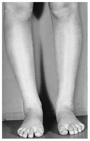
Beginning from the bottom, we see his left foot is turned in and his knee appears straight, He does appear to be banking a left turn, but we would expect him to probably have his toe out a bit for stability since he is really banking heavily into the frontal plane (he must have a great right gluteus medius because he is holding a fairly decent pelvic line into the bank), so our guess is he has a bit of internal tibial torsion (i.e., his foot points in when his knee is straight ahead. This is usually a condition from birth). Folks with internal tibial torsion tend to have their feet in supination more, so they are excellent levers, but it usually means they lack some shock absorption (so maybe we find a forefoot valgus in that shoe, who knows). Because his knee is not in line with his line of forward progression, there will be a conflict there, which could potentially hurt his knee, besides cutting down on his mechanical efficiency (the knee is like a door hinge, and works best when moving in one direction).

His lean to the left nicely complements his probable forefoot varus (or inversion of the forefoot), so at least he has that working for him. There is a little hip sag on the left, indicating a weak gluteus medius on the right; which further cuts down his mechanical efficiency; it is moving to the right as he is cutting to the left. Holding that bottle sure cuts down on his arm swing, and he looks a bit tense, as his left shoulder is higher than the right. Like he is trying to hide something. Finally, his head posture looks forward. This facilitates his flexors and inhibits his extensors, taking away from power to drive him forward. We could go on and on, and we have. Guess we told you how to build a clock and you only wanted to know what time it is. In short, whether he gets caught depends on the biomechanics of his pursuer.

We hope he doesn’t have internet access to our blog, we would hate for him to recognize his problems and improve upon them.  We would be contributing to improved “get-aways” in the future.  That wouldn’t be good, unless someone was chasing him with a .22 , then we might feel ok with it.

Hooch for the youngers, it’s not just for the back alley’s anymore !

- the gait guys

A different kind of Marathon: So you think you are tough ? This guy was tough. A marathon a day for over 120 days…..on one leg, with cancer. Today, a Tribute to Terry Fox.

Half of The Gait Guys grew up in Canada. We were barely a teenager when Terry began his plight.  His mission, 26 miles a day, every day, until he had crossed the expanse of Canada. He made it an amazing 120+ days in a row, 3339 miles, before his cancer returned. The whole country stood cheering watching him do something no mortal man would attempt, let along with one leg, and cancer.  Today we pay a tribute to this rockstar……. his candle went out long before it should have.  So, still think you are a tough runner ? This kind of stuff amazes us. RIP Terry.  You did more in 22 years of life than most of us will do in a lifetime.  We have neither not forgotten you nor what you stood for, and we never will.   Shawn & Ivo

A brief gait analysis of a pretty famous barefooter from a pretty famous study…

OK so we know this rather famous person is on a treadmill, so yes, there is a component of preload to the hip extensors, as well as an increased deceleration component (but those are topics for another post!), but there is some great stuff to look at here.

1st off, note the great technique: mid to forefoot strike, good toe dorsiflexion (although it could stand to be a bit increased to help prepare for even better tripod contact), and good ankle rocker. You can see his excellent shock absorption, through midfoot pronation, ankle dorsiflexion, knee flexion and hip flexion. Also check out the awesome action of his peroneals on his L leg, driving that 1st ray down to the ground for a great foot tripod and prelude to supinaion

But did you notice something else? How about the lack of hip extension? He barely gets past zero. How about the flexion at the waist? We bet his hip flexors are tight! Is some of this caused by the treadmill? Probably, but we would need to see some non-treadmill footage to be sure.

The Gait Guys….No gait is safe from us

Yesterday’s Video Case: The Gaits of Hell

We have received many emails on this case already. Overwhelmingly people are saying……. “Hey, this isn’t easy….. It’s easy when you guys tell us right away because we can see it."  
Yes, when we are all alone to solve these gait problems our heads can start to swim with all the variables. Gait analysis is not easy.  Even the video assessment computer programs do not give you the answers and diagnosis, they just give you variables and data.  The thinking still has to be  done at the end of the day.


I remember how much I struggled with this case back during my orthopedics residency. I remember even pulling out my undergrad notes from Univ. of Waterloo as a student of the famous Dr Stewart McGill and mapping out FBD’s (Force-Body Diagrams) on this case. Oh, the horror !!!  I still have occasional FBD nightmares, being asked to solve an equation in front of the whole class. Pure anxiety ! Holy night terrors ! But, it is amazing what a few decades of study will do for you, we can now look at this case and see things for what they are, see them quickly and know what is going on almost immediately.  It takes some time, so if you are new to this stuff, be patient…… it will come.

CASE REVIEW:

in this video we see the following:

  1. large step length off of the left foot abruptly onto the right, this step is sudden and he crashes down on to the right foot sooner than he normally would to catch his forward moving body mass. ( this will make more sense after reading #5).
  2. there is a delayed left heel rise and delayed left calf recruitment , actually, it’s not delayed, it’s absent. )
  3. the left foot remains supinated through the entire gait cycle. 
  4. the left foot shows extraordinary long toe flexor recruitment (seen on the end of the video during the foot close up)…….this point is important
  5. pelvic unleveling is apparent but a mirage for the most part. We really do not see a true Trendelenberg style gait (although it sort of looks like the left hip drops) rather, what you see is the result of the manufactured delayed left limb departure and subsequent impact at right limb load … but this is not a Trendelenberg gait, he had no Gluteus medius weakness.  Explained another way, he is having troubles departing off of the left foot (this diagnosis is the reason, he has compensated from a neurologic lesion affecting the strength of the calf) and so he extends ( behind him) the left leg longer and further than normal because he cannot push off, plus he hyperextends the left knee because of these factors. Normally, the calf fires after passive heel lift occurs. But with a lesion affecting the calf it has arrested the push off. So, in his case, the heel stays on the ground until it is dragged off from enough  forward body carriage. So, when you see this from a sagittal view the left hip will look like it is dipping as it does here, but it is not truly, he is just taking a long lurching step off of the left and onto the right, the longer left hip extension behind him sets up the illusion of a left hip drop.  Try this at home to feel this gait, walk down your hallway and try to delay the left heel rise for as long as you can.  You will find that you get into your left gluteals more, take a longer step on the left, and take a sudden lurching load onto the right limb to catch your forward progressing body mass. This is exactly what this chap is doing.  But why ? The left calf lesion. 
  6. continuing on #5, there is abrupt right frontal plane loading (because of the sudden transition from left foot to right the frontal plane is engaged longer than normal) and thus the pelvis is carried further to the right in the frontal plane.  He makes a  noble attempt to protect this range by turning out the right foot into the frontal plane (aka. increased right foot progression angle) to allow the quadricep muscles to assist the gluteus medius, abdominal obliques and lateral limb stabiliers in decelerating this frontal plane challenge.

Diagnosis:This doctor came to see me while I was completing my orthopedics residency and mid way through my course work in the neurology post doctoral program. He had been treated for mechanical low back pain with failed results ( well, to be accurate. his low back pain had resolved but pain had peripheralized into the left leg. To review, peripheralizing pain is rarely a good neurologic sign.)  After an examination showing an absent left S1 Achilles reflex it was highly suspicious we were dealing with a radiculopathy. An MRI confirmed a substantial left foraminal disc herniation obliterating the left S1 nerve root foraminally. The S1 nerve root expands into branches feeding input into the lower limb muscles.  In this case, the unfortunate group affected was the gastrocnemius almost exclusively. So in this case this makes sense to what is presented clinically and on gait evaluation. He is overutilizing his long toe flexors (fortunately untouched) as seen in the video because they are basically all that is available to him to plantarflex the foot ( create heel rise and push off).  They are certainly not well suited for this task but subconsciously the brain will use what is available to it, worthy or not. In this case they are a feeble attempt at best. There is no way the long toe flexors can lift his body mass into heel rise and propulse it forward, they are synergists of this task and not agonists / prime movers.
Sequencing Summary:So, this is a case of an aberrant or pathological gait pattern that will be permanent because the nerve damage was fixed by the time i had seen him.  Muscular wasting of the gastroc complex had already occurred.  The culprit was the space occupying lesion (disc in this case) in the left spinal vertebral foramina effacing and deforming the nerve root sufficient enough to create dennervation.  A surgical consult and EMG/NCV (as best as i can recall) confirmed this case was non-surgical at that time (no one wanted to touch the case).  The nerve damage disabled the calf so that push off was impaired.  He thus delays his ability to create adequate heel rise and propulsion so the long toe flexors are called to attempt the feat.  The foot supinates to maintain its rigidity ( it is also hard to pronate through the foot when the toe flexors are in an all out contraction). And because the heel does not rise on its own from muscular strategies, the foot waits to be lifted off of the ground by simple forward progression of the body.  This creates an increased left hip extension range and gives the appearance of a left hip drop which is a false appearance pseudo-Trendelenberg sign.  Due to the fact that he is on the left limb longer, he will be on the right limb for a shorter period.  This right stance phase is initiated abruptly as he falls from the delayed left stance phase. The abruptness of the load on the right challenges the right frontal plane as evidenced by the right foot turn out and right pelvis sway (subtle).  He then departs off the right to  begin the cycle once again.
PS: It is coming a little late, but thank you Dr McGill. Your teachings to a young undergrad set my biomechanical thinking on the right path very early in my studies of human kinetics. Thank you, Sincerely. 
Dr Shawn Allen…… The other half of The Gait Guys

New feature: “The Gaits of Hell - Heaven’s Gaits”.

__________________________________________________________

Something new here on The Gait Guys blog…….we are introducing our case of the month in the new feature for The Gait Guys………the feature is called “The Gaits of Hell - Heaven’s Gaits”.  A bit of a play on words, but the goal of the monthly or bimonthly feature will be to put up either a disasterous gait (such as this one) or a gorgeous gait.  We will provide detailed commentary on the cases, to let you inside our heads. 

Have a look at the video today……study it.  Dissect it.  Replay it over and over until you have it figured out. In some respects, you should have a list of lets say……. 3 diagnostic differentials on your list of possibilities.  If after several viewings you cannot figure out the limp, put it aside……..and do what we do……. mimic the gait pattern and figure out what they are doing by what you are feeling.  We just gave away one of our biggest secrets with this little tidbit.  Give it a try.  We will have the answers Tuesday. 

We were going to give the summary here today…….but then we figured you might not struggle with it enough to learn what you need to from this case.  The learning principles here are huge……. so spend some time with it……get out a piece of paper and a pencil and put it all down…….. ie. which side is the limp on, which planes of motion are being violated, what are the compensations.

We actually know the precise diagnosis in this mid 20 year old doctor, he was a patient of Dr. Allen’s back in his residency days almost 16 years ago.  Yes, if you look closely that young lad with the tie and all the hair is actually me…… man what i would do for a head of hair like that once again ! 

Enjoy, …… struggle with this one……it is a good one. 

(film quality, sorry kids….. this was on VHS to start with ! Then it was converted to AVI…..and then to quicktime.  So, yes……. its a bit grainy and not HiDef……. but this case is worth every second of its painful low quality !)

Dr. Allen

The Windlass Mechanism of the Plantar fascia. What is a Windlass anyway?
After yesterdays post, we thought we might provide more insight to the Windlass mechanism and low and behold; we found AN ENTIRE PAPER on it! Wow, were we thrilled since there …

The Windlass Mechanism of the Plantar fascia. What is a Windlass anyway?

After yesterdays post, we thought we might provide more insight to the Windlass mechanism and low and behold; we found AN ENTIRE PAPER on it! Wow, were we thrilled since there is not a ton of decent stuff out on this topic (yes, we are a little geeky, but then again so are you if you are reading this !).

A Windlass is the tightening of a rope or cable around a pivot point. The plantar fascia acts like a cable between the calcaneus and its distal insertion into the proximal phalanyx at the metatarsal phalangeal joints. When the toes are dorsiflexed (as in forefoot rocker from yesterdays post, see bottom diagram above), the heel and toes SHOULD become approximated, as the plantar fascia shortens from its winding around the metatarsal head, contributing to supination of the foot.

To be accurate, this concept of the Windlass mechanism is quite complex because the dorsiflexion of the great toe also shortens the length of the flexor hallucis brevis.  And we know that the sesamoid bones under the big toe are embedded in its tendon. Their repositioning as the Windlass engages will drive the sesamoids under the metatarsal, elevating it, and shift this joints eccentric axis.  But this is a complex story and post better left for another day.  Simply put, this is a complex joint, do not let anyone fool you otherwise.  Don’t beleive us ? Ask any bunion (or God forbid a fusion) surgery patient how they are doing.  You will get the point then. 

Here’s the link to the article (we know you want to read it ! )

http://www.ncbi.nlm.nih.gov/pmc/articles/PMC385265/

The Windlass Mechanism. Just another fascinating foot fact from The Gait Guys.

Forefoot Rocker and Premature Heel Rise:


Remember the rockers? We did a series on this a few weeks ago. Remember there are three: heel, ankle and forefoot. We are going to concentrate on the forefoot today. As a reminder, forefoot rocker occurs at the 1st metatarsal phalangeal joint (big toe knuckle) as the tibia progresses over the forefoot during forward movement. There are 4 things that SHOULD happen at this point to ensure the heel comes up :


1. continued forward momentum  of the body

2. the posterior compartment (primarily the gastroc/soleus group and tibilais posterior) contract to accelerate the rate of forward limb movement.

3. passive tension in the posterior compartment muscles

4  the windlass effect of the plantar fascia (see diagram)

Watch this slow motion video and what do you see? You should see some midfoot collapse and premature heel rise, especially on the right foot. Did you notice the little “bounce” in his step? How about the subtle adduction of his heel, L > R?  Watch it again until you see it. (The bounce is generated by the premature heel lift and premature firing of the calf compartment muscles.  Normally the body mass is further forward of the heel rise event, and thus contraction of the calf generates a more forward directed vector, however, when the heel rise is premature the body mass is still somewhat over the foot.  Thus, if the calf were to fire at this moment, it would cause a vertical body mass movement vector.  When this occurs bilaterally these clients will have a very “bouncy” vertically oriented gait strategy.  This is very inefficient gait when it occurs. Plus there is a dramatic reduction in the pronation phase of gait, so shock absorption is severely reduced.)

Does he have forward progression of the body mass? Yes

Do you think the posterior compartment is actively contracting? Probably too much. Remember the medial gastrocnemius adducts the heel at the end of terminal stance to assist in supination.

Does there appear to be increased passive tension in the posterior compartment? Yes, it appears to be the case !

How is his windlass mechanism? Good but not good enough. (see our next blog post regarding the Windlass)

Premature heel rise… Coming to a midfoot overpronator and people with loss of hip extension near you.

Telling it like it is. We are the Gait guys…..

The Cross-Over Gait: Slow Motion barefoot video analysis.

Thanks to Runblogger.com for once again providing materials on their blog for us to peck away at clinically.

Before we get into our findings from this clip we need to jump up on our soapbox once again on our typical rant on video analysis.  Here we go again, ……. What you see on video analysis is quite often not what is wrong with your client.  What you are seeing is their strategy to run or walk with the parts that are available for them to use.  You are seeing a compensation pattern.  If a gluteus medius is notably weak or inhibited you will not see its functional expression on the analysis, rather you will see your clients neuromuscular strategy with the parts that are available.  Sure you can hypothesize some things, but without proper muscle testing and evaluation, even if you are screening with “functional pattern tests”, there is no guarantee that your suspicions are correct.  Not until you can get them on the table and assess their anchoring musculature, stabilizers and prime movers with more specific muscle tests.  Without this component as part of our client evaluation, you must leave room for the cognitive reality that you are  basing your next thoughts and direction on some assumptions, and we all know what can happen when you do that.  It would be a crime to prescribe or train your client further into assumed patterns, not knowing that you are building skill, endurance and strength on an improper compensation/cheating pattern. Take these thoughts to your next gait evaluation expert, and ask these hard questions. 

(people like examples, so below you will see an example of this thought process)

OK, now, back to our visual findings (assumptions or concerns) in the video above. There is good midfoot strike for the most part. In keeping this simple for today, lets just focus on the stability problem in the frontal plane.  We can easily see a cross over gait here.  This chap is literally running on a line, it is an example of poor biomechanics… 1) it is not biomechanically correct or efficient and 2) it requires a ton of gluteus medius and abdominal core support to hold midstance correctly (which is far from what we see here).  We see evidence of this mechanical collapse and insufficiency here.  The foot should be under the knee, the knee should be under the hip, the opposite side pelvis should be horizontal or show a little hike and the pelvis should be quiet for the most part.  In this case, there is a suspicion (yes, an assumption that we would manually test) that he does not have enought gluteus medius and abdominal strength to stop the obvious lateral deviation of the hip/pelvis during stance phase. 

“But how can you say this Gait Guys ? We cannot even see the pelvis in the video ! ?”

Here is how. We know that the opposite hip has to be dropping, evidence of the lack of stance stability, because in everyone, when the swing phase hip drops it allows the limb to adduct…….. thus driving the swing phase knee towards the stance phase knee.  This drives the cross over gait , or “line runners” as we like to call them.  Just as we see here ! This one is a dead ringer for the need of hip/pelvis/ core evaluation.  Who is driving it ? We do not know, and you cannot know either, not without testing.  Perhaps it is the abdominal oblique as the primary, or maybe the primary is the gluteus medius, , maybe it is the adductors.  There is no way to know. And basing your training or homework or therapeutic exercises for this client from a gait evaluation is about as silly as prescribing a shoe for this foot that looks like it is pronating too much.  When the truth is that it could be doing so to make up for the suspected faulty mechanics above.

In this scenario, telling this guy he should not be in Vibrams is just about as silly as telling him he needs a stability shoe, even though the two thoughts are directed at the same thing.  That thing being the increased pronation appearance that you do not like.  In this case, we need to get to the root of the problem and that means you have to have the clinical skills to do so.  Perhaps this guy is pronating so much through the foot because he does not have enough internal hip rotation. And in order to complete the stance phase of gait the internal rotation has to occur somewhere to get his body mass past the foot so he can get to his glutes to propulse forward.

What you see is not what is wrong most of the time, as in this case.  This client should not be told he should not be in Vibrams, nor should he be told he should be in a stability shoe.  What he should be told is…….“ Look buddy, there may be some issues here you. You might want to have some of this stuff looked at a little deeper”

Video Gait analysis……. it is not just for breakfast, lunch or dinner anymore……..it is the fork you use to help you eat those meals.  Its a tool ! ……….. just like The Gait Guys…….. yup, 2 tools…….. Shawn and Ivo

A Case of Hip pain in a Young Runner: Perthes Disease

here is a nice little short video of a young girl with a healed Perthe’s Disease (full name, Legg-Calve-Perthes Disease) that came to see us a few years ago with right hip pain.  After an examination and a very brief treatment stint films were obtained and found an early stage Perthe’s Hip.  Early diagnosis is always important in this disorder that affects the vascularity of the head of the femur. Failure to make an early diagnosis is a disaster which leads to deformity and permanent disability for the patient.  Perthe’s affects mostly male boys under the age of 10. There is really no clear etiology but many studies point to a period of increased pressure within the joint from an inflammatory process. A term “Transient Synovitis” has been labeled by some.  In this case, the disorder was caught in its first stage and the hip revascularized, did not collapse and it is doing well.  Collapse is the most devastating outcome of this disease process, it is why you do not mess around with children with unresolving hip pain, obtain imaging early.  The main problem, as is seen here, is that she cannot get to her gluteal muscles to stablize the hip in the frontal plane.  Here you see a classic Trendelenberg Sign when she steps onto the right leg. 

When she steps onto the left hip the hip,knee and foot are well aligned in the frontal plane and the right hemipelvis rises above the left hip joint line.  Comparatively, when she steps on the right, there is a significant lateral pelvic and body mass shift beyond a line drawn up from the foot-knee line.  Consequently the left hip drops and she looks like she has a short right leg.  Measurements (as unreliable as they are)  do not show a leg length discrepancy.  However, this type of mechanical behavior can put undue stress on a healing femoral head.  Using a sole lift to help regain pelvic leveling during gait help maintain balanced femoral head pressures and cartilage coverage during the last stages of joint formation in this adolescent.  The problem is that there will be dependency on the lift so regular daily exercises with guaranteed compliance is imperative.  She must regain use of the glute in gait and stance or this hip will be a problem in later years, guaranteed.  So, this is a difficult case.  It is not for the faint of heart.  Bottom line, do not mess with kids with hip pain for long without imaging to rule out terrible problems like this.  There are so many gait problems that will ensue if the gluteal stability is not regained.  To name just a few, the right foot will always be supinated and this means risk for bunions (see last weeks Dr. Ivo video on bunions and the adductor hallucis muscle) and other disorders that are caused by an unanchored first metatarsal.  Additionally, the knee can degenerate the lateral compartment quickly not to mention the plethora of muscular problems (low back pain, knee pain etc) and strategies (ie. pelvic distortion patterns) that will ensue from such a gait.  There is so much more to Perthes Disease than we have mentioned here, but this is not the venue for such complicated topics.  The important thing is to beware of systemic problems that can compromise the integrity of the neuromusculoskeletal system that can have short and long term effects on one’s gait. Here is a link to some more info on Perthes Disease …… but even this is scant info (http://www.ncbi.nlm.nih.gov/pubmedhealth/PMH0002244/)….. make sure you do your reading if you are in the clinical world and see young patients. 

It is not always just about muscles and shoes and orthotics. You have to always be on your toes (no pun intended).

we are…….. so much more…….. than just Gait Guys.

Shoe Review: The Brooks Pure Project Line.
Ok, we have been meaning to get to this for months but are just getting around to it now. So for those of you who have been hounding us for the data, sorry, but thanks for keeping us on it.  Here are the sp…

Shoe Review: The Brooks Pure Project Line.

Ok, we have been meaning to get to this for months but are just getting around to it now. So for those of you who have been hounding us for the data, sorry, but thanks for keeping us on it.  Here are the specs for the EVA midsole thicknesses and ramp numbers. Remember, ramp angle can only be given if the length of the foot is known, so those numbers will not be given here.  What is good to know is that we have another shoe in the category of the Saucony Kinvara, the Brooks Pure Project line.  Below you will see the specs for all 4 in the line up.  All have a 4 mm forefoot to rearfoot rise, in other words……the heel is only 4 mm lifted compared to the plane the forefoot is resting on.  This still changes the biomechanics and neuromechanics that we were all given at birth that would really prefer the rear and forefoot to be on the same plane 1:1 ratio although a 4 mm rise is pretty darn close !  Our man beef with the Saucony Kinvara is that they did not use much black rubber outsole on the shoe other than the small thin layer glued to the traction lugs  throughout the mid and forefoot.  We have found that these shoes barely get 200 miles on them (give or take) and we and all our clients are already into the EVA midsole which wears down as fast as bubble gum might.  This is a serious design flaw in our opinion. We like this shoe and like it for many clients but we are having to explain that they will burn through them in under 350 miles most likely.  So, we are excited for the October Release of the Brooks Pure Project line……in the hopes that they have not made this same design choice.  Remember, if you are new to this line of shoes, the 4mm lift variety, wean down from your old 12-20mm rear-foot lift trainers and try these with your shorter runs until skill, endurance and strength are achieved in this new foot orientation.  It is gonna take some people some time to accomodate.  (remember, there is no substitute for a doctor’s exam and watchful eye to see if you can even entertain this shoe type with your foot type). (Do not be fooled into believing there is going to be much stability provided by these shoes.  They are all pretty neutral. If you have a  forefoot varus, you better look in another direction !)

Here is the data …….

Brooks Pure Connect

lightest and most flexible shoe in the line, the PureConnect puts as little as necessary between the runner and road. 7.2 oz men, 6.5 oz women – 14 mm heel:10 mm forefoot

Brooks Pure Flow

For runners who want to connect with the run without losing the comfort
of dynamic cushioning. 8.7 oz men, 7.5 oz women – 18 mm heel :14 mm forefoot

Brooks Pure Cadence

Runners who need more supportive features can still experience the feel
of a more natural stride. 9.5 oz men, 8.3 oz women – 18 mm heel:14 mm forefoot

Brooks Pure Grit

Trail runners will love the hug-your-foot upper, slim midsole, and pliable
yet protective outsole. 8.9 oz men, 7.6 oz women – 15 mm heel:11mm forefoot


__________


tumblr_loa5ivO33C1qhko2so1_400.jpg
tumblr_loa5ivO33C1qhko2so2_400.jpg

Photographs of a 6 yr. old.

What do you see ? How does the alignment look ? Are they developing normally ?
At what point should you intervene to ensure proper alignment and
development occurs in this child ? Can we standardize our visual
screen to fit all cases in all children ?  So many questions !

* this case goes very nicely with a prior case on the blog published on July 15th.

Click back and forth between the two photos above on the blog.  Youcan see that in the first photo that when the feet are parallel, the patellae point inward (normal compensation).

In photo 2, we can see that when we put the patellae in the saggittal plane (pointing
forward) the foot progression angle is severely positive (externally postured or out-toed).  

This is a severe case of FEMORAL ANTETORSION
with compensatory EXTERNAL TIBIAL TORSION.  The external tibial
torsion is an external spin along the long axis of the shaft in a
response to try to correct alignment.  You can see that in this case
there is no happy medium.  The torsion in the long bones is so extreme
that either the knees are positioned inwards when the feet are
normally aligned or the feet are spun outwards when the knees are
properly aligned.  Regardless, there is much abnormal stress on the
hip and knee joints and the labrum of the hip and menisci of the knees
not to mention the challenges into the foot mechanics and gait.

______________________________

Taking this into a gait perspective:

Patients with antetorsion are forced to externally rotate the limbs to bring the knee forward to a normal alignment from an internally rotated position.
This takes up some, occasionally all, of the lateral (external) rotation needed for the hips to function normally during gait causing compensatory rotation of the spine during single leg stance; the spine being really the only place the rotation can be achieved.

Normally, during gait the hip rotates laterally from toe off through to heel strike and is present during the initial loading response.
When this rotation does not occur in the hip, it must occur as a compensatory motion somewhere else in the kinetic chain and this is usually the pelvis or lumbar spinal joints.
Furthermore, the abnormal alignment of the femoral head in the acetabulum can repetitively irritate the hip joint and labrum let alone place abnormal torque on the tib-femoral joint (knee).
Antetorsion patients who compensate and realign the feet to a normal progression angle via tibial external torsion will force the femoral head anteriorly into the anterior hip capsule and this is a common source of pain.  It is a “hammocking effect” into the anterior capsule and it can leave the femoral head and it’s cartilage uncovered and uncompressed thus advancing arthritis degeneration. 
This is a similar symptom phenomenon as in “anterior femoral glide syndrome” as described by Shirley Sahrmann.  It is not uncommon to see a sway back lumbar posture in these clients.
These patients may have anterior contractile tissue deficits, namely stretch-weak iliopsoas as described by Kendall.
Remember to check for limb length symmetry in these clients who’s anteversion is not symmetrical. 
Watch for a future series on torsions and versions

We may be twisted, but the last time we looked, we are still …… The Gait Guys

1 + 1 usually = 2

take yesterdays tibialis posterior dialogue……. translate that to the resultant foot structure that can occur when there is complete insufficiency in that muscle (a valgus heel, midfoot collapse and an abducted forefoot) and then understand that this resultant foot will have a first ray complex insufficiency whereby 1st metatarsal head anchoring will be compromised. Now watch todays video by Dr. Waerlop and put the two together.  Yes, tib posterior insufficiency can be a cause of bunion-hallux valgus formation.

1 + 1 = 2   

not only are we foot geeks, but as you can see we missed our calling in mathematics as well !

Shawn and Ivo …….. 

More on Bunions: proof we know what we are talking about

[Sequential lateral soft-tissue release of the big toe: an anatomic trial].

Z Orthop Unfall. 2007 May-Jun;145(3):322-6. Roth KE, Waldecker U, Meurer A.Source: Abteilung für Orthopädie, Universitätsklinik Mainz. roth@orthopaedie.klinik.uni-mainz.de

___________

Summary:  Dr. Ivo in his brief video today discussed the altering of the origin/insertion effects on the adductor hallucis and the big toe.  When the first metatarsal is not anchored on the ground the lateral toes cannot be pulled towards the medial foot, instead the lateral foot acts as the anchor and the big toe/hallux is pulled laterally towards the anchor rendering the all famous bunion/hallux valgus.

This surgical study pretty much proves this principle.

This study showed that when the soft tissues (capsule, tendon and ligament) are surgically released, the contractile affects on the joint angle of the bunion/hallux valgus are released and the hallux valgus angle was predominantly and significantly improved.  A significant correction of the intermetatarsal angle did not take place however.

Kind of a radical procedure ultimately destabilizing the joint and medial foot structure…….but hey……whatever floats your surgical boat.  To each his own.  We suppose that on a case by case basis all options need to be considered.

……we’re still the gait guys…….. with no scalpels, but with big oars

tumblr_louwzePPmE1qhko2so2_1280.png
tumblr_louwzePPmE1qhko2so5_r1_250.jpg

Biomechanical and Clinical Factors Related to Stage I Posterior Tibial Tendon Dysfunction.     Rabbito M, Pohl MB, Humble N, Ferber R.

CONCLUSION:

The increased foot pronation is hypothesized to place greater strain on the posterior tibialis muscle, which may partially explain the progressive nature of this condition. J Orthop Sports Phys Ther, Epub 12 July 2011. doi:10.2519/jospt.2011.3545.


What the Gait Guys say about this article:

Do these results really surprise us? The Tibialis posterior (TP) is one of the more important extrinsic arch stabilizing muscles. It is a stance phase muscle that fires from the loading response through terminal stance. It ‘s proximal attachments are from the posterior aspect of the tibia, fibula and interosseous membrane and its distal attachments are the undersurface of all the tarsal’s except the talus and the bases of all the metatarsals except the first.

Since the foot is usually planted when it fires, we must look at its closed chain function (how does it function when the foot/insertion is fixed on the ground), which is predominantly maintenance of the medial longitudinal arch, with minor contributions to the transverse metatarsal and lateral longitudinal arches; flexion and adduction of the tarsal’s and metatarsals, eccentric slowing of anterior translation of the tibia during ankle rocker. It is also an external rotator of the lower leg and is the prime muscle which decelerates internal rotation of the tibia and pronation. As the origin and insertion are concentrically brought towards each other during early passive heel lift it becomes a powerful plantarflexor and inverter of the rearfoot.  There is also a  component of ankle stabilization via posterior compression of the tarsal’s and adduction of the tibia and fibula.

Alas, there is soooo much more than the typical open chain function of plantar flexion, adduction and inversion. Perhaps it is some of these other, closed chain functions, that cause the “progressive nature of the condition”?

We remain…The Gait Guys…Going above and beyond basic function and biomechanics.