Ancient Footprints Reveal Earliest Signs of Human-Like Gait

Recent trails found in Tanzania suggest fully upright walking began 3.7 million years ago.

The footprints they found suggest that the fully upright gait of present day humans existed about 2 million years prior than previously thought !

“The shape of the human foot is probably one of the most obvious differences between us and our nearest living relatives, the great apes. The difference in foot function is thought to be linked to the fact that humans spend all of their time on the ground, but there has been a lot of debate as to when in the fossil record these changes occurred. Our work shows that there is considerably more functional overlap than previously expected,” Bill Sellers, of the University of Manchester’s Faculty of Life Sciences, said in a University of Liverpool news release.

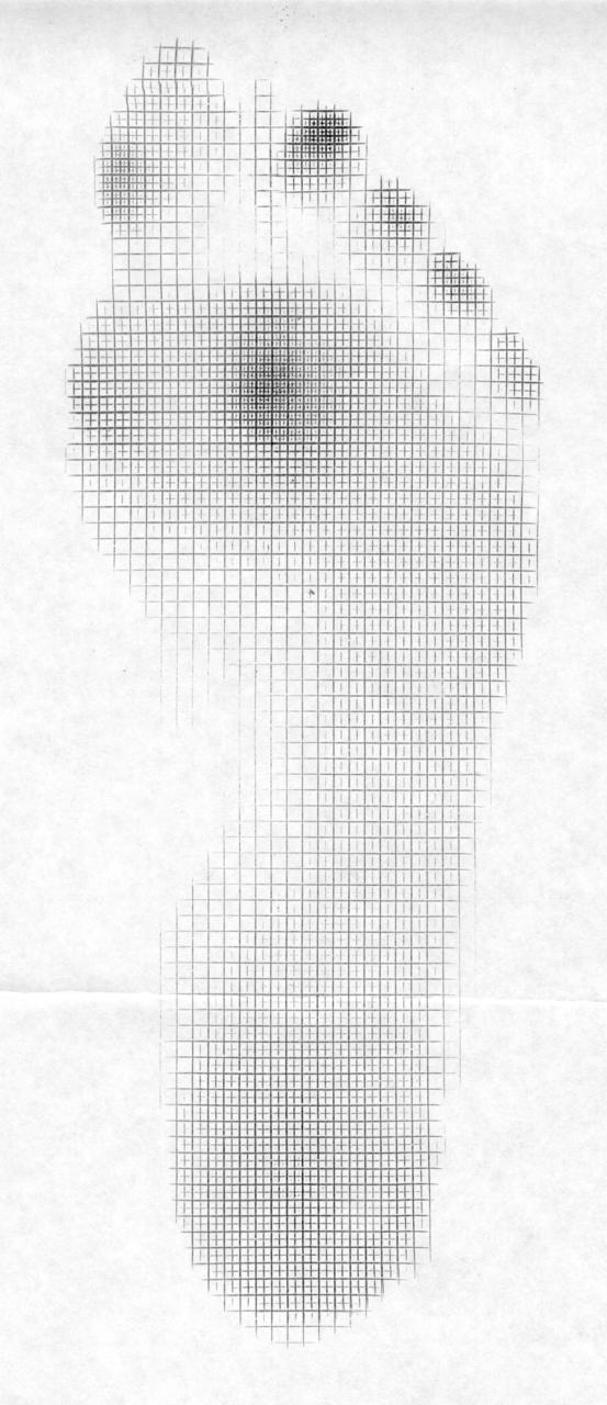
Known as the Laetoli trail, the 11 individual footprints found in Tanzania are the earliest known footprints made by human ancestors. The prints reveal features of a gait with more similarities to the way modern humans walk than the bipedal walking of chimpanzees, orangutans and gorillas.

The study found that instead of the crouched walking posture with mid foot push off of present day apes, the researchers found that these new found foot impressions categorize a bipedal upright walk driven off the front of the foot and largely the big toe highly representative of the  human gait of today.

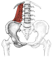
This study suggested that Australopithecus afarensis had a much different build than that of modern day humans.  Their build had a long torso and short legs.  A reverse of modern man’s physique.  Thus, this studies researchers proposed that this species could only ambulate bipedally for short distances. 

_______________________________

Click on the title link at the top  for the link to the copywrited article. Excerpts quoted from the article.

So, if you are a believer in evolution (or, have been watching “Ancient Aliens”, the Docu-series on The History Channel   {wry humor}) ……. then this is worthy of longer term memory.

Q.  Why is this article here and why do we deem it important to share with you ? 

A. Because history is full of clues. To be good in this field of gait and motion, we feel that you should have a decent foundation in anatomy, neurology and physiology, store facts, keep an open mind, study the research (both old and new), do not discount historical information merely because it is old, and consider all options and possibilities so that knowledge can follow from more current experiences).

(Fact: such memory items are encoded by your hippocampus, entorhinal cortex, and perirhinal cortex, but consolidated and stored mostly (we think) in your temporal cortex.

Tidbit on memory:

In gait, Procedural memory refers to movements of the body, such as how exactly to use use a phone, walk or ride a bicycle. This type of memory is encoded and probably stored by the cerebellum and the striatum.

have a good week everyone…….. The Gait Aliens……. Shawn and Ivo

tumblr_lohzpbv2Zj1qhko2so1_500.png
tumblr_lohzpbv2Zj1qhko2so2_250.jpg

The Mighty Quadratus: Part 2  The Quadratus and Gait

Acting unilaterally without the ipsilateral foot fixed on the ground, it can raise the ilia on the side of contraction (as in a pull up or side bend on a Roman chair). The quadratus lumborum was more active than other muscles during isometric side support postures where the body is held horizontally almost parallel to the floor as the subjects supported themselves on one elbow on the floor together with both feet. 

It is active during single limb support during stance phase of gait on the contralateral side (along with the external oblique) to elevate the ilium. This is coupled with the ipsilateral anterior fibers of the gluteus medius and minimus pulling the iliac crest toward the stable femur.

Sahrmann states “the QL is optimally situated to provide control of lateral flexion to the opposite side via its eccentric contraction to provide control of the return from lateral flexion via its concentric contraction. The muscle is also positioned to play a role in the rotation that occurs between the pelvis and spine during walking”. This makes you really think about the interplay of this muscle, and another stance phase stabilizer, the psoas major, which attaches opposite the QL on the anterior aspect of the vertebral body, IN FRONT of the transverse process.

Acting bilaterally, it extends the lumbar spine, deepening the lordosis and acting to limit anterior shear of the vertebral bodies.

It is able to stabilize the 12th rib during forced expiration, thus acting as an accessory muscle of respiration. This fixation is important when we need to superimpose pelvic movements upon it. Furthermore, it increased activation in response to increasing compression in static upright standing postures.

Bottom Line?:

Think of the QL, especially during gait abnormalities or recalcitrant low back pain. The more it is stressed, the more it is activated. If someone had mild weakness of the stance leg gluteus medius, it may be called into play to pick up some of the slack. Expect to see increased activity paraspinally, with particular attention paid to the 12 rib attachments.

In our flexor dominant society, the QL may play a role in generating unilateral shear forces on the lumbar spine (along with the ipsilateral psoas), especially in individuals with poor ankle rocker or decreased hip extension.

The QL: it’s not only for breakfast anymore…..

We still are…The Gait guys

do you know we are on FACEBOOK and TWitter ?

Do you know we are on facebook and twitter as well ?

sometimes things that are slightly less worthy, but still helpful, are put on our FACEBOOK PAGE…….see links here

feel free to LIKE us so you get the daily feeds and notices immediately so that you do not have to constantly check back to the blog here for updates.  All updates are immediately notified on FAcebook so you will get notification immediately there.

Do you like the work we are doing here?  Have a friend that might get something out of it as well ? Feel free to forward our links.

http://www.facebook.com/pages/The-Gait-Guys/169366033103080

tumblr_lohzj4iyha1qhko2so1_400.jpg
tumblr_lohzj4iyha1qhko2so2_250.jpg

The Mighty Quadratus: Part 1

Today we explore the Quadratus lumborum and its functional anatomy.

It is useful to think of the QL as having two divisions. Though they can’t act independently, it helps when thinking about it from a functional standpoint. The first, or lower division arises from the medial portion of the iliac crest and adjacent iliolumbar ligament, inserting onto the transverse processes of the lumbar vertebrae, running in the coronal plane from lateral to medial and posterior to anterior in the saggital plane. The second, or upper division arises from the lumbar transverse processes of the upper 4 lumbar vertebrae at their upper and lower corners and insert into the inferior border of the 12th rib, running in the coronal plane from medial to lateral and in the saggital plane from anterior to posterir. Approximately half of the fascicles of this second division act on the twelfth rib; the rest act on the lumbar spine.

The QL is primarily a coronal plane stabilizer. Acting unilaterally with the lower body fixed and feet on the ground, it laterally bends the lumbar spine. Normally, with lateral bending of the lumbar spine while in a lordotic posture, we see what is called type I coupled motion, or deviation of the spinous process to the side of lateral bending. The QL would oppose (or perhaps more correctly attenuate) this motion, having a moment of moving the spinous process to the opposite side of contraction. Perhaps it is when the QL become dysfunctional, pulling the ipsilateral transverse process outward (and thus moving the spinous to the opposite side) that we see aberrant (or Type II) motion in the lumbar spine. It is interesting that when the lumbar spine is flexed (as in sitting or forward bending) type II motion is normal, and now the QL becomes prime mover. Due to the angle of attachment here, it can create shear and potentially contribute to injury.

Whew!! Stay tuned for the QL and gait tomorrow!

Yup, if you are reading this, you are a gait geek too!!

Shawn and Ivo

A brief gait review from a youtube clip we found:

at :03 notice the shrugged shoulders and trapezius activation, forcing respirations to the upper lung fields. This also facilitates the scalene muscles in the neck (which is probably one of the reasons they flex their neck). Breathing from here is shallow and inefficient. This action (shrugging the shoulders) activates the upper trap and deactivates the lats (which are the functional link between the upper and lower extremities)

at 05: they begin to flex the lumbar spine

at :06 they flex at the waist as well as the neck. This rounds the spine and puts the glutes at a mechanical disadvantage for extending the hips and limiting some of the driving power. They then become hamstring dependent, which isn’t as efficient. Dropping the head defacilitates the extensor muscles neurologically, so they will have some power loss (as well as stiffness loss) as well. They keep their neck flexed till :07, where they really begin to pick up more speed. The torso remains flexed at the waist through most of the footage.

it appears at :07 that the left foot strikes the ground in eversion bottom of foot pointing away from camera) indicating some degree of forefoot pronation. A shot from behind would be helpful to confirm this

The arm swing appears asymmetrical from left to right, right being greater both forward and especially backward. I would wonder what they are hiding (biomechanically) there (so are they increased on the right or less on the left?. Here is where foot age from behind would be instructional).

Ok folks. Hope you enjoyed the ride!

we still are….The Gait Guys…..

The Risks for Forefoot Strike running. YOU NEED TO READ THIS ! YES, YOU !

OK, we are going to go on a rant here…… it is time.

We have been talking about problems of forefoot strike for some time now. We like a midfoot strike, and we have research-based, well founded logic to our opinion.  A Forefoot strike reduces the amount of pronation possible for shock absorption because  when the foot is plantarflexed it is in a supinated state which is reserved for a rigid propulsive mechanism.  At impact some degree of pronation is necessary otherwise force attenuation must occur elsewhere in the kinetic chain otherwise it creates bone, joint or soft tissue pathology/injury somewhere in the chain.  However, one of the major issues we have been pounding our fists on the table about, for years, are forefoot orientation anomalies.  A significant portion of the population have forefoot types of varus or valgus, some flexible and some more rigid, some compensated and some uncompenated (yes, this is difficult stuff……but if you are going to make orthotics or if you are going to be a runner or sell or make shoes or coach or even speak about running form styles…… you had better know this stuff or we will call you out on it). 

No one is talking about this stuff except The Gait Guys. 

Is this because no one knows about it ? Maybe.

It is because those in the running fields do not understand it well ? Likely. 

It is because it creates fear and anxiety about selling shoes ? Probably.

Is it because it complicates shoe fabrication? Likely. 

Does that make it right to just ignore it all together ? No ! 

With a forefoot strike into one of these “pathologic” forefoot types the anatomical variance is accentuated.  In this scenario, a varus foot type that lands and subsequently has not choice but to drive hyperpronation strain not only suffers from the increased pronatory collapse but they are unable to acquire a subsequent rigid toe off which in itself can drive further pathology.  And a valgus forefoot strike is even more rigid than a neutral forefoot strike impact and they are also at risk for inversion strain on the lateral foot.  A midfoot strike can reduce some of these consequences by setting the foot up for a preparatory transition. We know this, we see this everyday, it is what we do. These runners need to be categorized and educated as to why their injury is present or chronically persists, and why we insist a program to reteach a midfoot strike. 

As always, if the doctor knows what anatomy presents itself with the client, and adequately educates the athlete……..then a good relationship and outcome will ensue. Additionally, a change in shoe is not  uncommon when their strike mechanics change.


Here is what spurred our soapbox rant today…….. thanks Lower Extremity Review for bringing the June ASCM to light early !!!
_________________________________________________________________________________

from LER, Link is above:

“The frequency content of vertical ground reaction forces generated during running differ among forefoot strikers and rearfoot strikers, and this may have implications for injury risk, according to findings from the University of Massachusetts presented in June at the ACSM meeting.

Researchers assessed frequency amplitude and power in 10 natural rearfoot strikers and 10 natural forefoot strikers as they ran across a force platform.

At frequencies above 9 Hz, rearfoot strikers’ amplitude exceeded that of forefoot strikers. Similarly, above 22 Hz, power was significantly greater in rearfoot strikers. This is consistent with previous reports that only rearfoot strikers have an impact peak, which occurs between 10 Hz and 20 Hz.

But between 4 Hz and 7 Hz, amplitudes were higher in forefoot strikers. And for frequencies less than 6 Hz and between 9 Hz and 11 Hz, power was greater in forefoot strikers.

Because the body attenuates shock differently at different frequencies, the findings could suggest that even forefoot strikers (including most barefoot runners) may be at risk for certain injuries despite lacking an impact peak.”

tumblr_lo77y0LOcc1qhko2so1_1280.jpg
tumblr_lo77y0LOcc1qhko2so2_1280.jpg

Time for a quick pedograph case:

This person presented with arch pain and occasional forefoot pain.

Note the increased size (length) of the heel print with blunting at the anterior most aspect. The midfoot impression is  increased, revealing collapsing medial longitudinal arches. The forefoot print has increased pressures over the 2nd metatarsal heads bilaterally, and the 1st on the left. She claws with toes 2-4 bilaterally.

This demonstrates poor intrinsic stability of the foot (as evidenced by the increased heel impression and midfoot collapse) and well as decreased ankle rocker (as evidenced by the increased forefoot pressures).

We also see increased ink under the distal second digit (esp on the right). This suggests some possible incompetence of the first ray complex and big toe, which is represented by the medial ink presentation under the great toe (suggesting a pinch callus, which is seen when there is spin of the foot and insufficient great toe anchoring and push off).  When the great toe function is compromised, we tend to see increased activity of the 2nd digit long flexors, represented well here by increased ink under the 2nd toe.

The pedograph truly does provide a window to the gait cycle!

We remain: Gait Geeks

Even cadaver feet speak volumes...

We hope you are thinking about muscles in a CLOSED CHAIN fashion, rather than open chain. In other words, when the foot is on the ground, it becomes the fixed point and the more proximal portion is the moveable portion. This paper shows (as Dr Allen did in his most excellent youtube video about foot function   http://youtu.be/TyRE9dReVTE  ) that the abductor hallicus is a dynamic elevator of the arch, in addition to being a great toe dorsiflexor and rear foot inverter!

Yup..we are definitely Gait Nerds….

A brief note on internal hip rotation from a cyclists perspective.

Today is Stage 14 in the Tour De France. We are big fans and we treat tons of Triathletes. So, it seemed perfect to do a little bike fit and mechanics today.

On the subject of cyclists, we have noted many have these 3 anatomical traits: femoral retoversion (see recent blog posts this week), tibial varum, forefoot varus. If you look at these closely, they all tend to supinate the foot foot more and make it a better lever. These folks are way better cyclists than runners.

With a FF varus, they often compensate on the down stroke to make the foot flat on the pedal (to use the 1st MTP); if they are retroverted, they have limited hip internal rotation to begin with and now you are asking them to internally rotate more, which leads to hip pain and at times, labral injuries.

Look at the attached clip, R leg; note how it comes closer to the center bar and the position of the knee; also look at the forefoot.

Yup…The Gait Guys…We do bikes too!

Grab a Beer and Watch this Heel Strike Flick !

Wow, only one mid-forefoot striker in the whole bunch …….everyone else is a heavy heel striker. ……. and so the epidemic continues……… until this plague ends runners will always have injuries and guys like Ivo and myself will never be able to retire ! …….. until then…….. we remain dreamers of Boats, Beaches, Bars and Ballads (a great Jimmy Buffett box set !)

“Fins to the left, fins to the right, ….. We’re the only GAIT in town”

The Hip: Part 4....Putting it all together.


Correlating various foot problems to hip function and limitations. This is something we have been shouting about for years now.

One of the very first things we do after watching one’s gait on any patient visit is to have them supine on the table and check passive external and internal hip rotation (IR) ranges. At this time paying particular attention to the topic of INTERNAL HIP ROTATION, we do this particularly with the limb straight (lying flat on the table) to mimic what the range might be with them standing in midstance phase of gait/running (ie. the pelvis and body mass directly over the foot). We do this supine because checking IR in variations of hip flexion does not make much sense when it comes to gait pathology. We compare the ranges left and right. They should be symmetrical and sufficient. According to Michaud’s work, 4-6 internal rotation degrees is necessary, 45 is normal.
As we move through midstance the stance limb is converting from external hip rotation to internal rotation as the contralateral hemipelvis transitions forward during that leg’s swing phase. A major key for normal biomechanics moving downwards through the kinetic chain is that sufficient hip internal rotation is present. If there is a deficit functionally (and sometimes that is different from what the books say is necessary), the internal rotation has to occur somewhere. Quite frequently it occurs through more aggressive and faster internal tibial rotation which will challenge the amount of foot pronation that occurs (it usually causes more). [* please keep in mind if your solution for this local increased pronation at the foot is an orthotic to block the excess motion keep in mind that the body now has to find another place to put this internal spin and sadly it quite often has to go back to the hip and this can cause the hip labral problems that we have talked about all week. *Now you see why we set the week up this way preempting this discussion with the pathologies.]

Continuing our thoughts a bit further, if internal hip rotation is not sufficient as the body moves over the limb then the next succession, hip extension, is going to be compromised. This sets up a whole cascade of problems. here we go with some (but not all) …….

  • If you cannot get sufficient internal hip rotation you have to ask for it from tibial spin and thus possible increased pronation and arch collapse…..this could lead to a plethora of foot and ankle pathologies such as plantar fascitis, metatarsal pain syndromes, tendonopathies etc etc.
  • lack of subsequent hip extension will cause weakness of the gluteals, which will further compromise hip stability but also hip propulsion. This can cause a compensatory challenge to the calf muscles to do more in the department of forward propulsion which often has complications. Furthermore, if the calf becomes more dominant than it should, and we thus lose the relationship symmetry between the calf and the anterior compartment of the lower leg, then ankle rocker will be impaired. And loss of ankle rocker (dorsiflexion) has a whole host of pathologies that go with it (see numerous prior postings on this blog).
  • if the glutes are compromised then the glute-abdominal relationship is challenged and thus pelvic stability problems can occur. This disrupted relationship can allow anterior pelvic posturing which usually is accompanied by lower abdominal weakness. And you should know that the lower abdominals are the anchor for internal hip rotation (review the postings earlier this week).

And so …. if you follow this whole lineage, you will see a completion of a vicious cycle. So now, the entire gait pattern is disrupted. From internal hip rotation, to hip extension, to glute mediation, to pelvic instability, to impaired limb spin, to impaired pronation-supination cycle and thus …… gait pathology. And in time, but hopefully not, hip labral and knee meniscal pathologies to go with the gait pathology.

Remember what they always say…….. FORM FOLLOWS FUNCTION.

But here at The Gait Guys, we like to say……..FORM FOLLOWS DYSFUNCTION.

we ain’t no Gait Fools !

So many foot types.......

What have we been saying all  along ?………. everyone’s foot morphology is different…… no one just fits into the old pronator or supinator foot types.  There is so much more to it. 

From the brief newsreel……

“Ms Baxter’s PhD supervisor, Associate Professor Stephan Milosavljevic, of Otago’s School of Physiotherapy, says the study highlights that everyone’s foot morphology is different.

Previous studies have shown that Pacific Island and Maori people tend to have flatter, wider feet that are better designed for dexterity while bare-foot.

“So the ‘one-size-fits-all’ philosophy does not necessarily work because everybody’s feet are different, and this has implications for learning how best to protect against injuries down the line,” he says.”

The Hip, Part 3: More on Hip Rim Syndromes and Labral Tears

Tomorrow, in Part 4 (the last part), we will talk about functional hip problems in runners and cyclists but today we will finish up this little topic on some of the internal hip derangements. For tomorrow, remember our key words from the other day, INTERNAL HIP ROTATION range……. it is important stuff when we discuss gait and the hip problems that result from pathologic gait patterns.

________________________________

Labral detachments and tears are the most common clinically significant abnormalities to be identified.  To date it still seems that evaluation of the patient with chronic mechanical hip pain remains somewhat of a diagnostic dilemma for physicians.  The differential diagnosis is diverse including common entities such as osteoarthritis, fracture, and avascular necrosis, as well as less common entities including pigmented villonodular synovitis, synovial osteochondromatosis, snapping hip syndrome, and hemorrhage into the ligamentum teres.  Childhood disorders such as Perthes disease and dysplasia also need to be considered with adolescents. Similar to findings in the knee and shoulder, radiographs appear normal in the vast majority of patients with internal derangement as a cause for hip symptoms. In one study, labral lesions were identified at arthroscopy in 55% of patients with intractable hip pain. 

Imaging: As with other joints in the body, magnetic resonance (MR) arthrography of the hip has emerged as a technique for diagnosis of internal derangement of the hip.  In addition to depicting labral lesions, MR arthrography may also depict intraarticular loose bodies, osteochondral abnormalities, and abnormalities of the supporting soft-tissue structures. Radiographs in patients with labral tears are typically unremarkable. If early osteoarthritic disease is present, the pain is out of proportion to the radiographic changes.

Labral lesions have a strong correlation with symptoms such as:  anterior inguinal pain, painful clicking, transient locking. “giving way” of the hip. Pain may be reproduced with flexion and internal rotation of the hip. An audible click may also be present at times. The patient history usually does not reveal significant trauma. The onset of pain may be related to sports and may involve a mild twisting or slipping injury.  Major trauma such as dislocation may result in labral tear.


Patients with developmental dysplasia of the hip are at increased risk for labral tears and abnormalities of the labral rim. The Rim syndromes are categorized by two types of acetabular dysplasia;  one being the incongruent oval shaped acetabulum the other being the congruent, spherical acetabulum with poor lateral coverage of the femur head which leads to fatigue fractures of the acetabulum socket and articular and interosseous cyst formation. In patients with developmental dysplasia, the acetabular rim and the labrum are placed under increased stress. 
The possibility of a pathologic labral condition should be considered in individuals with developmental dysplasia of the hip in whom the pain is disproportionate to the radiographic changes, as well as in patients who have not experienced significant improvement after osteotomy. The fact that a detached labrum increases the risk of failure of treatment has been recognized.

Summary:
Mechanical hip pain can be a real enigma unless your doctor really knows their stuff. Not many studies talk about neuromuscular support, muscular function and movement patterns of the hip largely because the education in this area is poor, in our opinion.  Physician skill level with years of experience is also a real challenge when dealing with mechanical hip pain and the causes (as we have discussed here) of anatomic pathology that might occur when the normal hip mechanics are challenged.  Add an abnormal gait pattern to the mix and it is no wonder why some hip problems go undiagnosed in the early stages of problem.
A pathologic labral conditions, detachments or tears, are a common cause of chronic hip pain, and MR arthrography of the hip is the imaging procedure of choice for identifying an abnormal labrum.  Detachments are more common than tears and are identified on the basis of the presence of contrast material interposed at the acetabular-labral junction. 

_________________________________________________________
There is not a ton of literature out on the Rim syndromes, since some of you have been asking about it.  Here is an article we found. Link for article purchase is at the header of today’s blog.
J Bone Joint Surg Br. 1991 May;73(3):423-9.
The acetabular rim syndrome. A clinical presentation of dysplasia of the hip.
Klaue K, Durnin CW, Ganz R.
Abstract
The acetabular rim syndrome is a pathological entity which we illustrate by reference to 29 cases. The syndrome is a precursor of osteoarthritis of the hip secondary to acetabular dysplasia. The symptoms are pain and impaired function. All our cases were treated by operation which consisted in most instances of re-orientation of the acetabulum by peri-acetabular osteotomy and arthrotomy of the hip. In all cases, the limbus was found to be detached from the bony rim of the acetabulum. In several instances there was a separated bone fragment, or ‘os acetabuli’ as well. In acetabular dysplasia, the acetabular rim is subject to abnormal stress which may cause the limbus to rupture, and a fragment of bone to separate from the adjacent bone margin. Dysplastic acetabuli may be classified into two radiological types. In type I there is an incongruent shallow acetabulum. In type II the acetabulum is congruent but the coverage of the femoral head is deficient.

ARS: Acetabular Rim Syndromes.  Hip Pain.  
Some examples of MOI’s (Mechanism of Injury)
-An athlete complains of a gradual onset of pain deep within his or her anterior groin. -Forceful kicking a ball with the medial border of the foot may cause a …

ARS: Acetabular Rim Syndromes.  Hip Pain. 

Some examples of MOI’s (Mechanism of Injury)

-An athlete complains of a gradual onset of pain deep within his or her anterior groin.
-Forceful kicking a ball with the medial border of the foot may cause a sharp pain with a catching sensation.
-A case involving a ballerina with 10 months of left hip pain originated during a high kick in the abducted position; she felt a sudden catching sensation in the anterior left groin.
-A car accident with knee dashboard impact forcing femur posteriorly.
-A wrestler in a quadruped position forced back onto heels (buttock to heels)

Labral lesions have a strong correlation with
 anterior inguinal pain
painful clicking
transient locking
giving way of the hip
Pain may be reproduced with flexion and internal rotation of the hip
An audible click may also be present
The patient history usually does not reveal significant trauma
The onset of pain may be related to sports and may involve a mild twisting or slipping injury
Radiographs in patients with labral tears are typically unremarkable.
If early osteoarthritic disease is present, the pain is out of proportion to the radiographic changes.

While the pain is usually in the groin, it could also be in the trochanteric and buttock region. A significant trauma is not necessary to disrupt the labrum - twisting or falling may be causative. The injury is usually caused by the hip joint being stressed in rotation. The pain could be acute or insidious. The most common complaint is discrete episodes of sharp pain precipitated by pivoting or twisting. Clicking or catching is common but not always present. Activities that involve forced adduction of the hip joint in association with rotation in either direction tend to aggravate. The majority of labral tears (up to two-thirds) are located anteriorly.

Their hip pain is often nonspecific regarding symptoms.
Radiological findings may be negative.
It is important to rule out early any possibility of fractures, infections, inflammation or ischemic necrosis; laboratory tests of blood, urine and at times synovial fluid may be necessary.
Acetabular dysplasia, considered pre-osteoarthritic by some, is a valid clinical entity that must be considered.  Some references are calling this disorder dysplastic acetabular rim syndrome (DARS).

Early symptoms will occur due to overload of the acetabular rim caused by hip motions such as a combination of flexion, adduction and internal rotation.
Getting out of a car or doing the breast stroke are examples of this type of movement stress.
 Snapping, locking and clicking are common in ARS, causing the clinician to think of problems related to the labrum or a painless snapping iliopsoas.


Snapping hip complaints must be discriminated from functional hip problems such as anterior femoral glide syndrome and IT Band syndrome.
Symptoms due to hip instability may be related to ARS.
The patient may suffer unexplained falls or the feeling that his or her hip may give way.
With acetabular dysplasia, there may be excessive anteversion of the femoral neck, causing an increase in hip internal rotation on examination. The capsular pattern of the hip that indicates osteoarthritis is almost always a decrease in hip internal rotation. Therefore, as soon as osteoarthritis appears, decreased hip internal rotation will also appear.

We are going to leave things here for today…….we wanted to leave you with 3 words for the day……..INTERNAL HIP ROTATION.  Keep these 3 words in your clinical hat for the day, look for its loss and start thinking about your runners, your patients.  Look for this loss when the patient is supine and in the straight leg position.  Test the hip rotation from spinning the hip (from an ankle contact point) into internal rotation, compare side to side. 

More tomorrow ……but remember, sometimes it is not the part……but the anchor for the part….. hence why we will be talking about the lower abdominals as the week goes on. The amount of Internal hip rotation available is only as much as the abdominal wall can support or anchor (ie. a weak abdominal wall cannot support much functional internal hip rotation…….. why ? tune in tomorrow ! as we bring this full circle.)

……….. we are more than…….Just The Gait Guys

The Hip, Part 1: Hip Labral Tears & The RIM Syndromes

We have much to say on this topic. A few years ago I was doing some lectures on Hip Rim syndromes (ARS: Acetabular Rim Syndromes) for an imaging center and realized the lack of clinical knowledge on the topic.  Recently, we have been receiving some referrals and emails regarding  and we figured it was time to “hit the hip” topic for awhile. 

Here is an article to start with.  It has some basic info. If you want to be able to follow our progression of Rim Syndromes and labral issues and how to approach them clinically etc start here (and, if you are an athlete with hip issues, there will be understandable and usable info for you as well as the week progresses).  We have some nice powerpoint presentations on this stuff too, we are looking for a way to make them available for you as well.

____________________________________________________________________________________

**** Here is our main problem with the article, as admitted by the authors……… “

** "Clinical Relevance: Although this study does not include muscular forces across the hip joint, it does provide a clue as to the stresses about the labrum through the complete range of motions of the hip, which may help in providing a better understanding of the cause of labral tears and in the protection of labral repairs.”
from The Gait Guys……..“this is the problem with this study, and studies like it, particularly cadaver studies.  There is no way to accurately assess the muscular forces and function  at the joint.  We have taken many hip labral tear and Rim syndrome patients and resolved their pain by looking at the muscular dysfunction that is leading to the Rim syndrome, impingement, tears etc…….. remember, an MRI is a static  photo in a non-weightbearing state without muscular engagement.  A rather useless test for this problem if you ask us.  The information from the MRI’s regarding tissue pathology in the syndrome is nice and helpful, but you still have to fix the issues that allowed the problem to begin in the first place ! Repairing and debriding the labrum does not necessarily, and often does not,  resolve the causative issues.  Understanding normal gait and the implications of pathological gait patterns is paramount to fixing these issues. The tissue pathology is the tissue pathology, you still have to fix the problem that started the whole process ! ” …..The Gait Guys___________________________________________________________________________________

Study:

Am J Sports Med. 2011 Jul;39 Suppl:92S-102S.

Strains across the acetabular labrum during hip motion: a cadaveric model.

Safran MR, Giordano G, Lindsey DP, Gold GE, Rosenberg J, Zaffagnini S, Giori NJ.

Abstract

Background: Labral tears commonly cause disabling intra-articular hip pain and are commonly treated with hip arthroscopy. However, the function and role of the labrum are still unclear. Hypotheses: (1) Flexion, adduction, and internal rotation (a position clinically defined as the position for physical examination known as the impingement test) places greatest circumferential strain on the anterolateral labrum and posterior labrum; (2) extension with external rotation (a position clinically utilized during physical examination to assess for posterior impingement and for anterior instability) places significant circumferential strains on the anterior labrum; (3) abduction with external rotation during neutral flexion-extension (the position the extremity rests in when a patient lies supine) places the greatest load on the lateral labrum.Results: The posterior labrum had the greatest circumferential strains identified; the peak was in the flexed position, in adduction or neutral abduction-adduction. The greatest strains anteriorly were in flexion with adduction. The greatest strains anterolaterally were in full extension. External rotation had greater strains than neutral rotation and internal rotation. The greatest strains laterally were at 90° of flexion with abduction, and external or neutral rotation. In the impingement position, the anterolateral strain increased the most, while the posterior labrum showed decreased strain (greatest magnitude of strain change). When the hip is externally rotated and in neutral flexion-extension or fully extended, the posterior labrum has significantly increased strain, while the anterolateral labrum strain is decreased. Conclusion: These are the first comprehensive strain data (of circumferential strain) analyzing the whole hip labrum. For the intact labrum, the greatest strain change was at the posterior acetabulum, whereas clinically, acetabular labral tears occur most frequently anterolaterally or anteriorly. The results are consistent with the impingement test as an assessment of anterolateral acetabular labral stress. The hyperextension-rotation test, often used clinically to assess anterior hip instability and posterior impingement, did not show a change in strain anteriorly, but did reveal an increase in strain posteriorly. Clinical Relevance: Although this study does not include muscular forces across the hip joint, it does provide a clue as to the stresses about the labrum through the complete range of motions of the hip, which may help in providing a better understanding of the cause of labral tears and in the protection of labral repairs.”
Shawn and Ivo, ……… The Gait Nerds