


We have talked alot of gait this week, especially this past Wednesday night on our monthly teleseminar on Chirocredit.com. Suffice it to say, it has been a long week
Friday Follies could not be complete with at least one musical reference….
Get out there and Walk Like an Egyptian! (really)
Have a great Friday
Ivo and Shawn
The Secrets to Running Downhill Fast.→
/Last month we contributed to Jene Shaw’s article in Triathlete Magazine.
Please hit the link here for the entire great article by Jene Shaw. There is lots more here. LINK
http://triathlon.competitor.com/2012/05/training/the-secrets-to-running-downhill-fast_54031
Here were some of our Form tips used in Jene’s article for going fast downhill.
Lean forward from the hips, not the shoulders. Gravity naturally pulls you downhill. Avoid the urge to lean back and focus on keeping your body perpendicular to the ground. “As you increase speed, move your center of gravity forward with you; not enough and your feet are sliding out from under you, too much and you’re on your face,” Waerlop says.
Perfect Foot Position
Think of your foot as a tripod, with the three points being the heads of the big and little toes (at the ball line) and the heel. This tripod needs to be level for the foot to function optimally. If you are too much on your heel, your shins need to slow the descent of the foot, which can lead to shin splints. If you land too much on your forefoot, your calves have to work harder to lower your heel and will exaggerate any forefoot abnormality you have in your gait; this will place additional stress on your knees. —The Gait Guys, Drs. Ivo Waerlop and Shawn Allen
______________________________________
Here are some other things to remember when running down hill:
Be a drop of water: The Zen of watching water run downhill can teach us much. Taking the path of least resistance often is the least stressful for our bodies. Though terrain features like rocks, grass and dirt mounds can be useful to control speed, they also cause deceleration of our bodies, which means you need to reaccelerate them (remember Newtons 1st law?). This costs energy and wear and tear on our chassis. If you need to regulate speed on your descent, use the terrain options as described or angle your approach to one side or another zigzagging the descent. Taking the descent on an angle enables you to get 2 legs of your foot tripod on the ground almost immediately for added stability and it shifts your center of gravity to a more vertical or stable position, it also puts your body mass closer to the hill. A slip when descending on an angle is a better controlled slip. As you gain skill, you can point your feet more progressively down hill.
Do your homework: In the simplest explanation, muscles contract two ways: concentrically and eccentrically. Concentric contractions explain how the muscle shortens as it contracts, like picking up something. Eccentric contractions explain how the muscle lengthens as it contracts, like putting something you picked up back down. Eccentric contractions are much more costly from an energy and wear and tear perspective, as it takes more energy to break bonds between muscle fibers than make them. Running down hill requires lots of eccentric contraction of muscle, especially the quadriceps and muscles on the front of the shins, as there is a shift from glute drive to quadriceps loading. Running hills requires more (or extra) training particularly the eccentric phase for hill descents because of the increased demand.
The faster you go, the more perpendicular to the ground your body needs to be: Because of gravity, you are pulled down a hill. Our instinct may tell us to lean away (or backward) as we descend and pick up speed, but that could spell out disaster and perhaps your last run for quite some time. As you increase speed, you need to have your center of gravity move forward with you; not enough and your feet are sliding out from under you, too much and you are on your face. Again, if speed control is getting challenging, like in backwoods steep descents, zigzag your descent. Managing speed but covering more terrain safely is better than being the first down the hill while the last to get out of the first aid tent.
Look down the hill, not at your feet: Your brain works best pre-planning the next thing it should do, and works better when multitasking. Looking down at you feet actually facilitates your flexor muscles (the muscles which make you bend forward). This does two things: it makes you tend to fall forward and it turns off your extensor muscles (glutes, hams, back muscles) which are (or should be) the muscles in charge to keep you upright.
Engage your core: Think of your core as your engine and your legs as your transmission. An engine needs to drive the transmission. Your core muscles (abs, glutes) provide a stable platform for the other limb muscles to work upon. Not having the core engaged makes you more susceptible to injury, just like if your engine mount were broken.
Relax: Your mind is like a parachute; it works best when open. Stiffening up elicits protective reflexes that could be dangerous while running downhill. Imagine trying to drive your car with the airbag inflated. Let go and follow the flow.
External Tibial Torsion: A Video showing the effects on the knee, foot and hip.
Tibial torsion is measured as the angular difference between the tibial plateau and the distal malleoli. Here we have put 2 blue dots on the tibia, one at the middle of the tibial plateau and one at the half-way bisection of the malleoli (“ankle bones”). You can see that the dots clearly do not rest on the same vertical plane, they are nowhere close in this case ! This is tibial torsion. And since the lower dot is outside the upper dot, this is EXTERNAL TIBIAL TORSION. It is one of the factors which determine the progression angle of the foot (see our post here). It is easily seen here that, the foot will follow the lower dot because that is where the foot is attached to the ankle mortise joint. This is thus what is referred to as an “increased or excessive” Foot Progression Angle. Some will loosely, and humorously, refer to this as being “Duck Footed”. (But we have never seen a duck with external tibial torsion so who knows how this came about ! :-)
Normally, the angle is 0 degrees in an infant leaving the feet straight or slightly “in-toed”, and the tibia “unwinds” with growth, leaving the angle in adults at approximately 22 degrees. Angles in excess of 25 degrees are considered external tibial torsion; angles less than 15 degrees, internal tibial torsion.
In this case video it is critical to note a few things:
1- at the beginning we coached the client to straighten the feet forward so we could see the effects of the tibial torsion on the knees. In External Tibial Torsion, as in this case, the knees will always drift inwards (this is why these clients will always turn out the feet so that the knees and patella can track forward in the normal hinge progression that is necessary for gait. A case of external tibial torsion like this case will never see them walking with the feet straight forward (0 degree foot progression angle) because they will knock the knees together and the patella will track incorrectly and develop knee tracking pain.
2- you should be able to see that the client cannot dorsiflex the ankle sufficiently at the start because of the binding of the ankle into the torsioned distal tibia-fibula ankle mortise joint. The client gets “locked out” and cannot squat more than a few degrees. Be sure to notice this. These clients should not be pigeon-holed into how they do squats and lunges (“Straighten your feed lad!” should not be your recommendation, they just won’t be able to do much if they do. They will cheat !)
3. The second set of squats show them with the feet turned out excessively. They are able to get down further now, but the knees are now tracking too far outside and not forward. This was too much accommodation for the external tibial torsion.
4. The 3rd set are done (at 0:28 seconds into the video) with a more reasonable foot alignment. Reasonable for this client but far too much for someone who does NOT have external tibial torsion. You can see that the single planar hinge joint knee (the joint with the least tolerance) now moves nicely forward towards the camera. So, they will walk with the feet at this progression angle because this is where the external tibial torsion has left the knees to rest in the sagittal (forward) plane. Here the client will have minimal if any knee issues. However, one can only imagine what their ankles and feet and hips will think of all this !
To discern tibial torsions from femoral torsions, observe the orientation of the tibial tuberosity (the upper blue dot) with respect to the foot; in tibial torsion, there is a large difference. If this angle is withing the 15-25 degree range, then the torsion lies in the femur (femoral retro torsion and ante torsion…the subject of another post).
So, if you are training, coaching or rehabing a client are you aware of issues like these ? Are you attempting to drive skill, endurance and strength into your clients into a lower limb plane that is reasonable for their bony alignment? Maybe you were not even aware of these issues at all ? Lets hope not. You just cannot pigeon-hole all of your clients into a similar paradigm. It just isn’t that simple. Not if you do not want to injure someone. We get alot of these cases, sadly.
We discuss this in more detail in our Shoe Fit program that is soon to launch, but we also presented several 1 hour slide presentations on www.onlineCE.com in the recent months if you wanted to take those lectures.
Shawn and Ivo, two twisted fellas. Torsioned dudes. One is internally torsioned, the other external……… we cancel eachother out !
Case Quiz: Part 2: The Questions
Here was our reply:
She has a cross over gait pattern Right > Left; assumedly due to the amount of tibial varum on the Left; is it that prominent unilaterally? The lateral shift is compromising the LCL (lateral collateral ligament on the Left, combined with poor gluteus medius control. She appears to have an uncompensated forefoot varus bilaterally as well. I would question if she has an LLD (let length discrepancy) on the Right, with more pelvic glide/drift occurring to that side during stance phase of gait. Her arm swing is also greater on the right. With the reconstruction, she has a greater stride length on the Right, as she tries to unload the Left side. Does she look any better in the orthotics ?
Our suspicions are:
- LLD (leg length discrepancy), short on the right
- moderate Forefoot varus, uncompensated
- LCL (lateral collateral ligament) laxity
- weak Gluteus medius complex bilaterally
- crossover gait
What could be done?
- continued acupuncture for muscle facilitation
- “waddle walks” with theraband around legs (to challenge the gluteus medius), keeping them in some degree of abduction
- Single leg standing exercises on foot tripod
- foot intrinsic strengthening (lift, spread, reach exercise; EHB; FDB, EDL)
- Sole lift if indicated to help with limb length challenge
prolotherapy may help but you need to know WHY the leg translates laterally; otherwise you are just band aiding it
Hope that helps. Let us know how it goes and if she has an LLD (short leg, anatomically).
Ivo and Shawn: asking the tough questions….
Case Quiz: Part 1
Here is a case submitted by a friend of ours, Dr Lance Robbins in Florida. You can see the problem (and a description below). Rather than just give you the answers, we want you to come up with what questions to ask. Tune in later for what we think.
Ivo and Shawn
Dr Robbins notes on the client in the video:
Intermittent left knee pain with a painless limp while walking
Medical History is positive for an ACL reconstruction on the Left many years ago where they used part of the patellar tendon
Currently wears orthotics made by Xtreme Footwerks
Exam:
Gait showed a lateral knee deviation
Static exam findings showed a marked tibial varus on the left, bilateral external tibial torsion, along with Bilateral abducto-hallux valgus and mild bilateral forefoot varus.
There is a decrease in the right side ankle rocker, mid and forefoot motion is WNL (within normal limits).
She presents with unilateral right sided genu recurvatum. During the exam she explained that before her ACL reconstruction she had bilateral genu recurvatum and during the surgery they corrected the left side.
Static palpation reveals a tight hypertonicity in the posterior knee structure on the left. There is also a moderate a,out of swelling along the upper lateral side of the left knee around the insertion of vastus lateralis and the client indicates that this has been there for along time since the surgery. When she tried to reduce the swelling with a TENs unit her knee pain got worse.
Dynamic evaluation showed normal hip ROM (Range of Motion) and ankle ROM except for the decrease in ankle rocker noted above. The right knee ROM is WNL. The left knee has a very slight reduction in flexion compared to the other side but still falls within normal limits. There is a moderate amount of instability in the left knee during the Varus stress test indicating some LCL (lateral collateral ligament) laxity.
There is a decrease in the Left popliteus, biceps femoris, and glute medius muscle function.
After one session of CMT (chiropractic manipulative therapy) (L5, Left Sacroiliac joint), acupuncture to facilitate muscle function and kinesiotape to support ligament laxity she had an immediate reduction in the swelling around her knee without any occurrence of pain. This lasted for 4-5 days with a return of some swelling after.
The ligament laxity was not majorly effected by the treatment.
Prolotherapy is one alternative we are considering
My hunch is that this has developed as a post-surgical adaptation due to the change in structural orientation of the knee (unilateral correction of genu recurvatum).
Even with prolothery to tighten up ligament structure how do we proceed forward in order to prevent reoccurrence or early onset degenerative processes?

A case of the non-resolving ankle sprain. Things to think about when the ankle and foot just do not fully come around after a sprain.
Gait Guys,
A while back I had a severe ankle sprain while trail running. As I stepped on a rock my toes pointed downward, my ankle was rolled in and I felt a pop. This was follow by a lot of swelling and bruising both on the inside and outside of my ankle. Being experienced with ankle sprains, I jumped on the initial treatment immediately. The reduction in swelling and bruising lead me to believe that I was in for a 4-5 week recovery, then I would be back at what I love doing. I was proven wrong:
1. Initial treatment consisted of immobilization, icing, and a very high dose of Ibuprofen (3 days only). After a couple weeks of this I began stretching, massage and trying to get into some modified activities as the pain allowed me to. I was able to do some hiking but running was too painful.
2. After 6 weeks, I was still having pain in the posterior tibial tendon area as well as the deltoid ligament area. I tried running but, I was met with severe pain beginning in the middle of the gait cycle through the push off. I saw a PA at this time and was told to give it more rest. For the next few weeks I wore a soft brace and spent most of my time in a chair.
3. By week 9, there was no improvement. I could walk fine but, I had the same pain when I tried to run. I visited the PA again and was put in a walking cast and had an MRI. The MRI should a low grade deltoid and ATFL sprain as well as a bruised bone. I spent 2 weeks in the walking cast then returned to the soft cast for another week. During this time I did nothing besides give it rest.
4. At week 11, I did not see a noticeable improvement. I still had a sharp pain in my posterior tibial tendon area and deltoid area during the middle of my gait (when trying to run). At this time, I had another visit with the PA. After looking at my MRI more closely, he saw fluid buildup behind my talus. He thinks that I had an impact injury to my Os Trigonum. He also noticed that I had very limited dorsiflexion. He has advised me to stretch and give it a few more weeks. If it’s not going in a positive direction he recommended a cortisone shot.
As it stands today at week 12, in a dorsiflexion position, I have a sharp pain in what feels like my Achilles tendon and posterior tibial tendon area (the MRI shows these are intact). I also have a lot of tenderness in the deltoid area. Walking, I am almost pain free but as soon as I begin to run, the pain starts in the areas described above. This is the first injury I have ever had where I haven’t seen a steady improvement when recovering (maybe I am just getting old). The pain I am having now when trying to running is the same as it was at week 4. This really concerns me.
I guess my question is, where do I go from here? Do I keep doing what I am doing? Should I seek a second opinion? Any help or guidance you could provide would be greatly appreciated.
On a side note, your blog has helped me to get though the last 12 run-less weeks without losing my mind or falling into a deep depression. You guys do some great stuff. Keep up the good work!
Best Regards,
MR
____________________
Dear MR:
Somehow we missed this email. Sorry about that.
Whenever things are not resolving with reasonable intervention one must think of two things: either the injury was severe or the diagnosis is incorrect.
Without seeing you we are unable to determine either. But here are our thoughts.
The Os Trigonum syndrome is a good thought. It seems to be in the correct area of your complaint. These “Os” bones can be embedded in tendon or soft tissue and they can be fixed to the posterior talus by either bone or a cartilagenous bridge. It is possible for this to be your problem if the inversion event was severe enough although it is not that common in this described mechanism.
One must also be suspect of osseous compression of the medial talus against the medial calcaneus, which will bring thoughts of a posterior subtalar facet fracture. We pulled up an article we read a few years ago on this issue (click here), the article is entitled, “Pseudo os trigonum sign: missed posteromedial talar facet fracture”. Obviously this needs to be considered in your case since there are similar components in area and symptom of your complaints. Posteromedial talar facet fracture (PMTFF) is a rare injury, sparsely reported in the literature and it must be chased as a diagnosis of suspicion when all other clinical presentations have not panned out. Damage to the sustentaculum tali must also be assessed, as this too can be fractured. Osteochondral defects are also always on the list in violent inversion events; they are classically seen anteromedially and posteriolaterally at the ankle mortise joint.
Something else that is often missed in ankle inversion sprains is avulsion or rupture of the extensor digitorum brevis on the lateral foot. As the rearfoot inverts and forefoot plantarflexes the EDB is tensioned to the point of tearing. Although you seem to have no symptoms in this area it can never be overlooked. These are easy to discern from the lateral ligamentous structure damage because the areas are clearly separate from eachother. Look for tenderness down into the top of the metatarsals into the forefoot. Also test for weakness and pain of toe extension.
So, lots to consider here in this case. When things to not resolve you have to start looking for less common problems and damage. We would love to hear how you are doing MR. Drop us a line.
Shawn and Ivo……. also geeks of orthopedics. We paid the piper long ago.
Forefoot balance and forefoot variants. Are you a forefoot strike runner ? You had better read this.
So, what about the attached video ?
What do you see as this gentleman loads onto the forefoot. Watch the left hallux (big toe) and watch the long flexor strategy for the lesser toes. It is plain to see that this subject has flawed forefoot stability. The big toe does not even engage during forefoot loading ! The metatarsal head it taking it all, and that can mean risk to the metatarsal shaft, ligaments, sesamoids and soft tissues. Can you imagine this person running ? Without proper toe function, one of which is to help add stability and to offset metatarsal loading pressures, this person is at risk for pathologic loading responses in the forefoot. We see flawed patterns like this all the time in our runners, of all ages. Think this kind of educational information needs to be part of the form running classes and natural running courses being offered around the country ? We think so. Education does not mean you cannot do something, it merely helps the end user to be more aware of their limitations and risks. Education can lead to the adjustment of a behavior. But you often have to bring the behavior to a persons recognition.
The next time you decide to lace up your shoes, before you do it slip off your socks and do a double and a single leg forefoot heel risk like this fella in the video. What is your strategy? Do you clench the toes or do you PRESS them like you should with a balanced strategy with the long and short toe flexors and long and short extensors ? Do you load the big toe nicely? Do you have hammer toes ? How is your forefoot bipod stability ? Are you wobbling all over the place ? Remember, running is a single leg strategy. You are merely alternating one legged balancing when you run. You never have both feet on the ground. So, what is that single leg stability like? You may say that a static assessment like we have suggested is not reality. But we say that is incorrect. Sure it is different. But one main difference is that the forward speed of running allows momentum to blur the pathologies you might see in a quieter slower assessment. The forward momentum will surely blur frontal plane stabilities but we assure you, they are still there. One of our favorite lines is “speed kills”. But in this case speed will hide the tiny instabilities and flaws that exist. Just because you cannot see them or feel them when you run doesn’t mean they are not there. Kinda like the “Boogey Man”.
Work on your forefoot loading response every day. Get to the point that you can get to a single leg stance with good posture and stability with reduced sway. Then see if you can get to a clean quiet forefoot load on that leg as you lean forward into a wall.
The smallest of things can make the biggest difference. Especially if you are doing sometihng (like walking or running) thousands of times a day.
Don’t be a casualty. Do the work you need to do.
Here is some research to support or views.
Everyone is on the barefoot and minimalist running kick these days. Much of the time, justifiably so. But, if you have been reading our work here on our blog you will know that there are many issues that these same folks are just not talking about. We have tried to share our concerns about the forefoot load when there is a forefoot varus or forefoot valgus and the implications of faulty mechanics and injury resulting from asymmetrical forefeet. Not everyone can forefoot strike without heightened injury risk. This is why many times we suggest a midfoot strike since it dampens some of these risk factors when present. Manufacturers who promote a forefoot landing loading event need to be talking about these risks.
Today we share a research article giving a little more rooting to some of these concerns but from a slightly different angle. In this article,
Mickle et al discuss toe deformities such as hallux valgus and very likely hammer toes and their effects on balance and stability. In their abstract they focus more on older folks with hallux valgus and lesser toe deformities who displayed different gait, balance and plantar pressure characteristics compared to individuals without toe deformities.
Spatiotemporal gait parameters were measured as well as postural sway. Their results indicated that, although there were no effects of toe deformities on spatiotemporal gait characteristics or postural sway, older people with hallux valgus and lesser toe deformities were found to display altered forefoot plantar pressure patterns. These findings suggest that toe deformities alter weight distribution under the foot when walking, but that the relationship between toe deformities and falls may be mediated by factors other than changes in spatiotemporal gait parameters or impaired postural sway.
Make of this research what you will, but in our opinion you just cannot ignore the fact that faulty forefoot function will impact stability and performance. It may even be a predictor for injury, in this case falls from instability, but in our opinion other musculoskeletal injuries in the lower limbs. It is clear that altered biomechanics result in compensations and we all know that compensations are alternative strategies from the norm. And if you add enough miles to alternative strategies, injuries are not likely to be far behind.
Here is another article:
Foot Ankle Int. 2005 Jun;26(6):483-9. Gait instability in older people with hallux valgus. Menz HB, Lord SR. Musculoskeletal Research Centre, L Trobe University, Bundoora, Victoria.
In their study they determined that “subjects with moderate to severe hallux valgus were found to exhibit significantly reduced velocity and step length on both walking surfaces and less rhythmic acceleration patterns in the vertical plane when walking on the irregular surface compared to subjects with no or mild hallux valgus.”
They thus concluded that “hallux valgus has a significant detrimental impact on gait patterns that may contribute to instability and risk of falling in older people, particularly when walking on irregular terrain.”
___________________________________________
The smallest of things can make the biggest difference. Especially if you are doing something (like walking or running) thousands of times a day.
Don’t be a casualty. Do the work you need to do.
Shawn and Ivo


Saturday quickie:
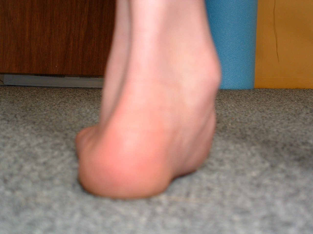
Hmmm…. Rearfoot Valgus
(Make sure to hover mouse over each image to examine more closely)
When the rearfoot is everted with respect to the fore foot. (wondering what this means? maybe you need to view our upcoming video course on foot types!)
Cardinal signs and pathomechanics
- Everted (heel is collapsed inward as in the pics above)
- midfoot/arch collapse: insufficient foot tripod
- due to midfoot collapse, the foot is in excessive pronation and is poor lever for toe off and propulsion
- Excessive internal rotation of the limb during gait cycle
Should you give up on fixing this? NO!
Should you put them in orthotics? Maybe
Should you sterilize them so they can’t reproduce? Definitely not!
Is there help for this? Of course!
What would you do? Think about that this weekend and tune in next week for some treatment ideas.
See you on the blog next week
Have a great Saturday.
Ivo and Shawn
OK, something different this Friday.
We admit computer models can only approximate human gait, and here is a perfect example. Watch the video a time or 2 and come back and read on….
Really….Did you watch it? Maybe you really should…
What do we see?
- A prime example of heel rocker but what is missing? How about midfoot pronation, a requisite for normal gait and one of the 4 shock absorbing mechanisms (pronation, ankle dorsiflexion, knee flexion and hip flexion)
- dip of the opposite hip with initial contact and loading response. looks like the computer model had built in gluteus medius weakness!
- what about that lack of anterior and posterior rock of the ilia?
- they do show g max activation (posterior view) during propulsion…nice!
- where are the abs initiating hip flexion?
- how about that forward head posture?
- we think there should be exaggerated torso rotation (contralateral to the side of strike) with no arms
So what does this prove?
This is a great attempt at simulating human gait, but gait being so complex and ….well….human, it is difficult to approximate with a computer modeling program.
We are the geeks of gait…The Gait Guys…Ivo and Shawn

We could have easily made this a blog post about shoe sizes or how to use the Brannock device. And maybe we will in time. But this picture, if you are really thinking, can give you more insight into the entire biomechanical flaw of a client. If you read our post today we bet you will forever look and compare the size of both feet of your clients … forever !
This is a picture of one of our patients. This person had a congenital “club foot” at birth also know as congenital talipes equinovarus (CTEV). It is a congenital deformity involving one or both feet. In this case it affected on the right foot (the smaller one). Multiple surgeries were performed at an infant to correct, and the correction is beautiful as these things go. TEV is classified into 2 groups: Postural TEV or Structural TEV.
That all aside, we have a smaller shorter right foot.
Where are we going with this ?
Foot size is often measured with the Brannock device in shoe stores, you know, the weird looking thing with the slider that measures foot length and width. In this case, the right heel:ball ratio, the length from the heel to the first metatarsal head, is shorter. The heel:toe length is also shorter, nothing like stating the obvious ! IF they are shorter then the plantar fascia is shorter, the bones are shorter, the muscles are smaller etc.
So, taking yesterday’s blog post in tow here (LINK to that posting), the maximal height of the arch on the right when the foot is fully supinated is less than that of the left side when also fully supinated (ie. during the second half of the stance phase of gait). Even with maximal strength of the toe extensors which we spoke of yesterday will not sufficiently raise the arch on the right to the degree of the left.
- Thus, this client is very likely to have a structural short leg. Certainly you must confirm it but you will likely see it in their gait if you look close enough.
- Also, you must remember that the shorter foot will also spend fractionally less time on the ground and will reach toe off quicker than the left. This may also play into a subtle limp.
- This client may have a mal-fitting shoe, the right foot will swim a little in a shoe that fits correctly on the left. You may be easily able to remedy all issues with a cork full length sole insert lifting both the heel and forefoot. This can negate the shoe size differential, change the toe off timing and remedy much of the short leg issue. * IMPORTANT: keep in mind, if you know your shoe anatomy (and you will if you get on board with our very soon to release “Shoe Fit Course”) you will know that the right foot at the metatarsal-phalangeal joint bending line will not be flexing where the shoe flexes on that right foot. The Right foot will be trying to bend proximal to the siping line where the shoe is supposed to naturally bend. This will place more stress into that foot. This brings up the rule for shoe fit: never size a persons shoe by pinching the toebox to see if there is ample room, the shoe should be fit to meet the great toe bend point to the flex point of the shoe.
- Strength of muscles is directly proportional to the cross sectional area of the muscle. With smaller muscles, this right limb is very likely to be underpowered when compared to the left.
- All of these issues can cause a failure of symmetrical hip rotation and pelvic distortion patterning.
- Altered arm swing (most likely on the contralateral side) is very likely to accommodate to the smaller weaker right lower limb. Do not be surprised to hear about low back pain or tightness or neck/shoulder issues.
- A shorter right leg, due to the issues we have discussed above, will place more impact load into the right hip ( from stepping down into the shorter leg) and more compressive load into the left hip (due to more demand on the left gluteus medius to attempt to lift the shorter leg during the right leg swing phase). This will also challenge the pelvic symmetry and can cause some minor frontal plane lumbar spine architecture changes (structural or functional scoliosis…… if you want to drop such a heavy term on it).
Gait plays deeply into everything. Never underestimate any asymmetry in the body. Some part as to take up the slack or take the hit.
Shawn and Ivo…….. far from symmetrical lads.
Gait Parameter: Ankle Rocker during the Squat as a predictor for Shin Splints.
Here is a brief video we shot in our clinic. One of the primary assessments we do with all clients is a basic squat. No a “potty squat” were the tibia remains vertical and the hips press backwards, just a basic squat where the knees come forward. We do this with toes down and toes up.
We shot this video so that we could have some visual to talk about a few things.
1. Why toes up ? You have read it here before on our blog. Raising the toes is done by use of the log and short toe extensor muscles (Extensor digitorum longus and brevis, EDL, EDB and of the hallux extensors EHL, EHB). When we activate the extensors the toes dorsiflex around the metatarsals and the toes elevate. This activates the windlass mechanism. This mechanism tightens the plantar fascia thus shortening the distance between the metatarsal heads and the heel. Thus, the arch is driven up. This is why we harp on gaining toe extensor strength in flat footed and hyperpronators. Go ahead, stand up, raise your toes and feel the arch lift. It is a solid biomechanical phenomenon.
So, why do the squat with the toes up ?
Because when the foot is weaker than it should be a squat can allow the arch to drop too much during the down-squat. If the arch drops the foot could pronate more than necessary. This can drive subtalar joint motion which can fake out the true squat determination and the true determination of available ankle rocker. The client will be able to get deeper into the squat but for assessment purposes this will be a fake out. We want to know true available functional range at the ankle mortise joint (tibial talar joint). With the toes up, the arch is maximized and cannot drop unless the toes drop. As you will see in this video, you can thus see the true ankle rocker in this client is barely sufficient however it is likely enough (100-110 degrees) for normal gait in the sagittal plane.
What if when they do this there is little if any rocker, less than this guy?
Then to get more (100-110, ie. 10-20 degrees past vertical) they will have to compensate. We talk about the strategies in this old video of ours (LINK HERE). One of the best ways to compensate is to pronate through the arch more than normal. This will drop the arch height and carry the tibia forward enough to allow for forward motion. Sadly, this increased pronation can do alot of things. One is to carry the knee medially and this can create patellar tracking issues or IT band tightness, to name just a few.
So, what is our point today ?
- You need to make sure your assessments are telling you what you need them to tell you.
- Sufficient toe extensor strength and range is critical in the gait cycle to ensure sufficient ankle rocker occurs at the tibial-talar joint and not somewhere else you do not want it ( a compensation). Any strength you put into a client who has insufficient true ankle rocker is strength into a compensation pattern. Can you say heightened eventual injury risk ?
- Ability to find the foot tripod is a skill. It needs to be developed in a simple skill like we show here and then the sensation can be carried forward into gait and running.
- A forefoot varus or forefoot valgus (please read our foot type blog posts over the past 3 weeks) can impair the foot tripod and thus the true ankle rocker.
- Make sure the knees hinges straight forward in this ankle rocker-squat test. If it is not a forward bend you must consider foot pronation excess, tibial torsion, hip version or torison, or simply the weak foot issues we are talking about here today.
- This is a form of homework for our clients, just want you see above in the video. We add layers to this as the gain strength. But that is a topic for another day.
This is a huge predictor and problem in chronic shin splints ? You bet ya it is ! It may be the main missed deficit we see in shin splints (both anterior and posterior shin splints). There is lots more to this topic, but we will stop here for today.
Shawn and Ivo…….. you have to know what you are seeing. And as Johnny Nash once said in his song
I can see clearly now, the rain is gone,
I can see all obstacles in my way
Gone are the dark clouds that had me blind
It’s gonna be a bright (bright), bright (bright)
Sun-Shiny day now that i understand ankle rockers better.“:-)


Retail Focus: Last (No, not the last retail focus, but a retail focus on lasts)
Lasts: What you need to know
Strictly speaking, a last is the mold or template for creating the shoe. It defines the shape of a shoe. We remember that men’s and women’s feet are shaped differently. Men (usually) have rectangular feet (the forefoot and heels are wider, or have less difference in width); Ladies (usually) have triangular feet (the forefoot is much wider than the heel). This is why it is important to know if the shoes you are fitting are a men’s or women’s specific last. Many thimes, the shoes come off the production line and the boy shoes are blue and the girls pink: both made from the same last.
The last determines whether a shoe is a high, medium or low volume shoe… Pretty important, if they have a high instep or flat foot. Companies like Altra have as many as 6 different, sex specific lasts. This results in a wide range of fit (and thus a bigger market share).
Take off your clients shoes and look at their feet. Note their shape and curve. Lasts need to match that “curve” so they can be relatively straight or curved (this refers to the shape of the “sole” of the shoe: see above). Turn a shoe over and look at the sole. Mentally bisect the heel with a line going to the front of the shoe. If the line bisects the front of the shoe, it is a straight lasted shoe (this corresponds to the axis of the 2nd metatarsal, or slightly lateral to it). If more of the shoe falls medial to this (more of the sole on the big toe side) it has a curved last.
Curved last shoes can vary in the degree of curvature. Curved last shoes are designed to help control pronation, as they provide medial support and slow its rate by causing a relative supination of the foot after heel strike (it weights the lateral border of the shoe for a longer period of time, theoretically allowing less pronation). Curved last shoes can put more motion into a foot, especially one with limited rearfoot motion (it still must pronate, but due to the lack of rearfoot motion, the forefoot must compensate and now must do so in a shorter period of time).
“Last” also refers to the material (or way that the material) overlays the midsole of the shoe. This “last” (look inside the shoe on top of the shank) is the surface that the insole of the shoe lays on, where the sole and upper are attached). Shoes can be board lasted, slip lasted or combination lasted.
A board lasted shoe is very stiff and has a piece of cardboard or fiber overlying the shank and sole (sometimes the shank is incorporated into the midsole or last). It is very effective for motion control (pronation) but can be uncomfortable for somebody who does not have this problem.
A slip lasted shoe is made like a slipper and is sewn up the middle. It allows great amounts of flexibility, which is better for people with more rigid feet.
A combination lasted shoe has a board lasted heel and slip lasted front portion, giving you the best of both worlds (theoretically).
A general rule of thumb is: You really can’t go wrong with a straight last. It will work for most feet, especially if you are using an orthotic. This is especially important with people with forefoot abductus, moderate to severe pronators and rigid feet (rear or forefoot). A forefoot abductus and severe pronator’s feet will move laterally in the shoe, often causing crushing, rubbing, cramping and blistering of the little toe against the side of the shoe. A rigid foot, because the foot needs to be able to pronate at the mid and forefoot, will have a similar problem. You can use a curved last with people with mobile or hypermobile feet, provided their pronation is not too severe (clinical judgment, trial and error).
We hope this clarifies some of the issues surrounding lasts, their shape, and usage. This will probably not be the last word on lasts, but hopefully will suffice some of the burning curiosities surrounding the subject.
We want to succeed as retailers. Consider taking our course and getting IFGEC certified, for your benefit as well as your clients.
Ivo and Shawn: The Gait Guys


What’s your foot type: Part 4
Forefoot valgus.
In this foot type, the fore foot is everted with respect to the rear foot and the little toe side of the tripod cannot gain purchase on the ground. This foot is a poor lever. The problem here is that in normal ambulation we progress our body mass lateral to medial, which engages normal biomechanics. In this foot, the body moves from medial to lateral, so we are unable to toe off from the bog toe side of the tripod. Lack of optimal toe off means poor propulsion strategies from the calf and gluteals. Consequently, patellar tracking is challenged, the limb is in a more relative external rotation, and the peroneal muscles are typically overburdened in an attempt to stabilize the lateral ankle area.
Missing something? Check out the last 3 weeks posts on foot types. Our shoe fit program is launching soon. You too can become certified and become “all that you can be” in shoe fitting.
Ivo and Shawn: Bald…Middle aged…Geeky…Good Looking….promoting foot and gait literacy here on a daily basis

Barefoot Versus Running Shoes: Which Is (Surprisingly) More Efficient?→
/Many folks extol the virtues of barefoot or minimal running shoes and or styles. We have contended that you often need to “earn the right” to be able to do this through our mantra “skill, endurance, strength”.
Here is an interesting take by Alex Hutchinson from Runner World and his review of Franz, Wierzbinski and Kram’s study published in Medicine and Science in Sports and Exercise, explaining why, metabolically speaking, shod running may be more efficient
The Gait Guys: sifting and surfing so you don’t have to…


Plantar Fascitis?
You’ve got plantar fascitis? We’ll try steroid injections. If that does not work, no problem, we’ll just cut it out….
Ah, yes…..Nothing like cutting one of the main stabilizing influences for the foot (via the windlass mechanism) to accomplish your goals. We sure are glad they used dead feet in this study!
And now, here is more evidence that those ligaments play a significant role (along, of course, with competent musculature) in stability of the foot.
The conclusion: “The data suggest that operations involving fasciotomy affect arch stability and should not be performed in patients with evidence of concomitant pes planus deformity, because of the likelihood of further deformation.”
The Gait Guys: Just the facts, so you can make more educated decisions..
Foot Ankle Int. 1997 Jan;18(1):8-15.
Mechanical behavior of the foot and ankle after plantar fascia release in the unstable foot.
Department of Orthopedics, Mayo Clinic, Rochester, Minnesota, USA.
AbstractThe change in position of the bones of the foot was studied in three dimensions after plantar fascia release in intact and destabilized feet. Fifteen fresh-frozen human foot specimens were used. Physiologic loads of 445 newtons were applied axially to simulate standing at ease, and the three-dimensional position of tarsal bones was determined with a magnetic tracking device. The positions were presented in the form of screw axis displacements, quantitating rotation, and axis of rotation orientation. After fasciotomy in the six intact feet, significant differences in rotation were observed at the talotibial and calcaneotalar levels. After fasciotomy in the four unstable feet with three supporting elements sectioned, significant differences in position were observed at the talotibial joint and a significant decrease in arch height was observed. After fasciotomy in the five unstable feet with five supporting elements sectioned, significant differences in rotation were observed at the talotibial joint (mean, 5.5 +/- 1.6 degrees; P = 0.001), calcaneotalar joint (mean, 6.1 +/- 2.1 degrees; P = 0.003), and metatarsotalar level (mean, 9.3 +/- 4.1 degrees; P = 0.007). The average decrease in arch height was 7.4 +/- 4.1 mm (P = 0.015). Displacement of all joints tested occurred after fasciotomy, with rotation about all three axes. These changes in displacement were more pronounced in unstable or destabilized feet. The data suggest that operations involving fasciotomy affect arch stability and should not be performed in patients with evidence of concomitant pes planus deformity, because of the likelihood of further deformation.


