Skeletal muscle in running.

Footnotes 7 - White and Red.jpg

"skeletal muscle may provide a fundamentally different mechanical function during running than during activities such as swimming,flying, or jumping. These activities require muscle contractions that produce mechanical work at high rates to overcome drag,produce lift, or accelerate the body. Because running requires negligible work against drag and the average mechanical energy of the body is constant over time (at a steady speed on level ground), the demands of support may be met most economically by muscles that produce force while minimizing mechanical work."- Roberts et al


Muscular Force in Running Turkeys:
The Economy of Minimizing Work
Thomas J. Roberts,* Richard L. Marsh, Peter G. Weyand,
C. Richard Taylor

A great paper on Hallux Limits

Don't let the title fool or dissuade you. 

Here is a great paper to support the post earlier this week on hallux limitus. 

Plantar pressure distribution in older people with osteoarthritis of the first metatarsophalangeal joint (hallux limitus/rigidus)

No surprise that the study found folks with osteoarthritis of the the 1st MPJ had greater maximum force and peak pressures under the hallux as well as the lesser toe than controls. 

BUT here is one of the gems from the study: " However, the plantar pressure changes observed in this study can be explained using the concept of high- and low-gear push off described by Bojson-Moller. This model suggests that there are two metatarsal axes through which propulsion may occur; a transverse axis connecting the first to second metatarsal heads, and an oblique axis connecting the second to fifth metatarsal heads. In the presence of normal first MPJ motion, a ‘‘high-gear’’ push- off occurs through the transverse metatarsal axis, resulting in an efficient transfer of bodyweight. In the presence of restricted first MPJ motion, propulsion through the transverse axis is not possible. Subsequently, a ‘‘low-gear’’ push-off occurs through the oblique axis, which subjects the lateral forefoot and toes to increased loading and results in hyperextension of the interphalangeal joint of the hallux prior to toe-off."

Zammit, G. V., Menz, H. B., Munteanu, S. E. and Landorf, K. B. (2008), Plantar pressure distribution in older people with osteoarthritis of the first metatarsophalangeal joint (hallux limitus/rigidus). J. Orthop. Res., 26: 1665–1669. doi:10.1002/jor.20700.    

link to FREE FULL TEXT: http://onlinelibrary.wiley.com/doi/10.1002/jor.20700/epdf


When the boot is the cause of your client's problems/pain.

Our Patreon site is LIVE.
Patreon Sampler video,
Here's the kind of exclusive stuff you'll get as a Patreon sponsor, even at the 1$ and 5$ tier levels. Stuff right from our clinics, right when we see them, as fresh as content can get.
Of course, the 10$, 20$ and 50$ tier contributors will get this stuff too, but also "juicier" stuff , so if you don't wanna miss a step, come on over to our Patreon page and join even the lowest tiers, become part of a more intensive advanced focus group with ivo and I weekly ! (And in the process you'll be helping us keep new, advanced stuff coming your way).

Asymmetry seems to matter with pathology.

image credit: https://commons.wikimedia.org/wiki/File:PSM_V46_D167_Outer_surface_of_the_human_brain.jpg

image credit: https://commons.wikimedia.org/wiki/File:PSM_V46_D167_Outer_surface_of_the_human_brain.jpg

When you have low back pain, your gait is apt to be asymmetrical

...And that is just what this study showed. It looked at 82 right leg dominant folks with slightly less than 1/2 of them havong low back pain. The folks with lower back pain spent more time on their non dominant leg at the beginning of a gait cycle and on their dominant leg at the end of it. Not surprising that they wanted to find a more stable base or center their COP over the weight bearing foot, especially in light to the fact that the back has such poor cortical representation.

Sung PS, Danial P. A Kinematic Symmetry Index of Gait Patterns Between Older Adults With and Without Low Back Pain. Spine (Phila Pa 1976). 2017 Dec 1;42(23):E1350-E1356. doi: 10.1097/BRS.0000000000002161.

Want to bring out gait pathology? Add something new into the mix...

image source: https://en.wikipedia.org/wiki/Walking

image source: https://en.wikipedia.org/wiki/Walking

We have talked about bringing out compensations and asymmetries in gait patterns by adding a novel stimulus to the exam, like having the client/patient put their hands over their heads, or close their eyes. Here is yet another tool for your toolbox: having the client walk backwards.

Both forward and backward walking share pattern generation control circuits in the brainstem, providing similar (though reversed) kinematic patterns. Backward walking requires different muscle activation sequences which can highlight subtle gait asymmetries, particularly in individuals that have cortical impairment (like the kids with cerebral palsy in this paper) or perhaps people with more subtle cortical impairments, like cerebellar dysafferentation from abnormal joint and muscle mechanoreceptor input and integration. Don’t believe us or what the study says, try it on yourself! It can be a humbling experience : )

In part, the study concludes: “The observed spatiotemporal asymmetry assessments may reflect both impaired supraspinal control and impaired state of the spinal circuitry.”

The next time you are having a difficult time seeing something in an evaluation, or are trying to bring out an asymmetry, in addition to your other tricks, have them walk backwards.

Cappellini G, Sylos-Labini F, MacLellan MJ, Sacco A, Morelli D, Lacquaniti F, Ivanenko Y. Backward walking highlights gait asymmetries in children with cerebral palsy. J Neurophysiol. 2018 Mar 1;119(3):1153-1165. doi: 10.1152/jn.00679.2017. Epub 2017 Dec 20.

Progressions and injuries

Progressions and injuries.
If your client cannot demonstrate a competent foot tripod, and they load eccentrically too long into the tib posterior, peroneus longus, and they load too much through the arch and blow into too much arch splay (loosely/slang meaning beyond reasonable pronation limits) and perhaps they evert the calcaneus a little too much.......

If . . . . if they cannot do these things properly, soundly and in a controlled fashion during a double support (standing on 2 feet) demonstration, through a simple standing knee and hip bend . . . if they cannot control their feet in this simple skill . . . .

Then, how in the heck can they do this standing on one leg,? how are they going to do it in a lunge? or as they step forward onto the foot ?How in the world will they do it walking ?
How in the world will they do it properly, soundly, running ? Squatting? Deadlifting? jumping ? or or or or . . . .

if you are wondering why your client has problems, maybe it is because they are doing things way beyond the pay grade of their foot's (limb's) abilities, skills, endurance, strength etc. If you do the simple stuff wrong, in a cheating corrupt fashion, you a will certainly do it that way when things really matter (running, lifting, playing sports).
Sometimes you have to start at the beginning, at the starting line. If your client is having pain and problems, more load doesn;t necessarily make it right. Proper loading, progressively introduced, might however.

Podcast 141: Deep dive podcast (lots of random gait and running thoughts).

Topics:

strength, asymmetry, gait, thegaitguys, gaitanalysis, achilles, windlass mechanism

Links to find the podcast:

Look for us on iTunes, Google Play, Podbean, PlayerFM and more.

Just Google "the gait guys podcast".

Our Websites:
www.thegaitguys.com

doctorallen.co

summitchiroandrehab.com

shawnallen.net

Our website is all you need to remember. Everything you want, need and wish for is right there on the site.
Interested in our stuff ? Want to buy some of our lectures or our National Shoe Fit program? Click here (thegaitguys.com or thegaitguys.tumblr.com) and you will come to our websites. In the tabs, you will find tabs for STORE, SEMINARS, BOOK etc. We also lecture every 3rd Wednesday of the month on onlineCE.com. We have an extensive catalogued library of our courses there, you can take them any time for a nominal fee (~$20).

Our podcast is on iTunes and just about every other podcast harbor site, just google "the gait guys podcast", you will find us.

Where to find us, the podcast Links:

iTunes page:

https://itunes.apple.com/us/podcast/the-gait-guys-podcast/id559864138?mt=2

Google Play:

https://play.google.com/music/m/Icdfyphojzy3drj2tsxaxuadiue?t=The_Gait_Guys_Podcast

Direct download URL: http://traffic.libsyn.com/thegaitguys/pod_141_real_final.mp3

Permalink URL: http://thegaitguys.libsyn.com/podcaset-141-a-deep-dive-podcast-lots-of-random-gait-and-running-thoughts-0

Libsyn Directory URL:http://directory.libsyn.com/episode/index/id/7670285

Topics and links:

A new neuron has been discovered and named. The rosehip cell
https://www.wired.com/story/meet-the-rosehip-cell-a-new-kind-of-neuron/

Strength does matter in our opinion, especially when it is stacked onto asymmetry and dysfunction.
https://www.ncbi.nlm.nih.gov/pubmed/26566993

Am J Sports Med. 2016 Jan;44(1):105-12. doi: 10.1177/0363546515611632. Epub 2015 Nov 13.
Is Hip Abduction Strength Asymmetry Present in Female Runners in the Early Stages of Patellofemoral Pain Syndrome?
Plastaras C1, McCormick Z2, Nguyen C3, Rho M2, Nack SH2, Roth D4, Casey E5, Carneiro K6, Cucchiara A1, Press J2, McLean J7, Caldera F8.

Positive Work Contribution Shifts from Distal to Proximal Joints during a Prolonged Run
SANNO, MAXIMILIAN1,2; WILLWACHER, STEFFEN1,3; EPRO, GASPAR1,4; BRÜGGEMANN, GERT-PETER1,2,3
Medicine & Science in Sports & Exercise: December 2018 - Volume 50 - Issue 12 - p 2507–2517
https://journals.lww.com/acsm-msse/Citation/2018/12000/Positive_Work_Contribution_Shifts_from_Distal_to.16.aspx

Influence of the windlass mechanism on arch-spring mechanics during dynamic foot arch deformation
Lauren Welte, Luke A. Kelly, Glen A. Lichtwark, Michael J. Rainbow
Published 15 August 2018.DOI: 10.1098/rsif.2018.0270
http://rsif.royalsocietypublishing.org/content/15/145/20180270

Med Sci Sports Exerc. 2018 Dec;50(12):2500-2506. doi: 10.1249/MSS.0000000000001710.
Reducing Impact Loading in Runners: A One-Year Follow-up.
Bowser BJ1, Fellin R2, Milner CE3, Pohl MB4, Davis IS5.
https://www.ncbi.nlm.nih.gov/pubmed/29975300/

Achilles tendon structure differs between competitive distance runners and nonrunners despite no clinical signs or symptoms of midsubstance tendinopathy
Todd J. Hullfish,Kenton L. Hagan, Ellen Casey, Josh R. Baxter
09 Aug 2018https://doi.org/10.1152/japplphysiol.00012.2018
https://www.physiology.org/doi/abs/10.1152/japplphysiol.00012.2018

Our obligatory disclaimer:
This podcast is for general informational purposes only. It does not constitute the practice of medicine, nursing, rehab, treatment, therapy recommendations or anything of the sort. This podcast should not replace proper medical advise that should only be attained through proper medical channels that would entail a full medical and/or biomechanical physical examination and/or appropriate diagnostic testing. No doctor-patient relationship is formed by listening to this podcast or any information gleaned from our writings or social media work.
The use of this information and the materials linked to the podcast is taken at the users own risk. This podcast and the content shared is not intended to replace or be a substitute for appropriate professional medical advise diagnosis or treatment. Users should not disregard or delay obtaining medical advice for any condition they have and should seek the advice and assistance from their providers for any such conditions.

More subtle clues..LLD's

IMG_5518.jpg
IMG_5515.jpg
IMG_5521.jpg

This gentleman presented to the office with left-sided knee pain at the medial collateral ligament following a cutting injury, moving from right to left with the left foot planted. As you can see, he has an anatomical leg length discrepancy with tibial and probable femoral length deficiencies on the left side. Can you see the subtle, increased tone of the long flexors of the toes on the left hand side as it evidenced by the increased prominence of the long extensor tendons to a greater degree on the shorter side? This is a common compensation seen in true leg length discrepancies with clawing of the toes in attempt to create stability on the shorter leg side. Often times, the progression angle on the shorter side will be increased as well.

Pincer toe nails.

Screen Shot 2018-11-11 at 8.47.23 AM.png

Pincer Toe nails: You've seen them; did you know what they were and how they got that way? Or, did you dismiss them?

We think Hitomi’s hypothesis is correct. Here is why (this is paraphrased from our blog post on subungal hematomas and our revolutionary thinking on why they occur and it seems to fit well with pincer nail formation as well).

… when the skin is pulled at a differential rate over the distal phalange (from gripping of the toes rather than downward pressing through the toe pad) there will be a net lifting response of the nail from its bed as the skin is drawn forward of the backward drawn phalange (there is a NET movement of skin forward thus lifting the nail from its bedding). For an at-home example of this, put your hand AND fingers flat on a table top. Now activate JUST your distal long finger flexors so that only the tip of the fingers are in contact with the table top (there will be a small lifting of the fingers). There should be minimal flexion of the distal fingers at this point. Note the spreading and flattening of the nail. Now, without letting the finger tip-skin contact point move at all from the table, go ahead and increase your long flexor tone/pull fairly aggressively. You are in essence trying to pull the finger backward into flexion while leaving the skin pad in the same place on the table. Feel the pressure building under the distal tip of the finger nail as the skin is RELATIVELY drawn forward.] This is fat pad and skin being drawn forward (relative to the phalange bone being drawn backward) into the apex of the nail. Could this be magnifying the curvature of the nail and not offsetting the “automatic curving and shrinkage” function of the nail ? We think it is quite possible.

We have more to say on this topic, the above is just an excerpt of our blog post. More here, in the link below

https://thegaitguys.tumblr.com/post/127638788139/pincher-nails-who-knew-written-by-dr-shawn?fbclid=IwAR06ol516n9WF2Qh5TadlKd8esXrH5pVviycT_7QiMeScL0UJ3H9r1FF_OQ

Do you have dorsal (top) foot pain? Think you are tying your shoes too tightly?

Do you have dorsal (top) foot pain, at the peak of the arch? Think you are tying your shoes too tightly and that is the cause? Do you have pain over the dorsal or plantar mid foot on heel rise or jumping/landing or going up stairs ?

Just because you raise your heel and load the ball of the foot does not necessarily mean you have adequately plantarflexed the 1st metatarsal and loaded it soundly/stable with the medial tarsal bone. Heel rise, and thus loading onto the medial foot tripod, must be met with ample, stable, durable, 1st metatarsal plantarflexion and the associated medial tarsal bones. Also, without this, loading of the sesamoids properly cannot occur, and pain may ensue.

The first ray complex can be delicate in people who are symptomatic. In some people who do not have a good tibialis posterior-peroneus sling mechanism working harmoniously, in conjunction with a competent arch tripod complex to achieve a compentent arch complex (ie, EDL, EHL, tib anterior and some of the other foot intrinsics) this tarsometatarsal interval can become painful and instead of the 1st ray complex being stable and plantarflexing as the heel departs and the 1st ray begins taking load, it may not do so in a stable plantarflexed posturing. In some people it can momentarily dorsiflex as the arch subtly collapses (when it should be stable and supinated in heel rise).

"Subtle hypermobility of the first tarsometatarsal joint can occur concomitantly with other pathologies and may be difficult to diagnose. Peroneus Longus muscle might influence stability of this joint. Collapse of the medial longitudinal arch is common in flatfoot deformity and the muscle might also play a role in correcting Meary's angle."-Duallert et al

Soon, I hope to show you a video of how to watch for this problem, how to train it properly, how we do it in my office.
Dr. Allen

https://www.ncbi.nlm.nih.gov/pubmed/27015031

Clin Biomech (Bristol, Avon). 2016 May;34:7-11. doi: 10.1016/j.clinbiomech.2016.03.001. Epub 2016 Mar 10.

The influence of the Peroneus Longus muscle on the foot under axial loading: A CT evaluated dynamic cadaveric model study.

Dullaert K1, Hagen J2, Klos K3, Gueorguiev B4, Lenz M5, Richards RG6, Simons P7.

Your center of mass in relation to foot strike position.

For those arm swing/pulsers/ COM and head over foot folks consider some more research below.
Let the CNS drive the show, it is what it is there for . . . The leg motor patterns are dominant, the arms are passive and "shape" and influence the leg swing as a balance and ballast effect. As we discuss in an upcoming podcast, to cross the arms in a pumping motion across the midline of the body means one has to have compromised scapular mechanics (mostly protraction) to afford that much humeral adduction. This means we are forcing thoracic rotation as well. This means we are reversing what we know is more true, that "arm motion is driven passively by rotation of the thorax (Pontzer et al., 2009), an idea which is supported by shoulder muscle EMG data" (and not thoracic rotation by arm swing). Why would we try to create more unnatural axial spin through the spine when we are actually trying to move forward in the sagittal plane? Why would we try to force more rotation through the spine when the function of the thoracopelvic canister (ie. the core) is to stabilize rotational /angluar momentum? Hmmmm, things to ponder.

"Previous modelling studies have clearly shown that motion of the arms effectively counterbalances the angular momentum of the lower extremities during running (Hamner & Delp, 2013; Hamner et al., 2010). It has further been suggested that arm motion is driven passively by rotation of the thorax (Pontzer et al., 2009), an idea which is supported by shoulder muscle EMG data, consistent with the shoulders as spring-like linkages (Ballesteros, Buchthal, & Rosenfalck, 1965). Our data are con- sistent with this idea, showing motion of the thorax to be in the opposite direction to that of the swinging leg. Pontzer et al. (2009) also suggested that motion of the thorax is driven passively by motion of the pelvis. However, our data shows that the thorax reaches its peak angular velocity earlier than the pelvis, indicating that thorax motion is not completely passively driven by pelvic movements."

-S.J. Preece et al. / Human Movement Science 45 (2016) 110–118

Fatigue matters. Today's article looks at pre and post exercise fatigue and how, on EMG, our body changes.

Photo credit: pixabay.com

Photo credit: pixabay.com

Even for those of us who do (and should) know better, "the problem is, we are all often knee deep into compensations before we are aware of it, so most of us are always working on adding strength and endurance into our compensations without even knowing it. Our workouts layer things deeper. Yes, almost all of us are on this bus. Don't deny it. The next time you feel that tightness in your shoulder, or in your hip, or feel that tightness or soreness on one side of the low back, or one side of the neck, stop, and ask yourself that honest question. Again, you are on the bus with the rest of us."

We have spent much time discussing our order of things when intervening between a person and what ails them. Namely, our order is to first restore proper skill and patterning, then add endurance (move well often), and then add load, namely strength, power, force, explosive movements and the like. So, Skill, Endurance, Strength. This is a neurologic order, there is good reason for the necessity of this order. We have spend many an hour listening to Dr. Ivo explain why the CNS dictates this is the order with good reason. Cheat this order and you lay down neuroplastic patterns that are anything but what you want for your client. Enough said.

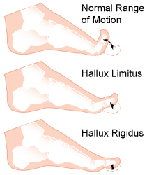
Today we introduce and article that the looks at the lumbo-pelvic-hip complex, a very complicated area, subject to large multi-planar movements and distortions (and hence, large complex multi-planar compensations). We must have good skill, endurance and strength in controlling this massive area safely, meaning, to avoid developing cheating compensatory patterns to negotiate around our days and activities and sports. The problem is, we are often knee deep into compensations before we are aware of it, so most of us are always working on adding strength and endurance into our compensations without even knowing it. Yes, almost all of us are on this bus. Don't deny it. The next time you feel that tightness in your shoulder, or in your hip, or feel that tightness or soreness on one side of the low back, or one side of the neck, stop, and ask yourself that honest question. Again, you are on the bus with the rest of us.

Today's article looks at pre and post exercise fatigue and how, on EMG, our body changes. Now keep in mind, and I will remind you of this again at the end of today's writing, keep in mind of the asymmetries, poor-skill, poor-endurance and poor strength in some areas that pre-exist, before even starting into our exercises. Imagine, assume, that these were there in all of this study's subjects, even prior to the exercise challenge. You should now fully grasp how layered things get for our clients.

Here is what the article said,

"fatigue may affect muscle recruitment, active muscle stiffness and trunk kinematics, compromising trunk stability".-Chang et al.

"The purpose of this study was to compare trunk muscle activation patterns, and trunk and lower extremity kinematics during walking gait before and after exercise. Surface electrodes were placed over the rectus abdominis, external oblique, erector spinae, gluteus medius, vastus lateralis, and vastus medialis of twenty-five healthy indviduals."

"The amplitude increased in the rectus abdominis during loading, midstance , terminal stance, and late swing after exercise. Amplitude also increased during swing phase in the erector spinae, vastus lateralis, and vastus medialis after exercise. There was less trunk and hip rotation from initial contact to midstance after exercise. Neuromuscular fatigue significantly influenced the activation patterns of superficial musculature and kinematics of the lumbo-pelvic-hip complex during walking. Increased muscle activation with decreased movement in a fatigued state may represent an effort to increase trunk stiffness to protect lumbo-pelvic-hip structures from overload."-Chang et al

What we found particularly notable was that they found less trunk and hip rotation from initial contact to midstance after exercise. And that, "neuromuscular fatigue significantly influenced the activation patterns of superficial musculature and kinematics of the lumbo-pelvic-hip complex during walking". As they concluded, increased muscle activation with decreased movement in a fatigued state plausibly indicates an effort to increase trunk stiffness as a protective measure. Translation, a protective compensation.

Here is what we have to say about that: do not leave the problem on the table and merely train your client around this. Resolve the underlying problem. The underlying problem may not, and likely will not, come out in a "functional screen". What will come out in the screen is how they are moving about with this existing compensation pattern(s). The screen shows WHAT they are doing with their limitations, not WHY Dive keep dear brethren. This is what it is all about, taking the time and diving deep. Find the "why".

So, as promised, here I am again, reminding you to keep in mind of the asymmetries, poor-skill, poor-endurance and poor strength in some areas that pre-exist, before even starting into our exercises. Imagine, assume, that these were there in all of this study's subjects, even prior to the exercise challenge. You should now fully grasp how layered things get for our clients.This is what can make, "helping someone get well", a difficult challenge, even on a good day.

*Muscle activation patterns of the lumbo-pelvic-hip complex during walking gait before and after exercise. Chang M1, Slater LV2, Corbett RO1, Hart JM1, Hertel J1.

Photo credit: pixabay.com Thank you for making such beautiful photos like this available for free use. Gorgeous photography !

How does hallux valgus and bunion formation cause toe hammering?

Screen Shot 2018-11-13 at 4.36.12 PM.png

Photo: you need to recognize this predictable pattern.

When the hallux begins to become incompetent, from perhaps pain, hallux rigidus, hallux limitus, bunion and in this case hallux valgus with bunion (in this case rotational instability) when this incompetence kicks in, we must find stability elsewhere. One will often, unconsciously, begin to increase the flexor tone and pressure to try and find stability since one cannot get it sufficiently from the hallux anymore.

Screen Shot 2018-11-16 at 6.28.27 PM.png


Here you see the tenting up of the 2nd toe, from increased long flexor activity (FDL) over time.
And here is the interesting thing you will notice, over time, the 3rd toe will start the same strategy, then the 4th. We see this often. It is not set in stone, but we see it a lot.
Notice it in your clients feet. Teach them why they are getting hammer toes, flexible ones at first, and possibly rigid ones over time. Hammer toes can have many causes, this is just one.
Solution: find a way to help your client re-find better hallux and medial forefoot stability to halt the progression.

How hallux valgus and bunions can affect the shoe toe box space.

Bunions and hallux valgus can change the toe box volume and shoe choice, so be careful, don't be fooled.
This photo shoes how a change in the forefoot width and length can be a result of a bunion or hallux valgus. Notice both feet are aligned the same, but the length of the foot is different in the hallux valgus foot.

Screen Shot 2018-11-16 at 6.23.31 PM.png


The old Brannock device use to help us all see this more clearly. You may recall that the device measured "heel to toe" (True foot length) and ALSO "heel to ball" length (the functional length and more important one. This length measured heel to the metatarsophlanageal joint line. This concept is important to know because we want the shoe "break point" or "bend point" at the forefoot to occur where the foot bends. Not all shoes have the flex lines (the creases on the bottom of the shoe were it is most likely to bend) in the same place, there is no standard. And if your client has shorter toes, longer toes or a long or short "heel to ball" length they man needs some help from a knowledgeable person like yourself making sure that their current forefoot complaints are not from a mis-fitted shoe.
Bottom line, the "heel to ball" length of a foot is far more important than the global foot length "heel to toe". So stop judging your shoe fit by pinching the front of the shoes to "make sure you have plenty of room"! Doh ! Face palm !

Because despite what many of the "experts" online are saying, that being "shoes don't matter". The fact is "sometimes they do". Period.

WAnt to learn this stuff? Got our website and buy the National Shoe Fit program. Hours of deep shoe, anatomy and biomechanics fun with ivo and shawn, in your own home over the holidays ! Give yourself the "gift" of ivo and shawn this year ! LOL

And for all of you who joined us last night on onlineCE.com for the 55 minute condensed nuclear version of the 3+ hours shoe fit program, we hope you have recovered with a good nights sleep !

GHS (generalized hypermobility syndrome) and foot loading.

GHS: Generalized Hypermobility Syndrome

Screen Shot 2018-11-16 at 6.20.23 PM.png

We have all seen these types of clients/patients. They have joints that have more than ample full range. It is easy for them to hyperextend their elbows and knees. They can fold over and touch their toes, everything seems lax and flexible. What they need is help gaining more control of their joints. But what about their gait ? Albeit a focal study, finally someone has looked at how these people interface the ground,

The forefoot region received higher loading in GHS clients.
So what could this mean? Does it mean they have challenges transitioning from rearfoot to forefoot? Does it mean their center of pressure is more foreward biased ? Does it mean they have to impart heavier loads through the forefoot during gait to feel stable? Are they premature heel raisers thus showing the forefoot bias increase? There are many questions here, too broad for this study, but they are the keys to understanding how the GHS body interprets movement. Regardless, it is highly suspect that these clients dominate their gait with the calf muscle complex doing plenty of extra work. We suspect they will be toe clenchers/grippers ( ie, have increased toe flexor dominance) and this can have long term impact on things like metatarsal loads, neuroma formation, bunions, lumbrical weakness, fat pad displacement, hammer toes and many other related issues that occur with premature or excessive forefoot loading. They may even have a little of that vertical bouncy gait we often discuss. We will keep our eyes open for this stuff and keep you in the loop.

J Back Musculoskelet Rehabil. 2018 Nov 2. doi: 10.3233/BMR-170973. [Epub ahead of print]
Generalized hypermobility syndrome (GHS) alters dynamic plantar pressure characteristics.
Simsek IE1, Elvan A1, Selmani M2, Cakiroglu MA2, Kirmizi M2, Angin S1, Bayraktar BA3.

Hip muscles may be smaller in unilateral OA.

Here, some "low quality evidence that specific hip muscles are smaller in unilateral hip OA".
Does this translate to osteoarthritis leads to muscular atrophy and thus weakness, or does this translate to muscle atrophy and weakness leads to OA ? Chicken or the egg ?
Sure, there is the whole trauma thought process, but that is not the discussion today.

Regardless, some possible implications here for targeted interventions in hip OA. This is why so many with arthritic joints improve with pointed, well thought out and target strengthening.

Muscle size and composition in people with articular hip pathology: a systematic review with meta-analysis

P.R. LawrensonEmail the author P.R. Lawrenson, K.M. CrossleyEmail the author K.M. Crossley, B.T. VicenzinoEmail the author B.T. Vicenzino, P.W. HodgesEmail the author P.W. Hodges, G. JamesEmail the author G. James, K.J. CroftEmail the author K.J. Croft, M.G. KingEmail the author M.G. King, A.I. Semciw

https://www.oarsijournal.com/article/S1063-4584(18)31512-7/fulltext?fbclid=IwAR2DTYxb2Azz4Df4I2Wy7CHst7egAg_x32DDH8DDfCbnAtYNf8h8iqlOQag

Adding strength to compensations and asymmetry.

FootNotes, with The Gait Guys.

Screen Shot 2018-11-13 at 4.36.12 PM.png

The CNS runs the show. Compensations are real, they are a calculated response, they are meaningful adaptive protective behaviors. Adding strength to a compensation makes them even more real, plastic, permanent. Fix the problem. Adding random strength is juvenile thinking. Letting one's client load/train/lift when in pain is juvenile (read below). Once we realize adding load to the problem can be helpful or hurtful, we are on the right path, we are winning. But it takes a deep understanding of how to add load safely, wisely, so that our client can benefit. We must try to understand adaptive behaviors, we must try to understand why our client's CNS made the choices it did.
Now, imagine a client with ankle pain, and resultant ankle dorsiflexion/ankle rocker loss. Now, imagine what their gait will look like as well with that premature heel rise and everything that adapts from that premature heel rise. Now, read below and understand one way how the CNS adapts. Why? So that the next time one chooses a stretch, mob, flossing, etc to gain a range of motion, without any additional meaningful measures, hopefully they will realize they are likely not addressing the deeper problem. Pushing a range of motion is not the same as safely re-earning a range of motion. Far from it. -Dr. Allen

*Effect of Achilles tendinosis on the agonist, synergist and antagonist muscles. Chang and Kulig

"In addition to the altered control system, the present study also observed an adaptive behaviour, as illustrated by the activity of agonist, synergist and antagonist muscles. This was seen during single-legged hopping, where the contribution from the triceps surae muscle to the plantar flexors was decreased and the co-contraction from the tibialis anterior muscle was also decreased on the involved side in individuals with Achilles tendinosis. This may be attributed to the protective mechanism shielding the already injured tendon from further injury or even rupture (Lund et al. 1991)."- Chang and Kulig

J Physiol. 2015 Aug 1; 593(Pt 15): 3373–3387.
Published online 2015 Jun 30. doi: [10.1113/JP270220]
PMCID: PMC4553058
PMID: 26046962
The neuromechanical adaptations to Achilles tendinosis
Yu-Jen Chang and Kornelia Kulig

Forget face recognition, gait recognition is here.

Gait recognition. Forget face recognition, your gait has its own neuroplastic pattern you have developed over the years. It is subconscious and like we repeatedly say, your gait is a representation of the parts that are working, not working and injuries you have compensated around. Sure, you can change your gait, but it is going to take retraining over 10+ weeks to adopt a new gait pattern and gait ID.

Remember this as well, coaching a gait form change does not fix anything, you are just teaching a new pattern on their existing patterns of ability and compensation. This is our main problem with basing diagnosis and corrective homework off of just a gait evaluation, they tell you nothing of what is wrong with your client, they only tell you what your client is presently doing.

https://abcnews.go.com/amp/Technology/wireStory/chinese-gait-recognition-tech-ids-people-walk-58988215?__twitter_impression=true&fbclid=IwAR2SvPXCBBya_P84THHtc6hdwvdjV47xn6JjFbRhaWZ_GCojarFxSrnnE_E