Movement, can it make us better humans ?

This will be the last blog post you read from us …  for 2017. Happy New year wishes to you all !

This is a rehash of some old stuff, and some new, it seems to bring together many good points and thoughts of our work this past year. We hope you agree. If this seems familiar for those who have been with us for the last 9 years, it is our typical year end post, but it is worth your time

We have an amazing video for you today, a testament to how amazing the human frame is and how amazing movement can be.  But first … . it has been an amazing year for both of us here at The Gait Guys. Through this year, we have bridged further chasms. Our podcasts went into high gear and our numbers continue to grow globally, we were blessed to know our voices spanned the miles into 90+ countries. The National Shoe Fit Certification Program continues to bring us deep gratitude emails from all professions. We blogged weekly , added some new videos and have made plans for more. We also made many new friends while learning much on our own end in our relentless research and readings. We appreciate every one of you who has followed us, and we thank you for your friendship.

As we find ourselves here at the end of another year, it is normal to look back and see our path to growth but to look forward to plan for ways to further develop our growth.  Many of you who read our blog are runners, but many of you are in one way or another involved with a sport or activity that incorporates running and gait. Hey, we all walk !  Even in the video above the dancers are seen running and walking. What we mean is that many of you are coaches or trainers or movement experts who develop those who run or move in one way or another in various sports, but many of you are also in the medical field helping those to run and move to get out of pain or improve performance.  And still yet we have discovered that some of you are in the fields of bodywork such as yoga, pilates, dance, martial arts and movement therapies.  It is perhaps these fields that we at The Gait Guys are least experienced at (but are learning) and like many others we find ourselves drawn to that which we are unaware and wish to know more in the hope that it will expand and improve that which we do regularly.  For many of you that is also likely the case. For example, since a number of you are runners we would bet to say that you have taken up yoga, pilates, lifting or cross training to improve your running and to reduce or manage injuries or limitations in your body. But why stop there ? So, here today, we will try to slowly bring you full circle into other fields of advanced movement. As you can see in this modern dance video above the grace, skill, endurance, strength, flexibility and awareness are amazing and beautiful. Wouldn’t you like to see them in a sporting event ? Wouldn’t you like to see them run ? Aren’t you at least curious ? Their movements are so effortless. Are yours in your chosen sport ? How would they be at soccer for example ? How would they be at gymnastics ? Martial arts ? Do you know that some of the greatest martial artists were first dancers ? Did you know that Bruce Lee was the Cha Cha Dance Champion of Hong Kong ? He is only one of many. Dance, martial arts, gymnastics …  all some of the most complex body movements that exist. And none of them are simple, some taking decades to master, if that, but most of which none of us can do. In 2018 we will continue to expand your horizons of these advanced movement practices as our horizons expand.  There is a reason why some of the best athletes in the NBA, NFL and other sports have turned to almost secret study of dance and martial arts because there is huge value in it.  Look at any gymnast, martial artist or dancer. Look at their body, their posture, their grace.  It is as if their bodies know something that ours do not.  And so, in 2018 The Gait Guys will dive even deeper into these professions to learn principles and bring them back to you. After all, everything we do is about movement. Movement is after all what keeps the brain alive and learning.

Below are excerpts from a great article from Kimerer Lamothe, PhD. She wrote a wonderful article in Psychology Today a few years ago  on her experience with McDougall’s book “Born to Run” and how she translated it into something more.  At some point, take the time to read her whole article.  But do not cut yourself short now, you only have a little more reading below, take the next 2 minutes, it might change something in your life.

We leave you now with our 2017 gratitude for this great growing brethren and community that is unfolding here at The Gait Guys. We have great plans for 2017 so stay with us, grow with us, and continue to learn and improve your own body and those that you work with.  Again, read Kimerer’s most excellent excerpts below, for now, and watch the amazing body demonstrations in the video above. It will be worth it.

Shawn and Ivo, the gait guys

_____________________

Can Running Make us Better Humans ?….. excerpts from the artcle by Kimerer LaMothe.

http://www.psychologytoday.com/blog/what-body-knows/201109/can-running-make-us-better-humans

“The Tarahumara are not only Running People, they are also Dancing People. Like other people who practice endurance running, such as the Kalahari Kung, dancing occupies a central place in Tarahumara culture. Or at least, it has. The Tarahumara dance to pray, to celebrate life passages, to mark seasonal and religious events. They dance outside where Father God and Mother Moon can see, in patterns consisting of steps and shuffles, taps and hops, performed in a line or a circle with others. And they dance the night before a long running race, while the native corn beer, or tesguino flows.

While McDougall notes the irony of “partying” the night before a race, he doesn’t ask the question: might the dancing actually serve the running? Might it be that the Tarahumara dance in order to run—to ensure the success of their run—for themselves and for the community?

At the very least, the fact that the Tarahumara dance when and how they do is evidence that they live in a world where bodily movement matters. They believe that how they move their bodies matters to who they are and to how life happens. They have survived as a people by adapting their traditional method of endurance hunting (running animals to exhaustion) to the challenges of fleeing Spanish invaders, accessing inaccessible wilderness, and staying in touch with one another while scattered throughout its canyons. As McDougall notes, they have kept alive an ancient genetic human heritage: to love running is to love life, for running enables life.

Yet McDougall is also clear: even the Tarahumara are not born knowing how to run. Like all humans, they must learn. Even though human bodies are designed to flourish when subject to the stresses of long distance loping, we still need to learn how to coordinate our limbs to allow that growth to happen. We must learn to run with head up, carriage straight, and toes reaching for the ground. We must land softly and roll inwardly, before snapping our heels behind us. We must learn to glide—easy, light, smooth—uphill and down, breathing through it all. How do we learn?

How do we learn to run? We learn by paying attention to other people, and taking note of the movements they are making. We learn by cultivating a sensory awareness of our own movements, noting the pain and pleasure they produce, and finding ways to adjust. We learn by creating and becoming patterns of movement that release our energy boldly and efficiently across space. We learn, in a word, by dancing.

While dancing, people open up their sensory selves and play with movement possibilities. The rhythm marks a time and space of exploration. Moving with another heightens the energy available for it. Learning and repeating sequences of steps exercises a human’s most fundamental creativity, operating at a sensory level, that enables us to learn to make any movement in any realm of endeavor with precision and grace. Even the movements of love. Dancing, people affirm for themselves and with each other that movement matters.

In this sense, dancing before the night of a running race makes perfect sense. Moving in time with one another, stepping and stretching in proximity to one another, the Tarahumara would affirm what is true for them: they learn from one another how to run.  They learn to run for one another. They run with one another. And when they race, they give each other the chance to learn how to be the best that they each can be, for the good of all.

It may be that the dancing is what gives the running its meaning, and makes it matter.

Yet the link with dance suggests another response as well. In order for running to emerge in human practice as something we are born to do, we need a culture that values movement—that is, we need a general appreciation that and how the bodily movements we make matter. It is an appreciation that our modern western culture lacks.

Those of us raised in the modern west grow up in human-built worlds. We wake up in static boxes, packed with still, stale air, largely impervious to wind and rain and light. We pride ourselves at being able to sit while others move food, fuel, clothing, and other goods for us. We train ourselves not to move, not to notice movement, and not to want to move. We are so good at recreating the movement patterns we perceive that we grow as stationary as the walls around us (or take drugs to help us).

Yet we are desperate for movement, and seek to calm our agitated senses by turning on the TV, checking email, or twisting the radio dial to get movement in a frame, on demand. It isn’t enough. Without the sensory stimulation provided by the experiences of moving with other people in the infinite motility of the natural world, we lose touch with the movement of our own bodily selves. We forget that we are born to dance and run and run and dance.

The movements that we make make us. We feel the results. Riddled with injury and illness, paralyzed by fears, and dizzy with exhaustion, our bodily selves call us to remember that where, how, and with whom we move matters. We need to remember that how we move our bodies matters to the thoughts we think, the feelings we feel, the futures we can imagine, and the relationships we can create with ourselves, one another, and the earth.

Without this consciousness, we won’t be able to appreciate what the Tarahumara know: that the dancing and the running go hand in hand as mutually enabling expressions of a worldview in which movement matters.”

Thanks for a great article Kimerer. (entire article here)http://www.psychologytoday.com/blog/what-body-knows/201109/can-running-make-us-better-humans

*oh, and want a little more of these performers in the video, check this out……. it will move you.

http://youtu.be/CvQBUccxBr4

Wishing a Happy New Year to you all, from our hearts……. Shawn and Ivo

The Gait Guys

Podcast 132: Arm swing, gait retraining and steroids.

Key Tag words:  thegaitguys, gait, gait analysis, arm swing, cortisone shots, corticosteroids, leg swing, running injuries

Links to find the podcast:

iTunes page: https://itunes.apple.com/us/podcast/the-gait-guys-podcast/id559864138?mt=2

http://traffic.libsyn.com/thegaitguys/pod_132f.mp3

http://thegaitguys.libsyn.com/podcast-132-arm-swing-gait-retraining-and-steroids

Libsyn Directory URL: http://directory.libsyn.com/episode/index/id/6087848

Our Websites:
www.thegaitguys.com

summitchiroandrehab.com doctorallen.co shawnallen.net

Our website is all you need to remember. Everything you want, need and wish for is right there on the site.
Interested in our stuff ? Want to buy some of our lectures or our National Shoe Fit program? Click here (thegaitguys.com or thegaitguys.tumblr.com) and you will come to our websites. In the tabs, you will find tabs for STORE, SEMINARS, BOOK etc. We also lecture every 3rd Wednesday of the month on onlineCE.com. We have an extensive catalogued library of our courses there, you can take them any time for a nominal fee (~$20).

Our podcast is on iTunes and just about every other podcast harbor site, just google "the gait guys podcast", you will find us.

Show Notes:

Corticosteriods and healing
https://www.eurekalert.org/pub_releases/2017-10/lu-cah101717.php

-steroid full text: https://www.nature.com/articles/s41598-017-12657-0

Can gait retraining prevent injuries ?
https://youtu.be/s8Og2bYsPTM

Exp Gerontol. 2002 May;37(5):615-27.
The reserve-capacity hypothesis: evolutionary origins and modern implications of the trade-off between tumor-suppression and tissue-repair.
Weinstein BS1, Ciszek D.

Arm swing:
Do you remember what Anti-phasic gait is ? If not, this study might not mean much to you. But we have written gobs about it on our blog over the years.

This study looked at "how arm swing could influence the lumbar spine and hip joint forces and motions during walking."

But, we have more to say on this, so, see you on the blog here . . .
https://www.thegaitguys.com/thedailyblog/2017/10/16/arm-swing-and-dynamic-stability-of-the-system

Effect of arm swinging on lumbar spine and hip joint forces
Lorenza Angelini et al. Journal of Biomechanics, Sept 2017
http://www.sciencedirect.com/…/article/pii/S0021929017304670

Want more stability, NOW?

balancing-stones.jpg

Try this...

While walking or running running (or watching a client walk, amble or run) you may be thinking  “I need to do something to improve my (their) proprioception, or they are going to fall (again)” If you were to increase your (their) surface area, and make yourself (theirself)vless top heavy, I (they) would be more stable. How can we accomplish that?

Here is what you can do:

First, spread your toes.; why not maximize the real estate available to your feet?

Next,  widen your stance (or base of gait). Spreading your weight over a larger surface area would be more stable and provide stability.

Third, raise your arms out from your sides (no don't try to fly) to provide more input from your upper extremities to your proprioceptive system (more input from peripheral joint and muscle mechanoreceptors = more input to cerebellum = better balance)

Lastly, Slow down from your blistering pace. this will give your (aging) nervous system more time to react.

All these actions were all “primitive” reactions of the nervous system when learning to walk. We did a post on that when my youngest son was learning to walk.

Want to have better balance?

  • Spread your toes
  • Widen your stance
  • Raise your arms
  • Slow down

Notice I didn’t say this would make you faster. Who is more likely to fall on a corner when being chased by a predator; the tortoise or the hare?

A little practical neurology for you this morning brought to you by the geeks of gait. Ivo and Shawn.

Something you can do NOW to help Parkinsons folks

199px-Sir_William_Richard_Gowers_Parkinson_Disease_sketch_1886.jpg

Anything that can help get more balance and coordination info to the higher centers is a plus for Parkinsons folks, and the texture of the surface they are walking on or the texture of the insole is no different. The more afferent info we can get in through the tactile receptors, joint mechanoreceptors and muscle receptors like the spindles and golgi tendon organs, the better. These all feed (eventually or sometimes directly) to the cerebellum, the king of balance.

In this study they placed little half spheres at the distal phalanx of the hallux, heads of metatarsophalangeal joints and heel. Theses are all areas of increased cortical representation when you look at the sensory homunculus.  They wore them for a week and plantar sensation and stride length both improved, but only the increased plantar sensation remained. Neuroplasticity takes time and we are willing to bet that if they wore them longer, the results would have been more profound.

Sometimes the simplest interventions can go a long way.


Lirani-Silva E, Vitorio R, Barbieri FA, et al. Continuous use of textured insole improve plantar sensation and stride length of people with Parkinson disease: A pilot study. Gait Posture 2017;58:495-497. [

Gait and the Autism issue.

One more possible piece to the autism issue.
 

In this study, researchers discovered that between the first and 2 years of age, the brain networks linked to walking change. 
"At 12 months, stronger connections between the brain’s motor and default-mode networks were associated with better walking and gross motor skills. By 24 months, brain networks linked to attention and task control also had become engaged in walking and gross motor skills, the research shows."

Scientists have identified brain networks involved in a baby's locomotion systems, and they feel this discovery may help predict autism risks. As this study indicates, building on prior research showing those infants who show skill development delays in coordination and movement are more likely to indicate risk for autism spectrum disorder. The researchers believe they have discovered a root cause in the "default-mode network", a network thought to be very involved in developing one’s own sense of self. The researchers feel it is possible that the brains of children who go on to develop autism are not as adept at making those network connections and processing that data.

“Walking is a huge gross motor milestone, and it’s associated with a child’s understanding of his or her own body in relation to the environment,” said first author Natasha Marrus, an assistant professor of child psychiatry at Washington University School of Medicine in St. Louis.

“Understanding the early development of functional brain networks underlying walking and motor function in infancy adds critically important information to our understanding not only of typical development but also of a key deficit that appears early in the development of a number of neurodevelopmental disorders, such as autism,” says Joseph Piven.

“When a child first learns to walk, a big breakthrough involves just putting one foot in front of the other and learning to control one’s limbs,” Marrus says. “As walking improves, it’s possible the child may begin to think, ‘Where, exactly, do I want to put my foot?’ Or, ‘Do I need to adjust my position?’ And by becoming more or less active, the default-mode network, along with other networks, may help process that information."

Read the original source article here,
http://www.futurity.org/learning-to-walk-autism-1626792-2/

The Future of Gait? Good but Scary...

"There is something scary about the 1st sentence of this abstract: "The wide spread usage of wearable sensors such as in smart watches has provided continuous access to valuable user generated data such as human motion that could be used to identify an individual based on his/her motion patterns such as, gait."

The conclusion is equally as scary:Based on our experimental results, 91% subject identification accuracy was achieved using the best individual IMU and 2DTF-DCNN. We then investigated our proposed early and late sensor fusion approaches, which improved the gait identification accuracy of the system to 93.36% and 97.06%, respectively."

This is good in many ways and shows us that gait is (almost) a fingerprint, and identification systems can help in forensics, as well as determining certain gait characteristics across groups.  The other side of the coin is that someone, somewhere is compiling this data and the question then becomes "Who owns this data?" and "How can I access my data?:

Good questions that we feel will probably be answered (though we may not LIKE the answer) in time. Perhaps sooner than we think...


Dehzangi OTaherisadr MChangalVala R. IMU-Based Gait Recogvition Using Convolutional Neural Networks and Multi Sensor Fusion Sensors (Basel). 2017 Nov 27;17(12). pii: E2735. doi: 10.3390/s17122735.

Headbonking and gait

A great article (see reference below) just came out looking at the gait changes that come along with a concussion. Basically it says that folks that have concussions have more coronal plane (i.e. side to side) sway and they walk slower. This reminded us of some of the "decomposition of gait" pieces that we have done and one post on proprioceptive clues in children gait that we did about 5 years ago. Having a concussion causes decomposition of gait, and we move toward a more primitive pattern, just like we see in kids. Here was the post:

We can learn a lot about gait from watching our children walk. An immature nervous system is very similar to one which is compensating meaning cheating around a more proper and desirable movement pattern; we often resort to a more primitive state when challenges beyond our ability are presented. This is very common when we lose some aspect of proprioception, particularly from some peripheral joint or muscle, which in turn, leads to a loss of cerebellar input (and thus cerebellar function). Remember, the cerebellum is a temporal pattern generating center so a loss of cerebellar sensory input leads to poor pattern generation output. Watch this clip several times and then try and note each of the following:

  • wide based gait; this is because proprioception is still developing (joint and muscle mechanoreceptors and of course, the spino cerebellar pathways and motor cortex)
  • increased progression angle of the feet: this again is to try and retain stability. External rotation allows them to access a greater portion of the glute max and the frontal plane (engaging an additional plane is always more stable).
  • shortened step length; this keeps the center of gravity close to the body and makes corrections for errors that much easier (remember our myelopathy case from last week ? LINK.  This immature DEVELOPING system is very much like a mature system that is REGRESSING.  This is a paramount learning point !)
  • decreased speed of movement; this allows more time to process proprioceptive clues, creating accuracy of motion

Remember that Crosby, Still, Nash and young song “Teach Your Children”? It is more like, “teach your parents”…

Proprioceptive clues are an important aspect of gait analysis, in both the young and old, especially since we tend to revert back to an earlier phase of development when we have an injury or dysfunction.


Manaseer TSGross DPDennett LSchneider KWhittaker JL1. Gait Deviations Associated With Concussion: A Systematic Review.  Clin J Sport Med. 2017 Nov 21. doi: 10.1097/JSM.0000000000000537. [Epub ahead of print]

Which foot exercises activate the intrinsics?

So, your goal is to strengthen the intrinsics. What exercise is best? Probably the most specific one, right? Well....maybe. These 4 exercises seem to all hit them.

This study looked at the muscle activation of the abductor hallucis, flexor digitorum brevis, abductor digiti minimi, quadratus plantae, flexor digiti minimi, adductor hallucis oblique, flexor hallucis brevis, and interossei and lumbricals with the short foot, toe spreading, big toe extension and lesser toes extension exercises with T2 weighted MRI post exercises (perhaps not the best way to look at it) and shows they all work to varying degrees.

"All muscles showed increased activation after all exercises. The mean percentage increase in activation ranged from 16.7% to 34.9% for the short-foot exercise, 17.3% to 35.2% for toes spread out, 13.1% to 18.1% for first-toe extension, and 8.9% to 22.5% for second- to fifth-toes extension."

Gooding TM, Feger MA, Hart JM, Hertel J. Intrinsic Foot Muscle Activation During Specific Exercises: A T2 Time Magnetic Resonance Imaging Study. Journal of Athletic Training. 2016;51(8):644-650. doi:10.4085/1062-6050-51.10.07.

link to full text: https://www.ncbi.nlm.nih.gov/pmc/articles/PMC5094843/

Club Foot, anyone?

Screen Shot 2017-11-27 at 9.47.14 AM.png

This gent came in to see us for a new orthotic prescription. As you can imagine, or are probably aware, these cases present a challange becuase of both the anatomy and pathomechanics, especially the plantar flexed foot and loss of ankle rocker.

Screen Shot 2017-11-27 at 9.47.38 AM.png
Screen Shot 2017-11-27 at 9.47.48 AM.png

This gent had his left foot is 20 degrees plantar flexion. He has bi-lateral rigid forefoot valgus, bi-lat. femoral retrotorsion and bi-lat. internal tibial torsion.  

We built him an othotic with a modified UCB (deep heel cup) with 20 degrees of plantar flexion (ie ramp delta or "drop") into the left orthotic as well as bilateral forefoot valgus posts.

If you would like to read up on clubfoot (or talipes equino varus, as it is called) , here is a nice, full text review.


Anand A, Sala DA. Clubfoot: Etiology and treatment. Indian Journal of Orthopaedics. 2008;42(1):22-28. doi:10.4103/0019-5413.38576.

link to free full text: https://www.ncbi.nlm.nih.gov/pmc/articles/PMC2759597/

The Tib Posterior...Revisited...

Posterior tibialis tendinitis is a primary soft tissue tendinopathy of the posterior tibialis that leads to altered foot biomechanics. Although the natural history of posterior tibialis tendon dysfunction is not fully known, it has mostly been agreed that it is a progressive disorder.(1)

148px-Tibialis_posterior.png

The tibialis posterior originates from the proximal posterior tibia and fibula and interosseous membrane; it is deep in the posterior compartment of the leg and changes its line of pull from the vertical to horizontal at the medial malleolus. The musculotendinous junction is in the distal third of the leg and the tendinous portion turns 90 degrees at the medial malleolus. It has a broad insertion into the plantar surface of the midfoot, largely into the navicular tuberosity with branches to the sustentaculum tali, and the remainder inserting into the entire plantar midfoot except for the 5th metatarsal(1-3).

1757-1146-2-24-4.jpg

 The tibialis posterior (TP) is one of the more important extrinsic arch stabilizing muscles. It is a stance phase muscle that fires from the loading response through terminal stance, acts eccentrically to loading response to mid stance to slow pronation and concentrically from mid stance to terminal stance to assist in supination.(4) Its recruitment seems to be increased with slower walking speeds (5).

Since the foot is usually planted when it fires, we must look at its closed chain function (how it functions when the foot/insertion is fixed on the ground), which is predominantly maintenance of the medial longitudinal arch, with minor contributions to the transverse metatarsal and lateral longitudinal arches (6) ; flexion and adduction of the tarsal’s and metatarsals and eccentric slowing of anterior translation of the tibia during ankle rocker. It is also an external rotator of the lower leg and is the prime muscle which decelerates internal rotation of the tibia and pronation. As the origin and insertion are concentrically brought towards each other during early passive heel lift it becomes a powerful plantarflexor and inverter of the rearfoot.  There is also a  component of ankle stabilization via posterior compression of the tarsal’s and adduction of the tibia and fibula.

Alas, there is much more than the typical open chain functions of plantar flexion, adduction and inversion. Perhaps it is some of these other, closed chain functions, that cause the “progressive nature of posterior tibial tendon dysfunction"(7)?

This muscle is easily accessed through the posterior compartment, lying deep in the midline to the gastroc and soleus (8,9). Care should be taken to avoid the sural nerve, often found between the heads of the gastroc and becoming superficial (and palpable) in the midline where it exits from the inferior junction of the 2 heads of the gastroc (10). The posterior tibial artery, vein and nerve lie on top of the tibialis posterior for the upper 2/3 of its route through the calf, becoming superficial (and palpable) in the distal 1/3 of the lower leg (11). I find starting laterally or medially and angling your fingers, instrument or the needle medially or laterally seems to work best. Make sure to count your layers!


1. Ling SK, Lui TH. Posterior Tibial Tendon Dysfunction: An Overview.  Open Orthop J. 2017 Jul 31;11:714-723. doi: 10.2174/1874325001711010714. eCollection 2017. link to free full text: https://www.ncbi.nlm.nih.gov/pmc/articles/PMC5620404/

2. Blake R.L., Anderson K., Ferguson H. Posterior tibial tendinitis. A literature review with case reports. J. Am. Podiatr. Med. Assoc. 1994;84(3):141–149. doi: 10.7547/87507315-84-3-141. [PubMed] [Cross Ref]

3. Supple K.M., Hanft J.R., Murphy B.J., Janecki C.J., Kogler G.F. Posterior tibial tendon dysfunction. Semin. Arthritis Rheum. 1992;22(2):106–113. doi: 10.1016/0049-0172(92)90004-W. [PubMed] [Cross Ref]

4. Semple R, Murley GS, Woodburn J, Turner DE. Tibialis posterior in health and disease: a review of structure and function with specific reference to electromyographic studies. Journal of Foot and Ankle Research. 2009;2:24. doi:10.1186/1757-1146-2-24.

link to free full text: https://www.ncbi.nlm.nih.gov/pmc/articles/PMC2739849

5. Murley GS1, Menz HB2, Landorf KB2.  Electromyographic patterns of tibialis posterior and related muscles when walking at different speeds. Gait Posture 2014 Apr;39(4):1080-5. doi: 10.1016/j.gaitpost.2014.01.018. Epub 2014 Feb

6. Kaye RA1, Jahss MH.  Tibialis posterior: a review of anatomy and biomechanics in relation to support of the medial longitudinal arch. Foot Ankle. 1991 Feb;11(4):244-7.

7. Rabbito M, Pohl MB, Humble N, Ferber R. Biomechanical and Clinical Factors Related to Stage I Posterior Tibial Tendon Dysfunction.  J Orthop Sports Phys Ther, Epub 12 July  2011.doi:10.2519/jospt.2011.3545.

 8. Howitt S, Jung S, Hammonds N. Conservative treatment of a tibias posterior strain in a novice triathlete: a case report. J Can Chiropr Assoc. 2009 Mar;53(1):23-31.   link to free full text: https://www.ncbi.nlm.nih.gov/pmc/articles/PMC2652628/

9.   Lou XF, Yang XD, Jiang SH, Sun CY, Zhang RF.[Study on angle and depth of needle insertion in acupuncture at Zusanli (ST 36)]. Zhongguo Zhen Jiu. 2006 Jul;26(7):483-6

10. https://functionalanatomyblog.com/2009/11/26/the-sural-nerve-anatomy-and-entrapment/

11. Enrique Ginzburg, ... Norman M. Rich VASCULAR ANATOMY OF THE EXTREMITIES in Current Therapy of Trauma and Surgical Critical Care, Mosby; Pages 467–472 2008

Imagining can make things better.

Screen Shot 2017-11-24 at 11.04.01 AM.png

Visualization is a key in most sports and activities if one wants to improve their skill and performance.  Gait retraining through visualization should thus work as well. This study which has yet to be executed, hypothesizes that we should be able to change and improve our gait through visualization of changes.  Motor imagery, envisioning motor actions without actual execution, has been used to improve gait in Parkinson's disease and poststroke. In this study subjects will be asked to specifically imagine walking, imagine talking and imagine walking while talking.  It will be interesting to see what they discover, but we suspect that this should be like improving any other motor task, that visualization improves the task.  Learning occurs on several levels.  One should also consider not only asking clients to do their prescribed corrective exercise homework and movements, but also visualize them even when actual physical execution is not feasible.

Neurodegener Dis Manag. 2017 Nov 22. doi: 10.2217/nmt-2017-0024. [Epub ahead of print]
Motor imagery of walking and walking while talking: a pilot randomized-controlled trial protocol for older adults. Blumen HM1, Verghese J1.

Labral tears and altered motion during loading.

Screen Shot 2017-11-22 at 8.37.23 AM.png

"One might argue, that we sit the majority of our days with our femur and thus our femoral head pressed forward into the anterior and roof of the acetabulum. This becomes particularly suspect when in a conforming chair, such as a "bucket" seat in a car." -Shawn Allen


This article follows nicely with yesterday's post about hip joint control and anterior hip pain.

The premise behind this study referenced below was to determine if contact forces and electromyography (EMG) muscle amplitudes were altered during lunging activities in clients with painful labral tears compared to hose who are symptom free.
The unsurprising conclusions of this study ("contact forces and EMG muscle amplitudes are altered during the lunge for patients with symptomatic labral tears") are mostly predictable. But one should, we would hope, propose the chicken or the egg theory here.  Are these clients having pain because they are loading into the labral tear, or is the pain from poor joint stabilitation (and thus possible impaired normal mobility and motion) which incidentally lead to the labral loading and thus tear ?  We propose this one all the time. Why? Because we get a decent population of clients with typical "suspect" anterior hip labral pain and after rehabbing them, the pain resolves. So in these cases, was it a labral tear? Labral irritation? Or just a faulty loading response?
*However, we also get enough clients who present with an MRI in hand that confirms a labral tear, and we take them through the same process, and many of them also stabilize and have pain resolved. This then proposes the end question from them "So, was my pain from the labral tear at all? Or was it because had a poor stabilization capability, which lead to the tear/irritation?" 
And that folks, is the big question that has to be asked in all cases, and that is the unanswerable question.  But, should the process change regardless? If your client is going to head into surgery for the tear, should they not be fully rehabbed in the first place? And if the rehab works, is surgery even necessary ?  In the successful cases, we just stare openly at the client and smile, we let them answer the question. After all, they know the answer anyways.

Make no mistake. not everyone responds to our, or your, care. And, not every labral tear is incidental. Not every labral tear is undamaging to the femoral head and to the longer term health of the joint.  But, taking a few weeks and dedicating some good work into your client's skill, endurance, strength, power and loading responses often either give your client answers or prepare them for a great outcome post-operatively. 

In a nut shell, these can be tricky challenging cases. People sit and use the glutes as a cushion all day. We sit the majority of our days with our femur and thus our femoral head pressed forward into the anterior and roof of the acetabulum (depending on our sitting posture and chair choice).  They load similarly in their cars in challenged ways. They do not move well or often enough. They have weak glutes and abdominals and their ability to control the pelvis in safe loading is poor.  So many patients, and non-patients are on this bus, in fact, the majority of us are on it as well.  It feels like we are seeing more and more of these anterior hip problems, and we are not surprised as the average human moves less, is getting weaker and less durable and robust physically, and they sit more, and drive more.  This anterior hip pain clinical entity should really be no surprise to anyone anymore.
To be thorough, this study did "surface electromyography electrodes were placed over the gluteus medius, gluteus maximus, adductor longus, and rectus femoris muscles of the patients' involved limb and matched limb of asymptomatic controls."  This makes this an incomplete study with incomplete conclusions. As we said yesterday, without information on the mighty psoas and iliacus to name a few other big players, this study is somewhat suspect, but overall, we do not thing the results would come out too terribly different.

-Shawn and Ivo, the gait guys


Do Neuromuscular Alterations Exist for Patients With Acetabular Labral Tears During Function?
Arthroscopy. 2016 Jun;32(6):1045-52. doi: 10.1016/j.arthro.2016.03.016. Epub 2016 Apr 27.  Dwyer MK1, Lewis CL2, Hanmer AW3, McCarthy JC4.

https://www.ncbi.nlm.nih.gov/pubmed/27129378

More anterior hip pain dialogue.

Screen Shot 2017-11-20 at 9.55.19 PM.png

On a recent podcast we discussed complex hip problems, particularly hip stability and mobility issues lending themselves to anterior hip pain.
We have often mentioned ankle rocker being important in the discovery of hip pain, insufficient rocker can cause some impairments and abilities to get to ample hip extension function and range.
Here, this slightly older article mirrors a discussion we had on a recent podcast. We discussed the need for balance in the hip. More so, that focusing only on the glutes and hip extension can get one into trouble. One needs to also consider hip flexion skill, endurance and strength. The glutes and the hip flexors are a team to help maintain hip stability, mobility, and centration of the opposing joint surfaces during roll and glide motions. This is some of Shirley Sahrman's work, and others of course. When these component parts are not in harmony, and a loading force potentiates the femoral head towards the anterior labrum, it is the job of the glutes and hip flexors, to name two of the big players, to centrate that femoral head and keep it from impinging, and applying a forward load especially when this occurs during end motion loading into hip flexion and extension. I came across an article a while back that suggested these anterior directed movement risks are greater when the limb is loaded from being externally rotated, such as when making a strong power move or "cut" off the stance leg into the contralateral direction (we are looking for that source).
The bottom line is pretty simple, create sufficient stability to endure the loading challenge, but have enough strength and skill to still enable safe mobility. That being said, it is the diagnostics and the remedy that can be the tricky and challenging part of this game.

Anterior hip joint force increases with hip extension, decreased gluteal force, or decreased iliopsoas force. Lewis CL1, Sahrmann SA, Moran DW. J Biomech. 2007;40(16):3725-31. Epub 2007 Aug 17.

"Abnormal or excessive force on the anterior hip joint may cause anterior hip pain, subtle hip instability and a tear of the acetabular labrum.

We found that decreased force contribution from the gluteal muscles during hip extension and the iliopsoas muscle during hip flexion resulted in an increase in the anterior hip joint force. The anterior hip joint force was greater when the hip was in extension than when the hip was in flexion."

Podcast 131: Managing your injuries and body mechanics

Key Tag Words: thegaitguys, gait, gait analysis, hallux rigidus, hallux limitus, calf strength, calf endurance


Links to find the podcast:

http://traffic.libsyn.com/thegaitguys/pod_131f.mp3

http://thegaitguys.libsyn.com/podcast-131

iTunes page: https://itunes.apple.com/us/podcast/the-gait-guys-podcast/id559864138?mt=2

http://directory.libsyn.com/episode/index/id/5958375

Our Websites:
www.thegaitguys.com

summitchiroandrehab.com   doctorallen.co     shawnallen.net

Our website is all you need to remember. Everything you want, need and wish for is right there on the site.
Interested in our stuff ? Want to buy some of our lectures or our National Shoe Fit program? Click here (thegaitguys.com or thegaitguys.tumblr.com) and you will come to our websites. In the tabs, you will find tabs for STORE, SEMINARS, BOOK etc. We also lecture every 3rd Wednesday of the month on onlineCE.com. We have an extensive catalogued library of our courses there, you can take them any time for a nominal fee (~$20).
 
Our podcast is on iTunes, Soundcloud, and just about every other podcast harbor site, just google "the gait guys podcast", you will find us.
 
Show Notes:

Researchers turn skin cells into motor neurons without using stem cells
https://futurism.com/researchers-turn-skin-cells-into-motor-neurons-without-using-stem-cells/

Immune cells release “red flag” to activate muscle stem cells in response to damage
http://scopeblog.stanford.edu/2017/09/25/immune-cells-release-red-flag-to-activate-muscle-stem-cells-in-response-to-damage/

Does structural leg-length discrepancy affect postural control? Preliminary study.
Eliks M, et al. BMC Musculoskelet Disord. 2017.

Evidence for Joint Moment Asymmetry in Healthy Populations during Gait. Rebecca L. Lambach  et al. Gait Posture. 2014 Sep; 40(4): 526–531.

J Phys Ther Sci. 2017 Jun; 29(6): 1001–1005.
Published online 2017 Jun 7. doi:  10.1589/jpts.29.1001
PMCID: PMC5468184

Does the weakening of intrinsic foot muscles cause the decrease of medial longitudinal arch height?
Kazunori Okamura, RPT, MS,1,* Shusaku Kanai, RPT, PhD,2 Sadaaki Oki, MD, PhD,2 Satoshi Tanaka, RPT, PhD,2 Naohisa Hirata, RPT, MS,3 Yoshiaki Sakamura, RPT, MS,4 Norikatsu Idemoto, RPT,1 Hiroki Wada, RPT,1 and Akira Otsuka, RPT, PhD5

A Tale of 2 Footies

Time for a pedograph, folks. What do we have here?

To review :

Let’s divide the foot into 3 sections: the rear foot, the mid foot and the fore foot.

First of all, are they symmetrical? Look carefully at the fore foot on each side. NO! the right foot looks different than the left, so we are looking at asymmetrical pathology.

Let’s start at the rear foot: The heel teardrop is elongated on both sides, slightly more on the right; this means incraesed calcaneal eversion (or rearfoot pronation) bilaterally, R > L. The right heel shows increased pressure (more ink = more pressure).

Next up, the mid foot. Similar shapes, more pressure and printing on the left. Did you notice the “tail” of the 5th metatarsal printing, giving it a wider print? This person is staying on the outside of their foot longer than normal, right (more ink) more than left.

How about the fore foot? Lots going on there.

Lets start on the left

Notice the mild increased printing of the 5th and 4th metatarsal heads. Force should be traveling from lateral to medial here, as the foot goes into supination. A relatively normal amount of pressure on the head of the 1st metatarsal.

Now look at the toes. Notice that space between the 2nd and 3rd? This gal had an old fracture and has an increased space between them.

Now how about the right?

Increased pressure on most of the heads with a concentration on the 1st metatarsal. Hmmm…what would cause that? this is typical of someone who has a 1st ray (cunieform and metatarsal) that is hypomobile, such as with someone with a forefoot valgus (as this person does) or a dropped 1st metatarsal head (which is usually rigid, as is NOT the case here).

Did you see that rpinting at the medial aspect of the proximal phalanyx of the hallux (ie. big toe)? This gal externally rotates the lower extremity to push off the big toe to propel herself forward. This is because the 1st metatarsal head hits the ground BEFORE the 5th (as we would normally expect to see, like in the left foot), and because the weight is now on the outside of the foot, she need to push off SOMETHING.

Getting better at this? We hope so. Keep reading the blog and look at some of our past pedograph posts here.

The Gait Guys. Teaching you about the importance of gait, each and every day!

What’s up, Doc?

Nothing like a little brain stretching and a little Pedograph action.

This person had 2nd metatarsal head pain on the left. Can you figure out why?

Let’s start at the rear foot:

  • limited calcaneal eversion (pronation) L > R. The teardrop shape is more rounded on the left. This indicates some rigidity here.
  • note the increased pressure at the  medial calcaneal facets on each side with the increased printing
  • very little fat pad displacement overall

Now let’s look at the mid foot:

  • decreased mid foot pronation on the L. See how thin the line is going from the rear foot to the forefoot along the lateral column? This indicates a high lateral longitudinal arch

Now how about the fore foot?

  • increased printing under the met heads bilaterally; L >> R
  • increased printing of 1st met head L >> R
  • increased printing at medial proximal phalynx of hallux  L >> R
  • increased printing of distal phalanges of all toes L >> R

 Figure it out?

What would cause increased supination on the L?

  • short leg on L
  • more rigid foot on L
  • increased pronation on the R

Did you notice the elongated 2nd metatarsals (ie: Morton’s toe) on each foot?

Here is what is going on:

  • there is no appreciable leg length deformity, functional or anatomical
  • The Left foot is more rigid than the Right, thus less rear, mid and fore foot pronation, thus it is in relative supination compared to the right foot

do this: stand and make your L foot more rigid than the right; take a step forward with your right foot, what do you notice?

  • Can you feel how when your foot is supinated
  • can you see how difficult it is to have ankle rocker at this point? remember: supination is plantar flexion, inversion and adduction
  • Can you feel the weight of the body shift to the outside of the foot and your toes curl to make the foot more stable, so you do not tip to the left?
  • now, how are you going to get your center of mass forward from here? You need to press off from your big toe (hallux)

Wow, does that make sense now?

What’s the fix?

  • create a more supple foot with manipulation, massage, muscle work
  • increase ankle rocker by training the anterior compartment (shuffle walks, lift/spread/reach exercise, heel walking, Texas walk exercise, etc)
  • have them walk with their toes slightly elevated
  • we are sure you can think of more ways as well!

The Gait Guys. Increasing your gait literacy with each and every post. I

The pedograph as a window to the gait cycle


Have you ever studied footprints on the beach or looked at the print left by a wet foot when you get out of the water? These are some of the most primitive types of pedographs.

The pedograph, 1st described by Harris and Beath in 1947 is a rubber mat surface with multiple protruding, small grid lines on one side, which, when covered with ink, imprints an underlying sheet of paper when weight (usually a foot) passes over it. Relative plantar pressures are indicated by the size and density of the inked area, creating a “footprint” reflecting passages of force through the foot at that instance in time.  They have fallen into and out of usage over the years, often discarded for more expensive technology such as pedobarographs, individual pressure sensors, and pressure sensitive mats, which have computer interfaces and can provide many useful measurements and calculations to assist the clinician with rendering a diagnosis. These systems, though more precise in some ways (provided a controlled, reproducible testing procedure) are often thousands of dollars, require a computer and the necessary skills, and have a substantial learning curve.  

The pedograph in contrast is simplistic, inexpensive, and reliable and only requires that the user have an intact visual pathway and cerebral cortex and knowledge of the events occurring in the gait cycle. With some practice and a good knowledge base, the subtle nuances detected by the sensitive pedograph (nuances that can be undetected with high end computer driven plantar pressure devices) can offer information critical to a precise diagnosis and give solid clues to gait flaws and compensations.  With minimal training using a pedograph, reproducible “prints” can be produced for analysis, in light of your findings clinically. They also make wonderful educational tools for your patients and clients!

An essential part of a comprehensive patient evaluation should include examination of the entire kinetic chain both in a static and dynamic fashion. Often what you see statically is either directly translated to or compensated for in the dynamic evaluation. (It is important to note that many of the available foot scan units available from orthotic companies scan a patient in a static standing position and give little information on how the feet and lower limb dynamically engage the ground during movement.) The pedograph is a useful visual tool representing a 2 dimensional image of tridimensional motion, and you are seeing the end product and compensation (or lack thereof) of the individuals mechanics at that point in time. Because of the specificity of what you are seeing refers to a particular point in time, technique and reproducibility are of paramount importance. Prints should be performed several times to insure what you are looking at is what you are looking at, and not movement artifact, because of the way the patient stepped on to or off of the mat.

With a pedograph, seeing is believing. When you have objective data about how an individual moves through space and how their joints and motor system help them to accomplish that, you have a better appreciation for the type or form of therapy which may be most appropriate. In the hands of a skilled clinician, seeing abnormal plantar pressures tells you where the biomechanical fault lies, and thus where manipulation may be appropriate, which muscles need strengthening and where neuromotor coordination is lacking and gait rehabilitation is needed.  

excerpted from the 1st edition of our Book “Pedographs and Gait Analysis: Clinical Pearls and Case Studies” Trafford Publishing

Supination, anyone?

Pronation gets all the press; but what about its counterpart, supination? There could not be one without the other. If anything, supination is at least as, if not more important to create propulsion.

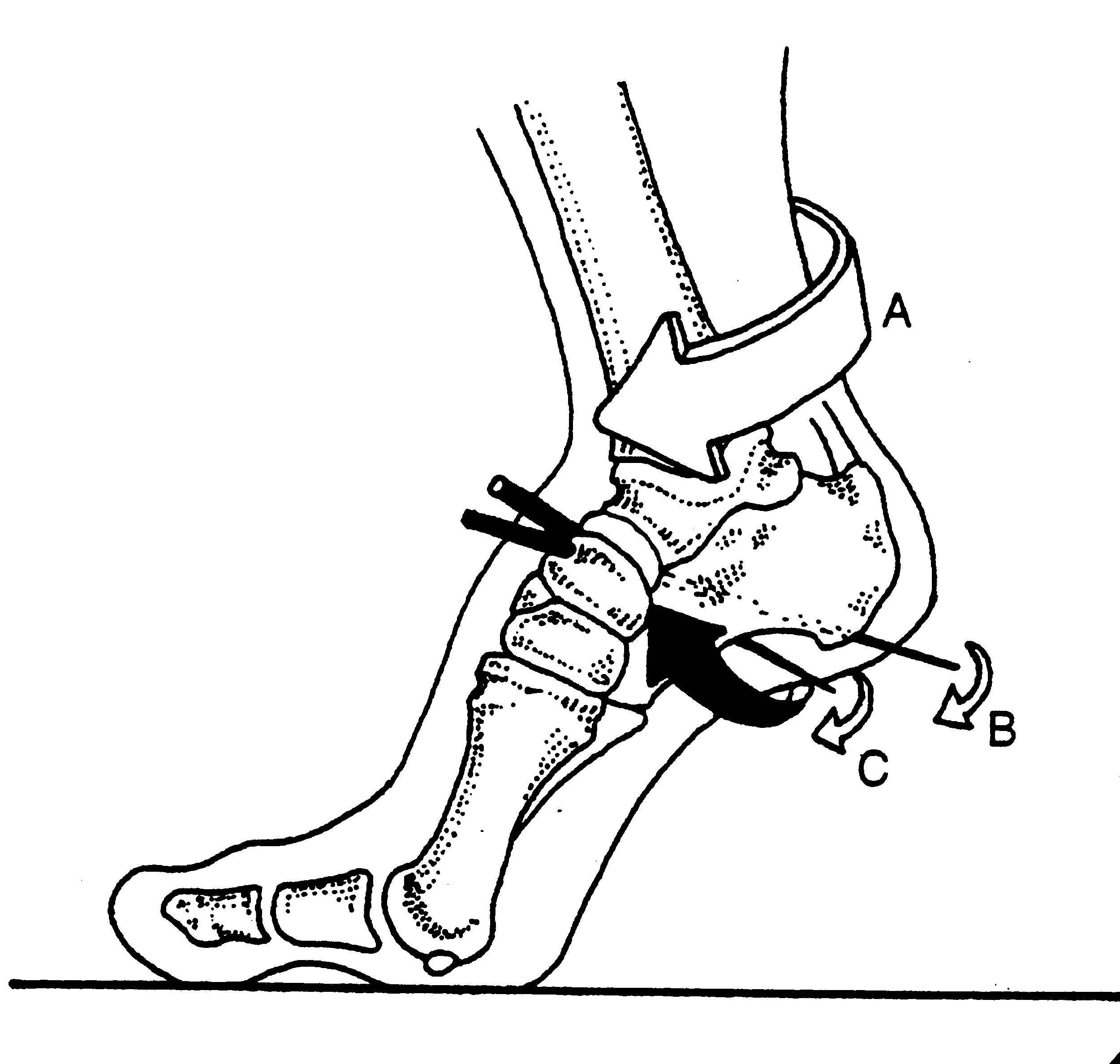
Pronation is dorsiflexion, eversion and abduction of the foot. It provides shock absorption. Supination is plantar flexion, inversion and adduction. It helps the foot become a rigid lever so we can GO (Like in Theo Selig’s “Go Dig Go” ).

external leg rotation supination.gif

Locking of the lateral column of the foot (4th and 5th metatarsal, cuboid and calcaneus) is a necessary prerequisite for normal force transmission through the foot and ultimately placing weight on the head of the 1st metatarsal for proper (high gear) toe off . Locking of the lateral column minimizes muscular strain as the musculature (soleus, peroneus longus and brevis, EHL, EDL, FDL and FHL) is usually not strong enough to perform the job on its own.

external rotary moment.gif

This process is initiated by the opposite leg going into swing phase, which initiates dorsiflexion, inversion and abduction of the talus

The peroneus longus tendon aids this process by wrapping around the cuboid (the brevis attaches to the base of the 1st metatarsal) on its way to insert onto the base of the 1st metatarsal. When the peoneus longus contracts, it dorsiflexes and everts the cuboid, which, along with the soleus (which plantar flexes and inverts the subtalar joint) allows dorsiflexion of 4th and 5th metatarsals and “locks” the lateral column. Without this mechanism, there is no locking. Without locking, there is no supination. Without supination, there is little rigidity and inefficient propulsion.

The calcaneo cuboid locking mechanism. Another cool thing you learned about gait today from The Gait Guys.

The Power of the 1st Ray?

PSM_V24_D673_Deforming_pressure_of_high_heels_on_the_foot_bone_structure.jpg

Does the 1st ray complex have super powers? Perhaps Marvel should consider a new superhero “Ray”? We are not sure but here is a story that gets us one step closer to the answer. 

While teaching a course this past weekend and doing a teaching case, we examined one of the participants who had high arches, a rigid rearfoot varus, internal tibial torsion, R > L, and foot pain R>L and a dorsal exostosis (growth of extra bone from stress at the base of her 1st metatarsal) where it articulated with the  1st cunieform on the right. No surprisingly, she also had a partially compensated forefoot supinatus on the right. She had increased wear on the lateral aspect of her shoes and a walking strategy which involved hiking the right side of the pelvis during stance phase on the left, and a pelvic shift to the right during stance phase on the right, as well as an inability to get the head of either 1st ray complex to the ground, R > L. It was also determined she had, not surprisingly, locking of the 1st metatarsal cunieform joint on the right and a loss of anterior and posterior shear at the superior tib fib articulation on the right, as well as hypomobility of the right sacoiliac joint. There was weakness of the abdominal external obliques bilaterally and posterior fibers of the left gluteus medius, along with the long toe extensors on the left and short toe flexors, a pattern that we often see clinically.

We then proceeded to treat her tib posterior, peroneus longus and flexor digitorum on the right, all of which have an effect on descending the 1st ray, along with the long extensors on the right, which would effectively raise the distal aspect of the 1st ray, but we thought may provide better eccentric control of the foot from initial contact to loading response, and again from the end of terminal stance and through swing phase.  We then mobilized the 1st met cunieform articulation only. Ideally, we should have reassessed after we made EACH change, but due to time constraints, AFTER we had done ALL these things. 

Rexamination had better 1st ray motion, restoration of tib fib motion and restoration of R sided SI mechanics. Her 1st ray descended much better, tib fib motion was normalized, L sided hip hiking strategy and R sided pelvic shift were greatly improved. For the 1st time in 10 years, the participant had no foot pain. Coincidence? Perhaps. Placebo? Maybe. You decide. 

Sometimes, doing a little of the right thing can be a good thing. Sometimes we overdo. I have to admit, because I am a chiropractor, I would have started with manipulation 1st of all 3 articulations with a recheck immediately post treatment AND THEN treated the other dysfunctions. For those of you who are manual therapists, I am sure you see miraculous things happen when we cavitate joints and change their instantaneous axes of rotation. I can thank Dr Ted Carrick and my good friend and colleague, Dr Paul Chille, for teaching me that. The students, in this case, were driving the bus and I went along with it.  I was surprised (though I shouldn’t have been) to see the pathomechanics resolve WITHOUT manipulation, but it got me thinking I should consider treating the muscular dysfunctions 1st, and then recheck and manipulate later. It makes sense that the receptor density of the lower extremity musculature has a much larger population of muscle mechanoreceptors, especially in the foot, since it has a greater cortical representation than the joint mechanoreceptors.

My students never cease to teach me something new...

A few minutes with Shawn, Episode 1

Pruning, Baking, & Corrective Exercise.

Just a few thoughts, rants, pearls, analogies and stories, once or twice a month . . . mostly leading towards a clinical point of view and things I find myself thinking about.

Where to download:

http://traffic.libsyn.com/thegaitguys/Minutes_with_Shawn_1.mp3

http://thegaitguys.libsyn.com/a-few-minutes-with-shawn-1

http://directory.libsyn.com/episode/index/id/5910367