The effects of aging on the proprioceptive system

When the nervous system breaks down, there are predictable patterns that we can see. Aging isn't that much different in the grand scale of things than some neurological disorders. Here is a brief video of a gentleman that presented to us with neck discomfort and limited range of motion. Step through it several times before proceeding.

Hopefully, you noted the following:

  • Increased arm swing on the right (or, decreased on Left)

  • Pelvic shift to the left on Left stance phase

  • Decreased step length on the left

  • Hip hike on Left during Right stance phase


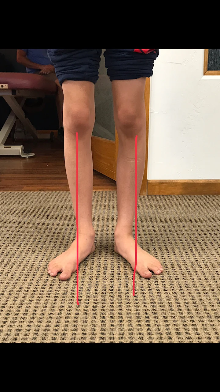
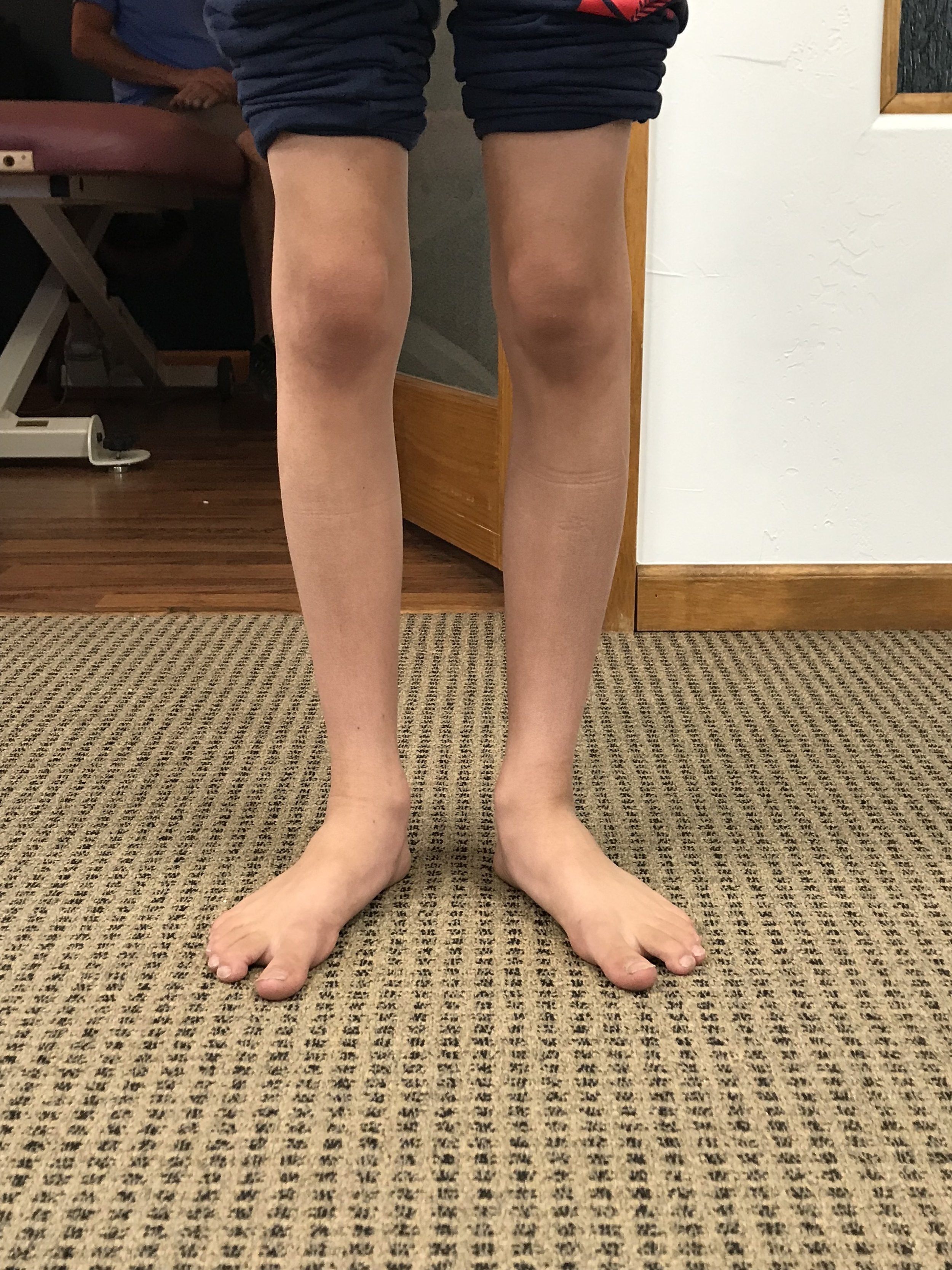
The patient DOES NOT have a leg length deficiency.

We remember that there are 3 systems that keep us upright in the gravitational plane:

1. vision
2. vestibular system

3. proprioceptive system

We also remember that as one of these systems become impaired, the others will usually increase their function to help maintain homeostasis. All these systems are known to decline in function with aging. So we have 3 systems breaking down simultaneously.

Did you also note the head forward posture, to move the center of gravity forward? How about the subtle head tilt to the right and “bobble” right and left? Motions which have to do with the head are functions of the vestibular system. He is attempting to increase the input to these areas (by exaggerating movements) to increase input.

How about the glasses? Presbyopia (hardening of the lens) makes it more difficult to focus. Movement (detected largely by rods in the eyes have a much higher density than cones, which are for visual acuity). By moving the head, he provides more input to the visual (and thus nervous system)

Amplified extremity movements provide greater input to the proprioceptive system (muscle spindles and golgi tendon organs (GTO’s), as well as joint mechanoreceptors).

Think of the cortical implications (and effects on the cerebellum, the queen of motor activity and important component for learning). You are witnessing the cognitive effects of aging playing out on the ability to ambulate and its effect on gait.


So what do we do?

  • Improve quality of joint motion, whether that is mobilization or manual methods to improve motion where motion is lost. Perhaps acupuncture to help establish homeostasis and improve muscular function. There are many options.

  • Postural advice and exercises

  • Core work

  • Proprioceptive exercises (like head repositioning accuracy, heel to toe and heel to shin)

  • Gait retraining


You get the idea. Providing some of that increased input for him and helping the system to better process the information will be the key to improving his function and helping to counteract and maybe slow the effects of aging on the locomotor system.

We are the Gait Guys; giving you the info so we can all make a difference, every day


We will be talking about some principals of proprioceptive rehabilitation along with 2 cases of neurological disorders Wednesday evening for our "3rd Wednesdays" talk on online.com: Biomechanics 321. 5 PST, 6MST, 7CST, 8EST

Special thanks to RM, who allowed us to use this video for this discussion.

When the nervous system breaks down, gait becomes more primitive.

Whether we are looking at an injury or a neurological disorder, when something goes awry, we can almost always predict that the gait pattern will start to decompose. We can learn a lot about gait from watching this kiddo walk. An immature nervous system is very similar to one which is compensating meaning there will often "cheat" around a more proper and desirable movement pattern; we often resort to a more primitive state when challenges beyond our ability are presented. This is very common when we lose some aspect of proprioception, particularly from some peripheral joint or muscle, which in turn, leads to a loss of cerebellar input (and thus cerebellar function). Remember, the cerebellum along with the upper brainstem is a temporal pattern generating center so a loss of cerebellar sensory input leads to poor pattern generation output.

Watch this clip several times and then try and note each of the following:

  • Wide based gait; this is because proprioception is still developing (joint and muscle mechanoreceptors and of course, the spino cerebellar pathways and motor cortex)

  • increased progression angle of the feet: this again is to try and retain stability. External rotation allows them to access a greater portion of the glute max and the frontal plane (engaging an additional plane is always more stable).

  • Shortened step length: this keeps the center of gravity close to the body and makes corrections for errors that much easier This immature DEVELOPING system is very much like a mature system that is REGRESSING. This is a paramount learning point !)

  • Decreased speed of movement; this allows more time to process proprioceptive clues, creating accuracy of motion

  • Sometimes we see increased arm and accessory movements, again to try and increase proprioceptive input and provide additional stability.


Proprioceptive clues are an important aspect of gait analysis, in both the young and old, especially since we tend to revert back to an earlier phase of development when we have an injury or dysfunction.

We will be talking about these principals along with 2 cases of neurological disorders and more this Wednesday evening for our "3rd Wednesdays" talk on online.com: Biomechanics 321. 5 PST, 6MST, 7CST, 8EST


Dr Ivo Waerlop, one of The Gait Guys

#gaitanalysis #decompositionofgait #proprioception #neurologicaldisorder #thegaitguys


What a difference a few months makes

Take a look at the pre-and post videos of this gal with a forefoot supinatus and impaired motor control of her feet and core. Shuffle walks, foot intrinsic exercises, core work and gait retraining can go a long way! The important thing to remember here is that the patient was very motivated and did what was required to make things happen. A testament to tenacity and dedication

Dr Ivo Waerlop, one of The Gait Guys

#beforeandafter #gaitretraining #gaitanalysis #forefootsupinatus

Yep, these shoes stink for this gal...

IMG_6882.jpg

Look at the left shoe and compare it to the right. See how the upper is canted on the outsole? This “varus cant” can create lots of problems or could actually be beneficial, believe it or not, depending upon the pathology.

In this particular persons story, it was NOT a good thing. They have an anatomical short leg on the left-hand side. If you remember from following us here in the past, generally speaking, the shorter leg tide tends to be more supinated and the forefoot tends to be in more varus. This means more of a “reach” with that foot during the contact phase of gait, Whether that’s running or walking. This generally means that the forefoot will pronate more on the long leg side.

This shoe “defect“ may actually be benefit for someone who has too much rear or mid foot pronation as it would “delay” pronation by starting to rearfoot in an inverted position at heel strike.

The Fix?

You could grind the sole into varus an equal amount to equal the varus cant. In our opinion, not a good idea.

You could return the shoe (that’s what this person is doing) and get another one

In addition, you could…

Give the person a 3 mm sole lift to correct for the leg length discrepancy

Make sure they have adequate range of motion in the first ray on the short leg side to be be able to plantar flex the 1st ray and reach the ground

Make sure they have adequate control of the core musculature as well as foot intrinsic musculature during stance phase.

Dr Ivo Waerlop, one of The Gait Guys

#badshoes #theshoeistheproblem #forefootvarus #leglengthdifference
#gaitproblem

Podcast 151: Gait and neurology of movement, including, Tightness? shortness? What’s the difference? It's the Neurology.

Truths about Stretching, a case of sesamoiditis, plus exercised induced muscle damage and impaired motor learning, central fatigue, POSE and Chi running and injuries. This is a good one gang, do not miss it !

Links to find the podcast:
Look for us on Apple Podcasts, Google Play, Podbean, PlayerFM, RADIO and more.
Just Google "the gait guys podcast".

Our Websites:
www.thegaitguys.com
Find Exclusive content at: https://www.patreon.com/thegaitguys
doctorallen.co
summitchiroandrehab.com
shawnallen.net

Our website is all you need to remember. Everything you want, need and wish for is right there on the site.
Interested in our stuff ? Want to buy some of our lectures or our National Shoe Fit program? Click here (thegaitguys.com or thegaitguys.tumblr.com) and you will come to our websites. In the tabs, you will find tabs for STORE, SEMINARS, BOOK etc. We also lecture every 3rd Wednesday of the month on onlineCE.com. We have an extensive catalogued library of our courses there, you can take them any time for a nominal fee (~$20).

Our podcast is on iTunes and just about every other podcast harbor site, just google "the gait guys podcast", you will find us.

Where to find us, the podcast Links:
Apple podcasts:
https://itunes.apple.com/us/podcast/the-gait-guys-podcast/id559864138?mt=2

Google Play:
https://play.google.com/music/m/Icdfyphojzy3drj2tsxaxuadiue?t=The_Gait_Guys_Podcast

Other links for today's show:

http://traffic.libsyn.com/thegaitguys/pod_151final.mp3

http://thegaitguys.libsyn.com/gait-and-neurology-of-movement-including-tightness-shortness-whats-the-difference-its-the-neurology

http://directory.libsyn.com/episode/index/id/11168369

Show notes and links:

We lose muscular Strength as we age.
Changes in supra-spinal activation play a significant role in the age-related changes in strength.
This motor system impairment can be improved by heavy resistance training
https://www.ncbi.nlm.nih.gov/pubmed/25940749

Age (Dordr). 2015 Jun;37(3):9784. doi: 10.1007/s11357-015-9784-y. Epub 2015 May 5.
Strength training-induced responses in older adults: attenuation of descending neural drive with age. Unhjem R1, Lundestad R, Fimland MS, Mosti MP, Wang E.

Osteoarthritis and running
https://journals.lww.com/acsm-csmr/Abstract/2019/06000/Running_Dose_and_Risk_of_Developing.5.aspx
Recent literature adds to a growing body of evidence suggesting that lower-dose running may be protective against the development of osteoarthritis, whereas higher-dose running may increase one's risk of developing lower-extremity osteoarthritis. However, running dose remains challenging to define, leading to difficulty in providing firm recommendations to patients regarding the degree of running which may be safe.

Can even experienced orthopaedic surgeons predict who will benefit from surgery when patients present with degenerative meniscal tears? A survey of 194 orthopaedic surgeons who made 3880 predictions
Non-surgical management is appropriate as first-line therapy in middle-aged patients with symptomatic non-obstructive meniscal tears.
https://bjsm.bmj.com/content/early/2019/08/12/bjsports-2019-100567

Sports Biomech. 2019 Jul 31:1-16. doi: 10.1080/14763141.2019.1624812. [Epub ahead of print]
Running biomechanics before and after Pose® method gait retraining in distance runners.
Wei RX1, Au IPH1, Lau FOY1, Zhang JH1, Chan ZYS1, MacPhail AJC1, Mangubat AL1, Pun G1, Cheung RTH1.

Plantar Plate Gait

This girl has a (healing) plantar plate lesion on the left hand side at the head of the second met. She also has an anatomical short leg on the same side. Her second metatarsal of both feet or longer than the first

A few things I hope you notice about the video:

  • Can you see how she “reaches“ to get to the ground with her left foot?

  • Can you see how her left foot is more inverted that strikes in the right, creating a greater amount of forefoot pronation that needs to be controlled?

  • Can you see how poor her motion control is of her pronation on the left foot with the sudden “crash” at impact?

  • Have you noticed her “crossover“ gait?


Does it make sense that because of her anatomy and running style, that the constant reach, increased forefoot inversion and lack of pronation control (which causes more abduction of the forefoot at toe off); this drives the force to the second metatarsal head which is longer and more prominent and is more than likely what led to her plantar plate lesion in the first place?


Remediation?

  • A 3 mm full length sole lift for the left foot

  • Foot intrinsic strengthening exercises

  • Hip abduction strengthening exercises/drills

  • Moving her more to a “midfoot strike” running gait with toes extended to engage the windlass


Dr Ivo Waerlop, one of The Gait Guys


#plantarplate #gaitanalysis #crossovergait #leglengthdifference #thegaitguys


External tibial torsion and lower back pain

How can external tibial torsion and lower back pain possibly be related? Let’s take a quick look at the anatomy and see how.

knees neutral, note external rotation of the right foot and decreased progression angle

knees neutral, note external rotation of the right foot and decreased progression angle

Remember the external tibial torsion is present if we drop a plumbline from the tibial tuberosity and it passes between the first and second metatarsals or more medially. This increases the progression angle of the foot. This occurs due to “over rotation" of the lower extremity during development, often exceeding the 1.5 degrees per year of external rotation per year up to age 15 or occurring for a longer period of time, up to skeletal maturity. It can be uni or bilateral.

note when the foot is neutral, the knee points inward

note when the foot is neutral, the knee points inward

Often, due to the increased progression angle, people will try to "straighten their feet" (ie, decrees their progression angle) to move forward in the sagittal plane. This places the knees to the inside of the sagittal plane which causes medial knee fall and sometimes increased mid and forefoot pronation. This results in increased medial spin of the thigh bilaterally which increases the lumbar lordosis. Combine this with a sway back or anterior pelvic tilt and you have increased pressure on the lumbar facet joints. The facets are designed to carry approximately 20% of the load put in these circumstances are often called upon to carry the much more. This often results in facet imbrication and lower back pain. You can strengthen the abdomen all you like but if you do not change the attitude of the foot, a will often develop lower back pain, especially when the abs fatigue. Now think about if the deformity is unilateral; this will often cause asymmetrical rotation of the pelvis in a clockwise or counter clockwise direction.

So, what can you do you?

Since external tibial torsion is a "hard deformity", we can influence how the bone grows before skeletal maturity but after that will not change significantly with stretching or exercise.

  • You can teach them to walk with an increase in progression angle (ie “duck footed”). This will often keep the knee in the sagittal plane and can be surprisingly well tolerated

  • You can use a foot leveling orthotic or arch support to bolster the arch and change the mechanics of the foot, causing external rotation of the tibia which will often result in a decrease in progression angle in compensation while still keeping the knee in the sagittal plane

  • You could place a full length varus wedge in the shoe which, by inverting the foot, externally rotates the tibia which the person will often compensates for by decreasing there progression angle to keep the knee and the sagittal plane


Dr Ivo Waerlop, one of The Gait Guys


#tibialtorsion #lowbackpain #LBP #progressionangle


Sometimes it’s OK for “toes in“ squats

We hear from folks and also read on a lot of blogs and articles about whether your toes should be in or out for squats or other types of activities. The real answer is “it depends”.

What it depends on is the patient’s specific anatomy. That means we need to pay attention to knees and hips and things like femoral and tibial torsion‘s. It’s paramount to keep the knees in the sagittal plane, no matter what the lower extremity orientation is.

When somebody has external tibial torsion (i.e. when you drop a plumbline from there to view tuberosity it passes medial to the line between the second and third or second metatarsal) then having your feet and externally rotated position places the knees in sagittal plane. Having the patient go “toes in” with this type of anatomy will cause both knees to for medially and create patellofemoral tracking issues.

Likewise, like the patient in the video, (Yes, I know I say “external tibial torsion“ at the beginning of the video but the patient has internal tibial torsion as you will see from the remainder of the video) when somebody has internal tibial torsion (I.e. when you drop a plumbline from the tibial tuberosity it passes lateral to the second metatarsal or a line between the second and third metatarsal) you would need to point the toes inward to keep the knees in the sagittal plane as demonstrated in the video. You can also see in the video when her feet are placed “toes out“ they fall outside sagittal plane laterally which creates patellofemoral tracking issues like it was in this particular patient.

So, knees in or knees out? It depends…

Dr. Ivo Waerlop, one of The Gait Guys

#internaltibialtorsion #externaltibialtorsion #kneepain #kneesin #kneesout #squats #thegaitguys

Podcast 150: Subtalar joint control? Plus Heel raise effects on low back pain

Links to find the podcast:
Look for us on iTunes, Google Play, Podbean, PlayerFM and more.
Just Google "the gait guys podcast".

Our Websites:
www.thegaitguys.com
Find Exclusive content at: https://www.patreon.com/thegaitguys
doctorallen.co
summitchiroandrehab.com
shawnallen.net

Our website is all you need to remember. Everything you want, need and wish for is right there on the site.
Interested in our stuff ? Want to buy some of our lectures or our National Shoe Fit program? Click here (thegaitguys.com or thegaitguys.tumblr.com) and you will come to our websites. In the tabs, you will find tabs for STORE, SEMINARS, BOOK etc. We also lecture every 3rd Wednesday of the month on onlineCE.com. We have an extensive catalogued library of our courses there, you can take them any time for a nominal fee (~$20).

Our podcast is on iTunes and just about every other podcast harbor site, just google "the gait guys podcast", you will find us.

Where to find us, the podcast Links:
Apple podcasts:
https://itunes.apple.com/us/podcast/the-gait-guys-podcast/id559864138?mt=2

Google Play:
https://play.google.com/music/m/Icdfyphojzy3drj2tsxaxuadiue?t=The_Gait_Guys_Podcast

Other links:
http://traffic.libsyn.com/thegaitguys/pod_1500final_-_81819_9.45_AM.mp3
http://thegaitguys.libsyn.com/subtalar-joint-control-plus-heel-raise-effects-on-low-back-pain

http://directory.libsyn.com/episode/index/id/10909609


Show notes


The HyProCure proceedure

https://images.search.yahoo.com/yhs/search;_ylt=AwrEeBmEH0RdlDUAiAUPxQt.;_ylu=X3oDMTByMjB0aG5zBGNvbG8DYmYxBHBvcwMxBHZ0aWQDBHNlYwNzYw--?p=hyprocure+sinus+tarsi+implant&fr=yhs-sz-001&hspart=sz&hsimp=yhs-001

https://www.ncbi.nlm.nih.gov/pmc/articles/PMC4621198/
https://www.ncbi.nlm.nih.gov/pubmed/21106413
https://www.ncbi.nlm.nih.gov/pubmed/29786228

High-heeled walking decreases lumbar lordosis.EdenyBaaklini et al.
https://www.sciencedirect.com/science/article/pii/S096663621730108X

The effect of high-heeled shoes on lumbar lordosis: a narrative review and discussion of the disconnect between Internet content and peer-reviewed literature. Brent S. Russell
https://www.ncbi.nlm.nih.gov/pmc/articles/PMC3206568/

Prolong Wearing of High Heeled Shoes Can Cause Low Back PainFarjad Afzal1* and Sidra Manzoor
https://pdfs.semanticscholar.org/afb4/641b8ed6450fcbdfa8ff99029d935c2bdc88.pdf

Relation between Wearing High-Heeled Shoes and Gastrocnemius and Erector Spine Muscle Action and Lumbar Lordosis. Cezar Augusto Souza Casarin
https://www.medscitechnol.com/download/index/idArt/892352

A flatter foot approach?
https://twitter.com/IzzyMoorePhD/status/1157034538192855041

Thoughts: titrate into speed work just like doing the same for longer and longer runs
Creating a "speed base"
https://www.fastrunning.com/?p=26410&preview=true

"monster walks"
Hip-Muscle Activity in Men and Women During Resisted Side Stepping With Different Band Positions. Lewis CL, et al. J Athl Train. 2018.
https://www.ncbi.nlm.nih.gov/m/pubmed/30615490/

Physical findings differ between individuals with greater trochanteric pain syndrome and healthy controls: A systematic review with meta-analysis.
Plinsinga ML1, Ross MH1, Coombes BK2, Vicenzino B3.
Musculoskelet Sci Pract. 2019 Jul 25;43:83-90. doi: 10.1016/j.msksp.2019.07.009. [Epub ahead of print]
https://www.ncbi.nlm.nih.gov/pubmed/31369906

The Fudge Factor

image credit: https://commons.wikimedia.org/wiki/File:Pieces_of_fudge_cut_from_a_slab,_April_2008_cropped.jpg

image credit: https://commons.wikimedia.org/wiki/File:Pieces_of_fudge_cut_from_a_slab,_April_2008_cropped.jpg

We know from experience that it is often easier to accomplish a task faster, rather than slower (like an exercise or skiing) because of the cortex “interpolating” or making its “best guess” as to what (based on past experience) is going to happen and in what order. There is a certain amount of guess work (or what we call “the fudge factor”) involved.

Walking at a slower speed (or performing an exercise at a slower speed for that matter) has increased muscular demands, than doing it more quickly. Here is one study that exemplifies that.

“These findings may reflect a relatively higher than expected demand for peroneus longus and tibialis posterior to assist with medio-lateral foot stability at very slow speeds”

Here, they thought muscular demands would be proportional to speed, increasing with increasing demands. Like many things, what we think is going to happen and what actually happens can be 2 different things : )


Dr Ivo Waerlop, one of The Gait Guys


#fudgefactor #corticalinterpolation #muscledemands #gait #gaitguys


Gait Posture. 2014 Apr;39(4):1080-5. doi: 10.1016/j.gaitpost.2014.01.018. Epub 2014 Feb 6.

Electromyographic patterns of tibialis posterior and related muscles when walking at different speeds.

Murley GS1, Menz HB2, Landorf KB2.

Botox for plantar fasciitis? Sounds like a bad idea to us....

image source: https://commons.wikimedia.org/wiki/File:Plantar_aponeurosis_-_axial_view.png

image source: https://commons.wikimedia.org/wiki/File:Plantar_aponeurosis_-_axial_view.png

Botox..For plantar fasciitis? Really?

We found this article (1) in one of our favorite journals, Lower Extremity Review , and were a little surprised. Let us get this straight: you are going to take one of the the most poisonous biological neurotoxins known (1) and inject it into your calf and foot?

The article in LER is well written and the results (thankfully) were inconclusive regarding its usage. They do cite 3 studies (with two by the same lead author) where it has been effective (2-4). Yes, it is better than saline (5) (but not as good as extracorporeal shock wave therapy (6)), and better than placebo (7-10) but considerably more risky.

So the premise is “if the muscle is dysfunctional, then let’s just take it out of the equation”. But this really doesn’t fix the problem, it just covers up the symptom. And what about the other potential side effects since botulinum toxin acts not only at the neuromuscular junction, blocking the release of acetylcholine, but also at the autonomic ganglia, postganglionic parasympathetic nerve endings, as well as the post ganglionic sympathetics that use acetylcholine (capillaries of skin, piloerector muscles and sweat glands) (11)?.

In our experience, most cases of plantar fasciitis are secondary to lack of forefoot rocker, lack of ankle rocker, lack of hip extension or in some cases, direct trauma. Wouldn’t it make more sense to strengthen the anterior compartment to reciprocally inhibit the posterior compartment, increasing ankle dorsiflexion and hip extension? We find, oftentimes, treating only the area of chief complaint and not what is "driving the bus" can offer temporary, symptomatic relief but not long standing pathmechanics or pathoanatomy.

Just like the road to enlightenment, there are no shortcuts in treating plantar fasciitis and if you are not going to treat the cause, then be prepared to reap what you sow.

Dr Ivo Waerlop, one of The Gait Guys

#botox #plantarfascitis #badideas #gaitproblem #thegaitguys

1. https://lermagazine.com/article/botox-injection-not-just-for-celebrities-furrows-and-wrinkles

2. Elizondo-Rodriguez J, Araujo-Lopez Y, Moreno-Gonzalez JA, Cardenas-Estrada E,
Mendoza-Lemus O, Acosta-Olivo C. A comparison of botulinum toxin A and intralesional steroids for the treatment of plantar fasciitis: A randomized, double-blinded study. Foot Ankle Int.
2013;34(1):8-14.

3. Díaz-Llopis IV, Rodríquez-Ruíz CM, Mulet-Perry S, Mondéjar-Gómez FJ., Climent-Barberá JM., Cholbi-Llobel F. Randomized controlled study of the efficacy of the injection of botulinum toxin type A versus corticosteroids in chronic plantar fasciitis: results at one and six months. Clin Rehabil. 2012;26(7):594-606.

4. Díaz-Llopis IV, Gómez-Gallego D, Mondéjar-Gómez FJ, López-García A, Climent-Barberá JM, Rodríguez-Ruiz CM. (2013). Botulinum toxin type A in chronic plantar fasciitis: clinical effects one year after injection. Clin Rehabil. 2013;27(8):681-685.

5. Ahmad J, Ahmad SH, Jones K. Treatment of Plantar Fasciitis With Botulinum Toxin. Foot Ankle Int. 2017 Jan;38(1):1-7. doi: 10.1177/1071100716666364. Epub 2016 Oct 1.1.

6. Roca B, Mendoza MA, Roca M. Comparison of extracorporeal shock wave therapy with botulinum toxin type A in the treatment of plantar fasciitis. Disabil Rehabil. 2016 Oct;38(21):2114-21. doi: 10.3109/09638288.2015.1114036. Epub 2016 Mar 1

7. Babcock MS, Foster L, Pasquina P, Jabbari B. Treatment of pain attributed by plantar fasciitis with botulinum toxin A: a short-term randomized, placebo-controlled, double blinded study. Am J Phys Med Rehabil. 2005;84(9):649-654.

8. Samant PD, Kale SY, Ahmed S, Asif A, Fefar M, Singh SD. Randomized controlled study comparing clinical outcomes after injection botulinum toxin type A versus corticosteroids in chronic plantar fasciitis. Int J Res Orthop. 2018;4(4):672-675.

9. Huang YC, Wei SH, Wang HK, Lieu FK. Ultrasonographic guided botulinum toxin type A treatment for plantar fasciitis: an outcome-based investigation for treating pain and gait changes. J Rehabil Med. 2010;42(2):136-140.

10. Ahmad J, Ahmad SH, Jones K. Treatment of plantar fasciitis with botulinum toxin. Foot Ankle Int. 2017;38(1):1-7.

11. Nigam PK, Nigam A. Botulinum toxin. Indian J Dermatol. 2010;55(1):8–14. doi:10.4103/0019-5154.60343

Low back pain and asymmetry.

Screen Shot 2019-04-07 at 10.00.54 AM.png

Do oarsmen have asymmetries in the strength of their back and leg muscles?
IF these oarsmen were more symmetrical would they not be in pain?

From the study below:
"Patterns of asymmetry of muscle activity were observed between the left and right erector spinae muscles during extension, which was significantly related to rowing side (P < 0.01). These observations could be related to the high incidence of low back pain in oarsmen."

Here we have a supported study of asymmetry and injury/pain. This is what we have been saying (asymmetry matters) in the last few days with our posts on asymmetry. This study eludes to a finding that strength can test normal and symmetrical, but EMG activity can show patterns of asymmetry that can result in problems/pain.

Have you ever rowed? I mean truly rowed, in a shell, on the water, not on land or on a Concept 2 rower? It is just not the same, especially if you have an unilateral asymmetrical loading arc, like an oarsman pulling from port or starboard. I have rowed on the water just like this, briefly, one summer in a camp for young teens. I rowed on my home town course, on the World famous Royal Canadian Henley Regatta. I was the 2nd seat, starboard, in an 8 man shell. 8 oars in the water, 8+1 guys, one oar a piece, alternating port and starboard. I was behind the stroke. I hated it. Perhaps the hardest thing I had ever done sport wise to that point, largely because this dude setting the pace was jacked on caffeine, or something else, I think. No one works harder than rowers if you ask me, they are some of the fittest athletes in the world. Why? because it is a whole body effort.
Ok, enough of the fluff.

Now imagine rowing like this for many years in high school, college and/or competitively. Forcefully pulling on one oar, across an arc of pull out one side of the boat, thousands of times a day for many years. If that isn't something that will develop asymmetry I do not know what might. Oarsman are under near constant high end effort pushing and pulling loads (push with the legs, pull with the arms). There are few, if any, sports with such high end constant effort than rowing.

From the Parkin et al study:
"The aim of this study was to establish whether asymmetry of the strength of the leg and trunk musculature is more prominent in rowers than in controls. Nineteen oarsmen and 20 male controls matched for age, height and body mass performed a series of isokinetic and isometric strength tests on an isokinetic dynamometer. These strength tests focused on the trunk and leg muscles. Comparisons of strength were made between and within groups for right and left symmetry patterns, hamstring: quadriceps ratios, and trunk flexor and extensor ratios. The results revealed no left and right asymmetries in either the knee extensor or flexor strength parameters (including both isometric and isokinetic measures). Knee extensor strength was significantly greater in the rowing population, but knee flexor strength was similar between the two groups. No difference was seen between the groups for the hamstring: quadriceps strength ratio. In the rowing population, stroke side had no influence on leg strength. No differences were observed in the isometric strength of the trunk flexors and extensors between groups, although EMG activity was significantly higher in the rowing population. Patterns of asymmetry of muscle activity were observed between the left and right erector spinae muscles during extension, which was significantly related to rowing side (P < 0.01). These observations could be related to the high incidence of low back pain in oarsmen."- Parkin et al.

Extra sauce:
I "caught a crab" many times when a novice oarsman and was nearly vaulted out of the boat on one fatal event. A crab is the term rowers use when the oar blade gets “caught” in the water. It is caused by a momentary flaw in oar technique and the paddle end of the oar is pulled into the depths instead of skimming just below the surface. Catching a crab has happened to anyone who has ever rowed. A crab may be minor, allowing the rower to quickly recover, or it may be so forceful that the rower is ejected from the boat as the handle end catches the oarsman under the arms lifting them out of the boat.

J Sports Sci. 2001 Jul;19(7):521-6.
Do oarsmen have asymmetries in the strength of their back and leg muscles? Parkin S1, Nowicky AV, Rutherford OM, McGregor AH.

Incorporating asymmetrical tonic neck responses into your rehab program

You may have heard about primitive reflexes. You may also have been taught that they don’t persist into adulthood, however, we can assure you, they persist into adulthood (1) and are modulated by both eye movement and muscular activity (2). When there is neurological compromise (think about physiological lesions, or shorts in the nervous system, not just anatomical lesions), the reflex can be more prevalent. They appear to arise from the joint mechanoreceptors in the neck and take advantage of the connection to the reticular formation of the brainstem (3), and may also be related to the cervicoocular reflex. It may modulate blood flow and cardiovascular activity too (4). 

tumblr_nfw1xxi7Yt1qhko2so1_250.jpg

The asymmetrical tonic neck reflex was 1st described by Magnus and de Kleyn in 1912 (5). Like in the picture above, when the head is rotated to one side, there is ipsilateral extension of the upper and lower extremity on that side, and flexion of the contralateral (the side AWAY from where you are rotating) upper and lower extremity. Take a few minutes to see the subtleness of the reflex in the pictures above. Now think about how this occurs in your clients/patients.  Don’t believe us, take an egg needle and put it into on of the upper or lower extremity extensors and rotate the head to the same side and watch the increased activity in that extremity.

tumblr_nfw1xxi7Yt1qhko2so2_540.png

So, how can we take advantage of this? We could follow in the footsteps of Berta Bobath (6) and incorporate these into our rehabilitation programs, which we have done, quite successfully. But rather than read a whole book, lets talk about how you could incorporate this into your stretching and exercise program. 

Let’s say you want to stretch the right hamstring:

  • actively rotating the head to the right (see reference 3) facilitates the right tricep and right quadricep AND facilitates the left bicep and left hamstring. You could easily incorporate this into your gait and exercise programs, using the same principal

  • through reciprocal inhibition, this would inhibit the right bicep and hamstring AND left tricep and left quadricep

  • To get a little more out of the stretch, you could actively contract the right tricep and quadricep (MORE reciprocal inhibition), amplifying the effect

We encourage you to try this, both on yourself and your clients. It really works!

Dr Ivo Waerlop, one of The Gait Guys

#gait #gaitanalysis #TNR #tonicneckresponse #tonicneckreflex #rehabilitationexercise #rehabexercises

  1. Le Pellec A1, Maton B. Influence of tonic neck reflexes on the upper limb stretch reflex in man. J Electromyogr Kinesiol. 1996 Jun;6(2):73-82.

  2. Michael D. Ellis, Justin Drogos, Carolina Carmona, Thierry Keller, Julius P. A. Dewal Neck rotation modulates flexion synergy torques, indicating an ipsilateral reticulospinal source for impairment in stroke Journal of NeurophysiologyDec 2012,108(11)3096-3104;DOI: 10.1152/jn.01030.2011

  3. http://www.worldneurologyonline.com/article/arthur-simons-tonic-neck-reflexes-hemiplegic-persons/#sthash.6QS3Eat3.dpuf

  4. Hervé Normand, Olivier Etard and Pierre Denise Otolithic and tonic neck receptors control of limb blood flow in humans J Appl Physiol  82:1734-1738, 1997. Bruijn SM1, Massaad FMaclellan MJVan Gestel LIvanenko YPDuysens J.

  5. Are effects of the symmetric and asymmetric tonic neck reflexes still visible in healthy adults?Neurosci Lett.2013 Nov 27;556:89-92. doi: 10.1016/j.neulet.2013.10.028. Epub 2013 Oct

  6. Berta BobathChartered Society of Physiotherapy (Great Britain)  Abnormal postural reflex activity caused by brain lesions Aspen Systems Corp. Rockville, MD, 1985 -

When you see this, you should be thinking one of 3 possible etiologies...

Cardinal sign of either a forefoot supinatus/forefoot varus or collapsing midfoot

I was hiking behind this young chap over the weekend along with my son and friends. Note the amount of calcaneal eversion present on the right side that is not present on the left. Also note the increased progression angle of the right foot and subtle circumduction of the extremity.

In my experience, you would generally see this much calcaneal diversion and one of three scenarios:

1. Moderate leg length discrepancy with the increased calcaneovalgus occurring on the longer leg side. This would support the amount of circumduction were seeing on the right side.

2. When there is a forefoot supinatus present and and inadequate range of motion available in the midfoot and/or forefoot. This is most likely the case here.

3. In moderate To severe midfoot collapse. This is clearly not the case as the medial aspect of the shoe is usually “blown out”.

Next time you see an everting rearfoot, think about these three possible etiologies.

Dr Ivo Waerlop, on of The Gait Guys

#evertedrrarfoot #calcanealvalgus #shortleg #forefootsupinatus #forefootvarus #gaitanalysis #thegaitguys

The amazing power of compensation. Coming to a patient in your office… Maybe today

This gal has had a right sided knee replacement. She has an anatomical right short leg, a forefoot supinatus, an increased Q angle and a forefoot adductus. So, what’s the backstory?

When we have an anatomical short leg, we will often have a tendency to try to “lengthen“ that extremity and “shorten” the longer extremity. This is often accomplished through pelvic rotation although sometimes can be with knee flexion/extension or change in the Q angle. When the condition is long-standing, the body will often compensate in other ways, such as what we are seeing here.

IMG_6736.jpg

The fore foot can supinate in an attempt to lenthen the extremity. Note how the right extremity forefoot is in varus with respect to the rearfoot, effectively lengthening the extremity. As you can see from the picture, this is becoming a “hard“ deformity resulting in a forefoot varus.

IMG_6740.jpg

Over time, the forefoot has actually “adducted “ as you can see, again in an attempt to lengthen the extremity. Remember that supination is plantar flexion, abduction and inversion, all three which are visible here.


You will also see that the Q angle is less on the right side (se above), effectively lengthening that extremity, but not quite enough as we can see from the picture :-)


Dr Ivo Waerlop, one of The Gait Guys

#forefootadductus #shortleg #kneereplacement #tkr #forefootvarus #gait #thegaitguys

External tibial torsion or femoral retrotorsion?

IMG_6727.JPG

This young lad presents to your office complaining of bilateral knee discomfort at the medial aspect, just below the patella, particularly when ascending and descending stairs and hills. You narrow it down to abnormal patellar tracking and 2 possibilities of who is driving the bus, but which is it?

Torsions of an extremity are said to exist when they measure two or more standard deviation‘s outside of normal. In external tibial torsion, the shaft of the tibia over rotates more than it’s 1.5° per year from zero at birth to greater than 19°. You are left with a foot that is has an increased progression angle and a center of gravity falls medial to the foot causing abnormal patellar tracking.

Femoral retro torsion is said to exist when the head of the femur over reduces from its 35° angle at birth to less than 8° resulting in severely limited internal rotation of the hips bilaterally. The lower extremity is often externally rotated to compensate.

An easy differential for the 2 is to drop a plumbline from the tibial tuberosity through the foot. This line normally passes through the second or between the second and third metatarsal‘s. If it falls medial to that it is eternal tibial torsion and lateral to that most likely internal tibial torsion or potentially a metatarsus varus or forefoot adductus.

IMG_6759.jpg


Another differential would be to perform “Craigs test” and measure how much internal and external rotation of the femur there is at the femoral acetabular articulation.

An easier way to put it is; those with femoral retrotorsion have less hip internal rotation and often increased amounts of external rotation; often they can’t even get past zero, never mind the requisite 4-6 degrees for normal gait. Those with increased internal rotation and diminished external rotation most likely have femoral antetorsion.

IMG_6726.jpg


So, Which is it? When his knees are Straightahead, his feet point out; when his feet are straightahead, his knees point inward. A plumbline from the tibial tuberosity passes medial to the second metatarsal. Looking at the pictures, you can see that he is external tibial torsion along with a sandal thong deformity that we talked about last week.

Dr Ivo Waerlop, one of The Gait Guys.

#externaltibialtorsion #outturnedfoot #increasedprogressionangle #kneepain #thegaitguys

When the big toes head...East? Whats the deal?

IMG_6721.JPG

What is this?

IMG_6722.JPG

A sandal gap deformity or hallux varus creates an expanded first interspace between the hallux and the rest of the toes. It is a likened to the gap caused by wearing a sandal but is actually a normal variant. It can occasionally be developmental. In the fetus, it can be a soft marker for other fetal anomalies such as Downs syndrome, an amniotic band or ectrodactyly. It’s considered benign, however in this individual could have been developmental.

IMG_6727.JPG

Notice how he has external tibial torsion (when his knees are pointing forward his feet point to the outside). External tibial torsion generally, because of the orientation of the foot, causes the center of gravity to fall medially thus the need for something to push and stabilize you more laterally, such as toes two through five abducting : )

Dr Ivo Waerlop, one of The Gait Guys

#halluxvarus #strangelookingfeet #hallux #thegaitguys #sandalgapdeformity


Barp EA, Temple EW, Hall JL, Smith HL. Treatment of Hallux Varus After Traumatic Adductor Hallucis Tendon Rupture. J Foot Ankle Surg. 2018 Mar - Apr;57(2):418-420.

https://radiopedia.org/articles/sandal-gap-deformity?lang=us

Munir U, Morgan S. Hallux Varus. StatPearls [Internet]. Treasure Island (FL): StatPearls Publishing; 2019 Jan-.
2019 May 6.

Ryan PM, Johnston A, Gun BK. Post-traumatic dynamic hallux varus instability. J Clin Orthop Trauma. 2014 Jun;5(2):94-8. doi: 10.1016/j.jcot.2014.05.005. Epub 2014 Jun 15.

Sixth toe disease...That growth on the outside of your foot… Or on somebody’s foot is coming to see you…

IMG_6704.JPG

You know what we’re talking about. That extra growth on the lateral aspect of the foot that happens way too often and many of your clients. A Taylor’s bunion or sometimes referred to as a “bunionette”. 

What is the usual fix?

Usually in a ski boot or hiking boot, they blow out the lateral side of the shoe. This is usually not a good fix because most of these folks have internal tibial torsion and somewhat of a forefoot supinatus/varus.

IMG_6706.JPG
IMG_6707.JPG

The internal tibial torsion places the knee outside the saggital plane and an arch support without a forefoot valgus post will just push it further out, creating a conflict at the knee. The forefoot supinatus and/or varus places them on the outside of the foot as well. Remember, most of these folks are ALREADY on the outside of the foot and the foot wants to migrate laterally...so creating more space just means it migrates farther. Good thought, doesn’t work that way.

IMG_6710.JPG

So what did we do?

  • We created a valgus post for the forefoot (see picture above) tapering from lateral to medial and to help “push“ the distal aspect of the first ray down (because there was motion available that was not being used)

  • We gave him exercises to help descend the first ray like the extensor hallucis brevis exercise, toe waving as well as peroneus longus exercises

  • We gave him plenty of balance and coordination work

    Dr Ivo Waerlop, one of The Gait Guys


#6thtoe #internaltibialtorsion #forefootvarus # forefootsupinatus #gaitanalysis #thegaitguys


Podcast 149: A runner's podcast. Many things running and biomechanics.


Links to find the podcast:
Look for us on iTunes, Google Play, Podbean, PlayerFM and more.
Just Google "the gait guys podcast".

Our Websites:
www.thegaitguys.com
Find Exclusive content at: https://www.patreon.com/thegaitguys
doctorallen.co
summitchiroandrehab.com
shawnallen.net

Our website is all you need to remember. Everything you want, need and wish for is right there on the site.
Interested in our stuff ? Want to buy some of our lectures or our National Shoe Fit program? Click here (thegaitguys.com or thegaitguys.tumblr.com) and you will come to our websites. In the tabs, you will find tabs for STORE, SEMINARS, BOOK etc. We also lecture every 3rd Wednesday of the month on onlineCE.com. We have an extensive catalogued library of our courses there, you can take them any time for a nominal fee (~$20).

Our podcast is on iTunes and just about every other podcast harbor site, just google "the gait guys podcast", you will find us.

Where to find us, the podcast Links:
Apple podcasts:
https://itunes.apple.com/us/podcast/the-gait-guys-podcast/id559864138?mt=2

Google Play:
https://play.google.com/music/m/Icdfyphojzy3drj2tsxaxuadiue?t=The_Gait_Guys_Podcast

Other links:
http://traffic.libsyn.com/thegaitguys/pod_149_-_71319_7.44_AM.mp3
http://thegaitguys.libsyn.com/a-runners-podcast-many-things-running-and-biomechanics
http://directory.libsyn.com/episode/index/id/10506122


Show notes:


Exercise matters
3 months of exercise training reprogrammed the epigenetics of sperm DNA in healthy young men. Exercise silenced genes in sperm DNA involved in schizophrenia, Parkinson's disease, cervical cancer, leukemia, and autism
https://www.ncbi.nlm.nih.gov/pubmed/25864559?dopt=Abstract
Epigenomics. 2015 Aug;7(5):717-31. doi: 10.2217/epi.15.29. Epub 2015 Apr 13.
Genome-wide sperm DNA methylation changes after 3 months of exercise training in humans.
Denham J1, O'Brien BJ2, Harvey JT2, Charchar FJ

Footstrike doesnt matter?
https://www.outsideonline.com/2397214/foot-strike-running-study

Adaptation of Running Biomechanics to Repeated Barefoot Running: A Randomized Controlled Study - Karsten Hollander, Dominik Liebl, Stephanie Meining, Klaus Mattes, Steffen Willwacher, Astrid Zech, 2019
https://journals.sagepub.com/doi/full/10.1177/0363546519849920
Is There an Economical Running Technique? A Review of Modifiable Biomechanical Factors Affecting Running Economy.
Moore IS. Sports Med. 2016.
https://www.ncbi.nlm.nih.gov/m/pubmed/26816209/

Running Technique is an Important Component of Running Economy and Performance.
https://www.ncbi.nlm.nih.gov/m/pubmed/28263283/
Folland JP, et al. Med Sci Sports Exerc. 2017.
https://journals.sagepub.com/doi/full/10.1177/0363546519849920

Important to note though than less vertical oscillation is associated with better economy within groups of distance runners, eg ncbi.nlm.nih.gov/m/pubmed/28263… ; ncbi.nlm.nih.gov/m/pubmed/26816… compliant tendons but greater leg stiffness is the goal for distance runners, correct?

This video shows how end. runners compliance & economy are achieved by greater vertical excursions vs. sprinters who hit hard, get off the ground fast and burn more energy.
https://www.nytimes.com/video/sports/100000004379956/identifying-the-best-way-to-run.html

typically have peak vertical forces of 2.5-3.0 times body weight to offset gravity during contact portion of the stride.
https://www.youtube.com/watch?v=hEnIbklXOiU

Effects of footwear midsole thickness on running biomechanics
Sports Medicine and Biomechanics
Mark H.C. Law, Eric M.F. Choi, Stephanie H.Y. Law, Subrina S.C. Chan , Sonia M.S. Wong, Eric C.K. Ching
https://www.tandfonline.com/doi/abs/10.1080/02640414.2018.1538066?journalCode=rjsp20

Knee muscle forces during walking and running in patellofemoral pain patients and pain-free controls.
Thor F.Besiera, Michael Fredericsona, Garry E.Gold, Gary S.Beaupréd, Scott L.Delp
Journal of Biomechanics Volume 42, Issue 7, 11 May 2009, Pages 898-905
https://www.sciencedirect.com/science/article/abs/pii/S0021929009000396

The Gracilis: Have you considered it in your medial knee pain patients?

The gracilis is the 3rd, seldom mentioned contributor to the pes anserine when it comes to knee pain. It lives in the medial compartment of the thigh and helps to add stability to the medial stabilizing complex of the knee.

image credit: https://en.wikipedia.org/wiki/File:Slide3rrr.JPG

image credit: https://en.wikipedia.org/wiki/File:Slide3rrr.JPG

During an ideal gait cycle, the gracilis tonically contracts throughout stance phase with bursts from terminal swing through initial contact and again from pre swing to initial swing; it is very similar to the adductors in this respect

The gracilis is a superficial muscle on the medial thigh, running from the the pubic symphisis and upper pubic arch to the pes, sandwiched between the sartorius and semitendinosis. The muscle adducts, medially rotates (with hip flexion), laterally rotates, and flexes the hip , and also aids in flexion of the knee. It can be called upon as a thigh flexor (as can the sartorius) when the abs (particularly the external obliques) do not initiate thigh flexion and the TFL, rectus femoris and iliopsoas groups are dysfunctional. Gracilis dysfunction may contribute to medial knee pain when the thigh needs help flexing. We see this particularly on people with excessive mid foot pronation when the foot remains on the ground too long and need some “help” or a “jump start” to initiate thigh flexion.

Think about this “unsung hero” next time you have a recalcitrant medial knee pain patient.


Gupta, Aman & Saraf, Abhinesh & Yadav, Chandrajeet. (2013). ISSN 2347-954X (Print) High-Resolution Ultrasonography in PesAnserinus Bursitis: Case Report and Literature Review. 1. 753-757. 

Gray H:  Anatomy of the Human Body  Lea and Febiger, Phildelphia and New York 1918

 Michaud T: in Foot Orthoses and Other Forms of Conservative Foot Care Williams & Wilkins, 1993 Pp. 50-55

 Michaud T: in Human Locomotion: The Conservative Management of Gait-Related Disorders 2011