And why does this guy have hip pain?

line up the center of the heel counters with the outsoles, and what do you see?

line up the center of the heel counters with the outsoles, and what do you see?

can you see how the heel counter is centered on the outsole, like it is supposed to be

can you see how the heel counter is centered on the outsole, like it is supposed to be

notice how the heel counter of the shoe is canted medially on the outsole of the shoe, creating a varus cant

notice how the heel counter of the shoe is canted medially on the outsole of the shoe, creating a varus cant

Take a guy with lower back and left sided sub patellar pain that also has a left anatomically short leg (tibial) and bilateral internal tibial torsion and put him in these baby’s to play pickleball and you have a prescription for disaster.

Folks with an LLD generally (soft rule here) have a tendency to supinate more on the short leg side (in an attempt to make the limb longer) and pronate more on the longer leg side (to make the limb shorter). Supination causes external rotation of the lower limb (remember, we are trying to make the foot into a rigid lever in a “normal” gait cycle). this external rotation with rotate the knee externally (laterally). Folks with internal tibial torsion usually rotate their limb externally to give them a better progression angle (of the foot) so they don’t trip and fall from having their feet pointing inward. This ALSO moves the knee into external rotation (laterally), often moving it OUTSIDE the saggital plane. In this case, the knee, because of the difference in leg length AND internal tibial torsion AND the varus cant of the shoe, has his knee WAY OUTSIDE the saggital plane, causing faulty patellar tracking and LBP.

Moral of the story? When people present with a problem ALWAYS TAKE TIME TO LOOK AT THEIR SHOES!

On the subject of manual muscle work…

image credit: https://commons.wikimedia.org/wiki/File:Muscle_spindle_model.jpg

image credit: https://commons.wikimedia.org/wiki/File:Muscle_spindle_model.jpg

Here is an older article that may seem verbose, but has interesting implications for practitioners who do manual muscle work with their clients. We would invite you to work your way through the entire article, a little at a time, to fully grasp it’s implications.

Plowing through the neurophysiology, here is a synopsis for you:

Tactile and muscle afferent (or sensory) information travels into the dorsal (or posterior) part of the spinal cord called the “dorsal horn”. This “dorsal horn” is divided into 4 layers; 2 superficial and 2 deep. The superficial layers get their info from the A delta and C fibers (cold, warm, light touch and pain) and the deeper layers get their info from the A alpha and A beta fibers (ie: joint, skin and muscle mechanoreceptors).

So what you may say

The superficial layers are involved with pain and tissue damage modulation, both at the spinal cord level and from descending inhibition from the brain. The deeper layers are involved with apprising the central nervous system about information relating directly to movement (of the skin, joints and muscles).

Information in this deeper layer is much more specific that that entering the more superficial layers. This happens because of 3 reasons:

  1. there are more one to one connections of neurons (30% as opposed to 10%) with the information distributed to many pathways in the CNS, instead of just a dedicated few in the more superficial layers

  2. the connections in the deeper layers are largely unidirectional and 69% are inhibitory connections (ie they modulate output, rather than input)

  3. the connections in the deeper layers use both GABA and Glycine as neurotransmitters (Glycine is a more specific neurotransmitter).

Ok, this is getting long and complex, tell me something useful...

This supports that much of what we do when we do manual therapy on a patient or client is we stimulate inhibitory neurons or interneurons which can either (directly or indirectly)

  • inhibit a muscle

  • excite a muscle because we inhibited the inhibitory neuron or interneuron acting on it (you see, 2 negatives can be positive)

So, much of what we do is inhibit muscle function, even though the muscle may be testing stronger. Are we inhibiting the antagonist and thus strengthening the agonist? Are we removing the inhibition of the agonist by inhibiting the inhibitory action on it? Whichever it may be, keep in mind we are probably modulating inhibition, rather than creating excitation.


Semantics? Maybe…But we constantly talk about being specific for a fix, not just cover up the compensation. Is it easier to keep filling up the tire (facilitating) or patching the hole (inhibiting). It’s your call


Yan Lu Synaptic Wiring in the Deep Dorsal Horn. Focus on Local Circuit Connections Between Hamster Laminae III and IV Dorsal Horn Neurons J Neurophys Volume 99 Issue 3

March 2008 Pages 1051-1052 link: http://jn.physiology.org/content/99/3/1051

Music to my ears. Movement to my steps...

https://en.wikipedia.org/wiki/Music

https://en.wikipedia.org/wiki/Music

"The applicable contribution of these novel findings is that music tempo could serve as an unprompted means to impact running cadence. As increases in step rate may prove beneficial in the prevention and treatment of common running-related injuries, this finding could be especially relevant for treatment purposes, such as exercise prescription and gait retraining."

Van Dyck E, Moens B, Buhmann J, Demey M, Coorevits E, Dalla Bella S, Leman M. Spontaneous Entrainment of Running Cadence to Music Tempo. Sports Med Open. 2015;1(1):15. Epub 2015 Jul 14.

link to free full text: https://www.ncbi.nlm.nih.gov/pmc/articles/PMC4526248/

image credit: http://pressplay.pbworks.com/w/page/82954552/Loebner%20Keith%20HW%203

Podcast 139: STEM &PRP therapies for athletes

Topics:
We are all over the board today folks, topics like PRP but more so we go down some paths that are more fundamentally clinical and neurologic with a sprinkling of orthopedics to round things out. Sometimes these are our best shows.

Links to find the podcast:

iTunes page:

http://traffic.libsyn.com/thegaitguys/pod_139finalfull_-_92218_7.16_AM.mp3

Google Play:

https://play.google.com/music/m/Icdfyphojzy3drj2tsxaxuadiue?t=The_Gait_Guys_Podcast

Direct Download: http://traffic.libsyn.com/thegaitguys/pod_139finalfull_-_92218_7.16_AM.mp3

Permalink URL:
http://thegaitguys.libsyn.com/podcast-139-stem-prp-therapies-for-athletes

Libsyn URL: http://directory.libsyn.com/episode/index/id/7077174


Our Websites:
www.thegaitguys.com

doctorallen.co

summitchiroandrehab.com

shawnallen.net

Our website is all you need to remember. Everything you want, need and wish for is right there on the site.
Interested in our stuff ? Want to buy some of our lectures or our National Shoe Fit program? Click here (thegaitguys.com or thegaitguys.tumblr.com) and you will come to our websites. In the tabs, you will find tabs for STORE, SEMINARS, BOOK etc. We also lecture every 3rd Wednesday of the month on onlineCE.com. We have an extensive catalogued library of our courses there, you can take them any time for a nominal fee (~$20).

Our podcast is on iTunes and just about every other podcast harbor site, just google "the gait guys podcast", you will find us.

Is there a need for "Gait Retraining'?...We think so

photo source: https://commons.wikimedia.org/wiki/File:Severe_(Tönnis_grade_3)_osteoarthritis_of_the_hip.jpg

photo source: https://commons.wikimedia.org/wiki/File:Severe_(Tönnis_grade_3)_osteoarthritis_of_the_hip.jpg

There seems to be some controversy with regards to gait retraining. Some folks seem to believe that it should be “left to itself” and they are fully compensated already (1). Perhaps this is true…or not. We have not seen any studies that compare gait retraining vs non gait retraining as a whole, but there seems to be plenty for specific conditions (2). We all see folks AFTER THE FACT and seek to correct the problems and reverse, halt or slow the progression of further pathology. That seems to be what many of us do.

This recent study (3) looks ate altered loads and muscle recruitment patterns in patients with osteoarthritis. they conclude:

“This study documents alterations in hip kinematics and kinetics resulting in decreased hip loading in patients with hip OA. The results suggested that patients altered their gait to increase medio-lateral stability, thereby decreasing demand on the hip abductors. These findings support discharge of abductor muscles that may bear clinical relevance of tailored rehabilitation targeting hip abductor muscles strengthening and gait retraining.”

There is substantial evidence that hip pathomechanics lead to osteoarthritis (4, 5). Wouldn’t it make sense to assist in altering motor patterns and correct those biomechanical faults before it becomes a problem? Lets change our focus (if we haven’t already) and concentrate on skill, endurance and strength, in that order for the betterment of ourselves, our patients and humanity.

  1. Nigg BM, Baltich J, Hoerzer S, Enders H. Running shoes and running injuries: mythbusting and a proposal for two new paradigms: “preferred movement path” and “comfort filter” Br J Sports Med. 2015 Jul; doi: 10.1136/bjsports-2015-095054. bjsports - 2015-095054. 

  2. Davis IS, Futrell E. Gait Retraining: Altering the Fingerprint of Gait. Physical medicine and rehabilitation clinics of North America. 2016;27(1):339-355. doi:10.1016/j.pmr.2015.09.002. FREE FULL TEXT

  3. Meyer CAG, Wesseling M, Corten K, Nieuwenhuys A, Monari D5, Simon JP, Jonkers I, Desloovere K. Hip movement pathomechanics of patients with hip osteoarthritis aim at reducing hip joint loading on the osteoarthritic side. Gait Posture. 2018 Jan;59:11-17. doi: 10.1016/j.gaitpost.2017.09.020. Epub 2017 Sep 22.

  4. Christian Egloff, Thomas Hügle, Victor Valderrabano: Biomechanics and pathomechanisms of osteoarthritis Swiss Med Wkly. 2012;142:w13583 FREE FULL TEXT

  5. https://www.the-rheumatologist.org/article/get-out-of-your-oa-box/?singlepage=1&theme=print-friendly

People who are injured move differently

Like we have said before, often times when folks are injured they often lose cortical function (afferent input) from a particular area, and their gait becomes more primitive, often taking a broader base, slower movement, increased amplitude of movement and sometimes requiring assistance or something to help them balance, like our post here

"Findings suggest that movement variability in those with a musculo-skeletal injury differs from uninjured individuals. Interestingly, there was an overall trend toward greater movement variability being associated with the injured groups, although it should be noted that this trend was not consistent across all subcategories (eg, injury type). "

Baida SR, Gore SJ, Franklyn-Miller AD, Moran KA. Does the amount of lower extremity movement variability differ between injured and uninjured populations? A systematic review. Scand J Med Sci Sports. 2018 Apr;28(4):1320-1338. doi: 10.1111/sms.13036. Epub 2018 Feb 14. (

Zonas vs K Tape

image source: https://commons.wikimedia.org/wiki/File:Kinesio_Taping_for_Soleus_and_Achilles_tendon.jpg

image source: https://commons.wikimedia.org/wiki/File:Kinesio_Taping_for_Soleus_and_Achilles_tendon.jpg

In this case, flexibility and an elastic component (K Tape), which adds proprioception, rather than rigid (Zonas), which takes it away, seems to work better. 

"Compared to Athletic Tape, Kinesio Tape (KT)  provides a flexible pulling force that facilitates foot eversion during early stance, while not restricting normal inversion in late stance during walking. KT may be a useful clinical tool in correcting aberrant motion while not limiting natural movement in sports."


Yen SC, Folmar E, Friend KA, Wang YC, Chui KK. Effects of kinesiotaping and athletic taping on ankle kinematics during walking in individuals with chronic ankle instability: A pilot study. Gait Posture. 2018 Aug 28;66:118-123. doi: 10.1016/j.gaitpost.2018.08.034. [Epub ahead of print]


What specific movement pattern(s) does a person with chronic ankle instability have?

image source: https://en.wikipedia.org/wiki/Ligament

image source: https://en.wikipedia.org/wiki/Ligament

...it is unique and depends on their compensation


"The researchers concluded that multiple distinct movement patterns were found in a high percentage of CAI subjects and each person likely incorporates unique positions and loads that contribute to the chronic nature of instability. Additionally, the data revealed distal joint stiffness was lower in those with CAI than controls generally, while proximal joint stiffness was greater than controls. These data support the theory that the hop plays a vital role in controlling lower extremity movement in CAI subjects."


Hopkins JT, Son SJ, Kim J, et al. Joint Stiffness Alterations, Grouped by Movement Strategy, in Chronic Ankle Instability.

http://lermagazine.com/special-section/conference-coverage/identifying-cai-through-specific-movement-patterns


Muscle Spindles and Proprioception

image source: https://en.wikipedia.org/wiki/File:Fusimotor_action.jpg

image source: https://en.wikipedia.org/wiki/File:Fusimotor_action.jpg

And what have we been saying for the last 6 years? 

Connected to the nervous system by large diameter afferent (sensory) fibers, they are classically thought of as appraising the nervous system of vital information like length and rate of change of length of muscle fibers, so we can be coordinated. They act like volume controls for muscle sensitivity. Turn them up and the muscle becomes more sensitive to ANY input, especially stretch (so they become touchy…maybe like you get if you are hungry and tired and someone asks you to do something); turn them down and they become less or unresponsive.

Their excitability is governed by the sum total (excitatory and inhibitory) of all neurons (like interneuron’s) acting on them (their cell bodies reside in the anterior horn of the spinal cord).

Along with with Golgi tendon organs and joint mechanoreceptors, they also act as proprioceptive sentinels, telling us where our body parts are in space. We have been teaching this for years. Here is a paper that exemplifies that, identifying several proteins responsible for neurotransduction including the Piezo2 channel as a candidate for the principal mechanotransduction channel. Many neuromuscular diseases are accompanied by impaired  muscle spindle function, causing a decline of motor performance and coordination. This is yet another key finding in the kinesthetic system and its workings. 

Remember to include proprioceptive exercises and drills (on flat planar surfaces, like we talked about here) in your muscle rehab programs


Kröger S Proprioception 2.0: novel functions for muscle spindles. Curr Opin Neurol. 2018 Oct;31(5):592-598. 

Woo SH, Lukacs V, de Nooij JC, Zaytseva D, Criddle CR, Francisco A, Jessell TM, Wilkinson KA, Patapoutian A. Piezo2 is the principal mechanotransduction channel for proprioception.Nat Neurosci. 2015 Dec; 18(12):1756-62. Epub 2015 Nov 9.

Fusimotor control of proprioceptive feedback during locomotion and balancing: can simple lessons be learned for artificial control of gait?

Hulliger M. Fusimotor control of proprioceptive feedback during locomotion and balancing: can simple lessons be learned for artificial control of gait? Prog Brain Res. 1993; 97:173-80.

All that creaks may not be pathological...

image source: https://commons.wikimedia.org/wiki/File:Runners-knee_SAG.jpg

image source: https://commons.wikimedia.org/wiki/File:Runners-knee_SAG.jpg

Gal with creaky knees? Patellar crepitus? Does all that noise mean something?

Well, it means that knee function is suboptimal and more than likely, there is abnormal patellar tracking. But is that clinically significant? The answer is ....maybe.

This study (1) looked at over 300 women, about 1/2 with patellofemoral pain and half without looking at the following outcomes: 

  • the knee crepitis test
  • anterior knee pain scale
  • self reported knee pain in the last month
  • knee pain after 10 squats 
  • knee pain after climbing 10 stairs

They found that if you had patello femoral pain, you were 4 times more likely to have crepitus than not, but there was no correlation of crepitus with  Knee crepitus had no relationship with function, physical activity level , worst pain, pain climbing stairs or pain squatting. 

We would have loved to have seen any correlation in this group with knee valgus angles (i.e. "Q" angles 2 ) and how much tibial or femoral torsion was present (as these things change pressure and contact area 3), but that will hopefully be found in the literature elsewhere. 


1. , Pazzinatto MFPriore LBDFerreira ASBriani RVFerrari DBazett-Jones DAzevedo FM. Knee crepitus is prevalent in women with patellofemoral pain, but is not related with function, physical activity and pain. Phys Ther Sport. 2018 Sep;33:7-11. doi: 10.1016/j.ptsp.2018.06.002. Epub 2018 Jun 6.

2. Emami MJ1, Ghahramani MHAbdinejad FNamazi H. Q-angle: an invaluable parameter for evaluation of anterior knee pain. Arch Iran Med. 2007 Jan;10(1):24-6.

3. Thay Q. Lee, PhD, Garrett Morris, BS, Rick P. Csintalan, MDThe Influence of Tibial and Femoral Rotation on Patellofemoral Contact Area and Pressure Orthop Sports Phys Ther 2003;33:686-693.

Recalcitrant medial knee pain? Have you heard about the "Problematic Pes"...

image source: https://commons.wikimedia.org/wiki/File:Slide2DADE.JPG

image source: https://commons.wikimedia.org/wiki/File:Slide2DADE.JPG

Recalcitrant knee pain just below the medial tibial plateau? Worse with sprints, hills and after running a while? It may be the pes anserine insertion(s).

Made up of the tibial insertions, from anterior to posterior, of the sartorius, gracilis and semitendinosis which lie superficial to the distal tibial insertion of the superficial medial collateral ligament. This structure is named from the way it looks, like a goose's foot (anserine pes), rather than its anatomical location. The pes anserine bursa lies below it and between the MCL and hamstring tendons and can be subject to compressive forces if compromised in some way, by injury or pathomechanics

The muscles of the pes anserine arise from three different compartments in the thigh. The sartorius originates from the anterior compartment,  the gracilis from the medial compartment and the semitendinosus, the posterior compartment. Their varied origins, paths, and actions, as these muscles approach their insertion all add stability to the medial aspect of the knee.

During an ideal gait cycle, the sartorius fires from toe off through nearly terminal swing, the semitendinosus from mid swing through nearly loading response, with a brief firing at toe off  and gracilis tonically throughout stance phase with bursts from terminal swing through initial contact and again from pre swing to initial swing.

image source: Tom Michaud, with permission

image source: Tom Michaud, with permission

We remember that the abdominals should initiate thigh flexion with the iliopsoas, rectus femoris, tensor fascia lata and sartorius perpetuating the motion. Sometimes, when the abdominals are insufficient, we will substitute other thigh flexors, often the psoas and/or rectus femoris, but sometimes sartorius, especially in people with excessive midfoot pronation. Think about all of the medial rotation occurring at the knee during excessive midfoot pronation and when overpronation occurs, the extra compensatory external rotation that must occur to try and bring the knee back into the sagittal plane. The sartorius is positioned perfectly for this function, along with the semitendinosus which assists and external rotation and closed chain with the innocent pes anserine bursa directly beneath. This is complemented by the compressive forces of this gracilis firing because of the increased coronal plane motion occurring at the pelvis.

Call it pes anserinus bursitis or pes anserine tendinitis but they both add up to medial knee pain when the thigh needs help flexing.

Look to this troublesome trio the next time you have recalcitrant medial knee pain.


Gupta, Aman & Saraf, Abhinesh & Yadav, Chandrajeet. (2013). ISSN 2347-954X (Print) High-Resolution Ultrasonography in PesAnserinus Bursitis: Case Report and Literature Review. 1. 753-757. 

https://www.anatomy-physiotherapy.com/knee/articles/systems/musculoskeletal/lower-extremity/knee/test-your-knowledge-the-pes-anserinus

 Michaud T: in Foot Orthoses and Other Forms of Conservative Foot Care Williams & Wilkins, 1993 Pp. 50-55

 Michaud T: in Human Locomotion: The Conservative Management of Gait-Related Disorders 2011

Podcast 138 (for real). Are you fighting your own gait/running neurology?

Topics:
1. Running with the extensors. Convergence and divergence of neurons.
2. Fighting your gait neurology. The lies about the Bird dog rehab exercise.
3. ACL and ACL rehab. Surgery or no sugery. Wise? Risks ? How social media discussions might just be getting it wrong.
4. Cross over gait and lateral heel strike and ensuing problems at great toe off. A failure to medial foot tripod high gear toe off ?
5. Are the hip flexors actually hip flexors in gait ? what are your high knee drills doing? Anything good?

Key words: acl, analysis, cross, extensor, flexors, gait, heel, hip, instability, knee, over, plri, pools, problems, running, strike, surgery

Links to find the podcast:

iTunes page: https://itunes.apple.com/us/podcast/the-gait-guys-podcast/id559864138?mt=2

Direct Download:http://traffic.libsyn.com/thegaitguys/pod_138_real_-_82818_2.12_PM.mp3

Permalink URL:http://thegaitguys.libsyn.com/podcast-138-for-real

Libsyn URL: http://directory.libsyn.com/episode/index/id/6978817

Our Websites:
www.thegaitguys.com

summitchiroandrehab.com

doctorallen.co

shawnallen.net

Our website is all you need to remember. Everything you want, need and wish for is right there on the site.
Interested in our stuff ? Want to buy some of our lectures or our National Shoe Fit program? Click here (thegaitguys.com or thegaitguys.tumblr.com) and you will come to our websites. In the tabs, you will find tabs for STORE, SEMINARS, BOOK etc. We also lecture every 3rd Wednesday of the month on onlineCE.com. We have an extensive catalogued library of our courses there, you can take them any time for a nominal fee (~$20).

Our podcast is on iTunes and just about every other podcast harbor site, just google "the gait guys podcast", you will find us.

Perhaps we need to change how we are are rehabbing X (insert your favorite weight bearing joint)

image credit: https://en.wikipedia.org/wiki/StrongBoard_balance

image credit: https://en.wikipedia.org/wiki/StrongBoard_balance

We have recently run across some research that has changed the way we look at some of the rehab we do, especially proprioceptive rehab. Perhaps it will do the same for you.

Traditionally, we present increasing balance requirements to the weight bearing structure by changing one or more of the three parameters that keep us upright in the gravitational plane: vision, the proprioceptive system (which include the muscles, joints and ligaments) and the vestibular system (the utricle, saccule and semicircular canals). We have discussed them extensively in multiple articles here on the blog. We generally would make the rehab task more difficult by removing a stimulus (closing your eyes, having someone stand on foam) or challenging (standing on one leg, putting someone on a wobble board, BOSU, extending the head, etc) the to make it more durable and "educated". More difficult task + better balance = more stable joint and better outcomes. 

The importaat thing is to think about how much of each system is apportioned; we often (wrongly) assume it is pretty equally divided between the three. It turns out, that it really depends on the surface you are standing on and the circumstances.

On flat planar surfaces, the division of labor looks something like this:

  • proprioceptive system 70%
  • vestibular system 20%
  • visual system 10 %

On uneven or unstable surfaces (like a BOSU, dynadisc, foam, Swiss ball, etc), it looks like this:

  • vestibular system 70%
  • visual system 20%
  • proprioceptive system 10%

So, if we are rehabbing an ankle, it would make the most sense to do most of the rehab (and additional challenges) on a flat planar surface, perhaps incorporating things like forward, backward and side lean, toe and heel work and closed chain strengthening. WE could also close the eyes to make them more dependent on the proprio system, or extend the head 60 degrees to dampen the influence the lateral semicircular canals. We can put them on a BOSU or unstable surface but we need to remember that in that case, we will be rehabbing the vestibular system AND PERHAPS teaching THAT SYSTEM to compensate more, than the "broken" system. Yes, they get better BUT we are not fixing the system that is injured. 

You could make the argument, that your athletes/clients run/walk/exercise on uneven surfaces and use their vestibular system more.Maybe so, but is the actual injury to the vestibular system or to the musculoskeletal one?

Armed with this information, try and think of the system that is compromised and focus your efforts on that system, rather than the other two. Yes, people have vestibular dysfunction and refractive errors and need therapy, exercises and/or corrective lenses, but many of us are not vestibular or opticokinetic therapists (kudos to those of you who are!)


Peterka RJ, Statler KD, Wrisley DM, Horak FB. Postural Compensation for Unilateral Vestibular Loss. Frontiers in Neurology. 2011;2:57. doi:10.3389/fneur.2011.00057.

Horak FB. Postural Compensation for Vestibular Loss. Restorative neurology and neuroscience. 2010;28(1):57-68. doi:10.3233/RNN-2010-0515.

Podcast Shorts # 137.1 Arm Swing in Sport

This is a small clip on arm swing from podcast 137.. For the full podcast, head over to our website or iTunes or anywhere that you choose to download your podcasts.

Key words: arm swing, gait, gait analysis, thegaitguys

Links to find the podcast:

iTunes page: https://itunes.apple.com/us/podcast/the-gait-guys-podcast/id559864138?mt=2

Direct Download: http://traffic.libsyn.com/thegaitguys/pod_138.1_arm_swing_-_82718_5.18_PM.mp3

Permalink URL: http://thegaitguys.libsyn.com/podcast-shorts-1381-arm-swing-in-sport

Libsyn URL: http://directory.libsyn.com/episode/index/id/6975092


Our Websites:
www.thegaitguys.com

summitchiroandrehab.com doctorallen.co shawnallen.net

Our website is all you need to remember. Everything you want, need and wish for is right there on the site.
Interested in our stuff ? Want to buy some of our lectures or our National Shoe Fit program? Click here (thegaitguys.com or thegaitguys.tumblr.com) and you will come to our websites. In the tabs, you will find tabs for STORE, SEMINARS, BOOK etc. We also lecture every 3rd Wednesday of the month on onlineCE.com. We have an extensive catalogued library of our courses there, you can take them any time for a nominal fee (~$20).

Our podcast is on iTunes and just about every other podcast harbor site, just google "the gait guys podcast", you will find us.

What do you know about pronation and Supination?

We have talked many times here on TGG about pronation, supination, overpronation, asymmetrical pronation, and more. 

When most people think of pronation, they think of midfoot pronation, or pronation about the subtalar or transverse tarsal joints. Pronation can actually occur about any articulation or bone, but with respect to the foot, we like to think of rearfoot (ie. talo-calcaneal), midfoot (talo-navicular) and forefoot (transverse tarsal). The question is why does this matter?

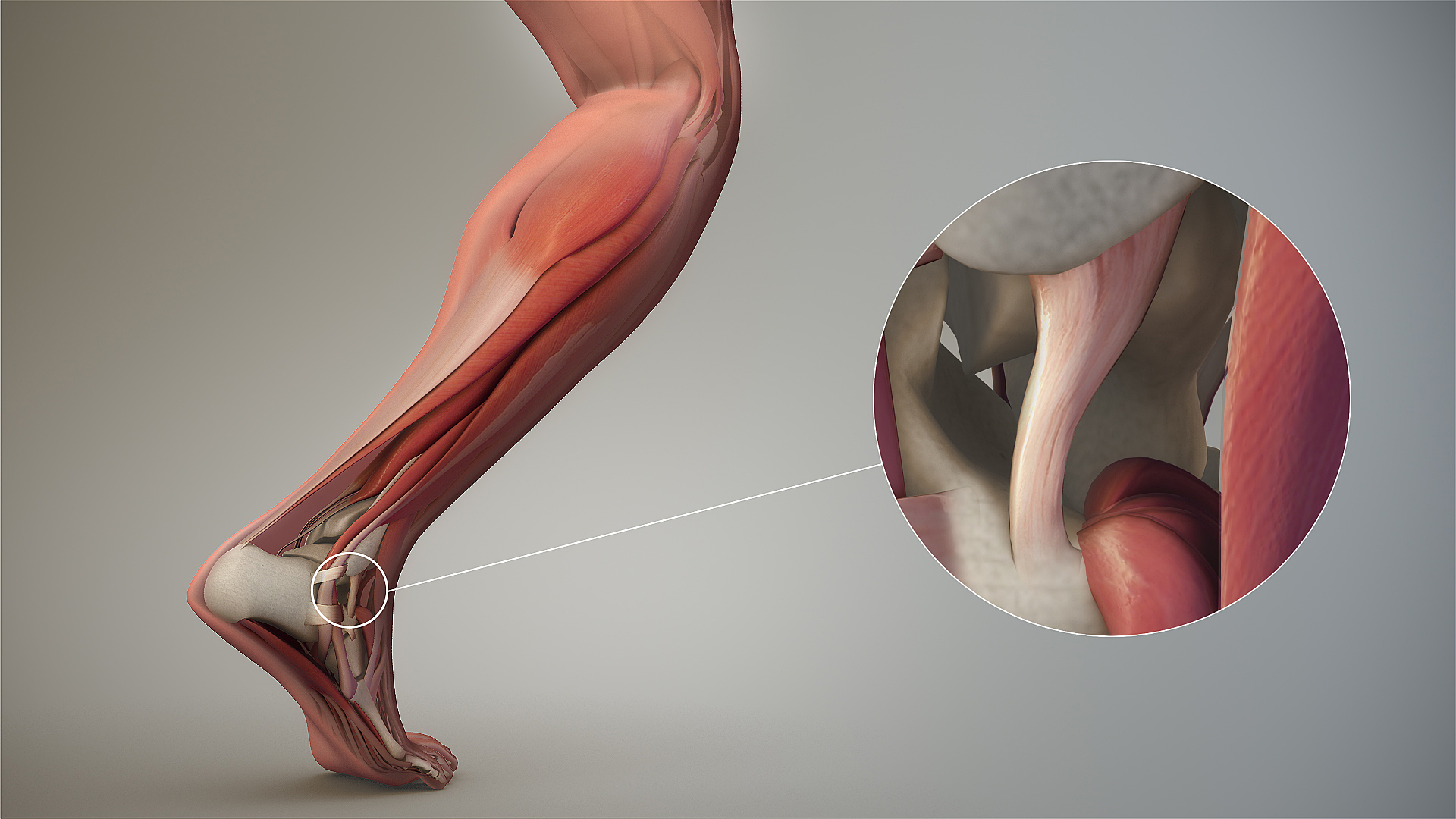
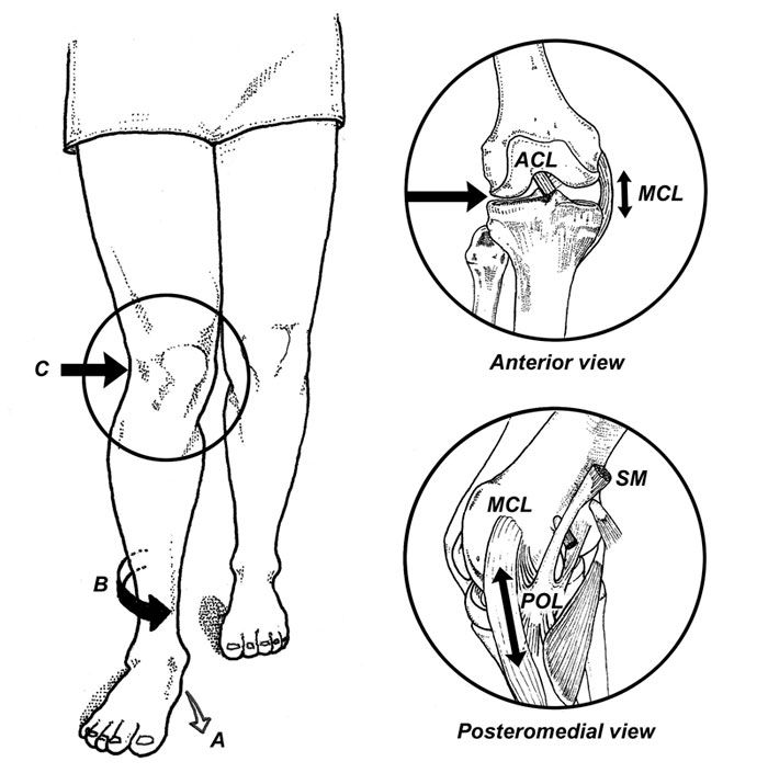
Pronation, with respect to the foot, is defined as a combination of eversion, abduction and dorsiflexion  (see picture attached) which results in flattening of the planter vault encompassing the medial and lateral longitudinal arches. In a normal gait cycle, this begins at initial contact (heel strike) and terminates at midstance, lasting no more than 25% of the gait cycle.

In a perfect biomechanical world, shortly following initial contact with the ground, the calcaneus should evert 4-8 degrees, largely because the body of the calcaneus is lateral to the longitudinal axis of the tibia. This results in plantar flexion, adduction and eversion of the talus on the calcaneus, as it slides anteriorly. At this point, there should be dorsiflexion of the transverse tarsal (calcaneo-cuboid and talo-navicular joints). Due to the tight fit of the ankle mortise and its unique shape, the tibial rotates internally (medially). This translates up the kinetic chain and causes internal rotation of the femur, which causes subsequent nutation of the pelvis and extension of the lumbar spine.  This should occur in the lower kinetic chain through the 1st half of stance phase. The sequence should reverse after the midpoint of midstance, causing supination and creating a rigid lever for forward propulsion.

Pronation, along with knee and hip flexion, allow for shock absorption during throughout the 1st half of stance phase. Pronation allows for the calcaneo-cuboid and talo-navicular joint axes to be parallel making the foot into a mobile adaptor so it can contour to irregular surfaces, like our hunter gatherer forefathers used to walk on before we paved the planet. Problems arise when the foot either under pronates (7 degrees valgus results in internal tibial rotation), resulting in poor shock absorption or over pronates (> 8 degrees or remains in pronation for greater than  50% of stance phase).

This paper talks about how foot and ankle pathologies have effects on other articulations in the foot. They looked at stance phase of gait in 14 people without pathology at 3 different walking speeds. they found

  • coupling relationships between rear foot inversion and hallux plantar flexion and rear foot eversion with hallux dorsiflexion

When the rear foot everts (as it does as discussed above) during pronation from initial contact to mid stance , the hallux should be extending AND when the rear foot everts, as it should from mid stance to terminal stance/pre swing, the hallux should be plantar flexing to get the 1st ray down to the ground

  • medial (internal) rotation of there leg was accompanied by mid foot collapse (read pronation) and lateral (external) rotation with mid foot elevation (read supination)

Because of the shape of the talar dome and shape of the talo calcaneal facet joints, the talus plantar flexes, everts and adducts from initial contact to mid stance, and dorsiflexes, inverts and adducts from mid stance to terminal stance/ pre swing

  • walking speed significantly influenced these coupling relationships

meaning that the faster we go, the faster these things must happen and the greater degree that the surrounding musculature and associated cortical control mechanisms must act

 So, when these relationships are compromised, problems (or more often, compensations) ensue. Think about these relationships and the kinetics and kinematics the next time you are studying someones gait. 

Here is a fun video talking about some of these relationships. 


Dubbeldam R1, Nester CNene AVHermens HJBuurke JH. Kinematic coupling relationships exist between non-adjacent segments of the foot and ankle of healthy subjects.Kinematic coupling relationships exist between non-adjacent segments of the foot and ankle of healthy subjects.Gait Posture. 2013 Feb;37(2):159-64. doi: 10.1016/j.gaitpost.2012.06.033. Epub 2012 Aug 27.


Lessons in Gait from Autistic Kids

“Additionally, there is the potential for the cerebellum, which receives sensory information and regulates movements, to have a level of dysfunction as well. Viewed collectively, the potential key contributors for gait asymmetry originate in the brain and specifically, the motor-controlling functions of the brain.” 

“While there is still little known regarding gait impairments in children with ASD, our findings illustrate that gait descriptors may provide insight into furthering working knowledge of ASD and may even enable gait-related symptoms to be treatable through therapies and interventions” 

“Alternative hypotheses suggest that children with ASD exhibit dysfunctional segregation of the motor cortex, which may be the key to uncoordinated movements” 

We often say that "gait is a fingerprint". Gait symmetry is often considered a window to neurologic function. We like to think "normal" gait has minimal asymmetries, while pathological gait does not. 

These are two landmark studies of gait in children with autism spectrum disorder. There were significant kinetic and kinematic differences in gait patterns in the 3 cardinal planes (saggital, coronal and transverse)  in ankle, knee and hip mechanics: The "pattern" is that there is no pattern, only changes. If you have a little time, check out this free, full text article here.

What this article says to us is that

  • We should be looking more carefully at gait asymmetries realizing that
    • These asymmetries are most likely cortical/cerebellar phenomena implying
  • Gait dysfunction equals cortical/cerebellar dysfunction

As clinician's, we should be thinking of altered gait as a window to what is going on north of the feet, knees, hip and pelvis. We remember that the joint and muscle mechanoreceptors feedback to the cerebellum and cortex via the spinocerebellar and dorsal column pathways which feed forward to the lower extremities via the anterior spinous cerebellar pathway as well as cortical spinal, rubrospinal had vestibula spinal pathways. The cortex, particularly the motor portion, has the capacity to alter gait just as abnormal mechanoreception has the capacity to alter cortical and cerebellar function. The two are interrelated and inseparable. Changes over time will altered pathways due to neural plasticity and adaptations will occur.

We need to be prudent and examined people fully and be very careful as to the modalities and exercises that we utilize and prescribed as ultimately they will shape that patients neural architecture.


Eggleston JD, Harry JR, Hickman R, Dufek JS. Analysis of gait symmetry during overground walking in children with autism spectrum disorder. Gait Posture 2017;55:162-166. 

Dufek JS, Eggleston JD, Harry JR, Hickman R. A comparative evaluation of gait between children with autism and typically developing matched controls. Med Sci 2017;5:1.  link to free full text: https://www.ncbi.nlm.nih.gov/pmc/articles/PMC5635776/


Sometimes, it doesn't matter whether it is long or short.

Achilles tendinopathy (AT) .. there are many factors that can contribute.

Have you considered leg length inequality? Generally speaking, People have a tendency to overpronate on the longer leg side and under prone only shorter leg side with strain on the medial and lateral aspects of the Achilles tendon respectively. It would make sense that this could be a contributing factor.

 "The mean inequality in length of legs (ILL) was 5 +/- 4 mm. Among the 48 patients with ILL > or = 5 mm, the side affected with ruptured tendon was longer in 48% of cases and shorter in 52%. " (1)

Age and pathology can play a role with younger, healthy tendons having greater compliance, with compliance being considered a risk factor for AT.(2)

Proprioception is impaired on the affected side of folks with Achilles tendinopathy (3). This is a "chicken and the egg" scenario. Did impaired proprioception cause the tendinopathy or is the tendinopathy causing the impaired proprioception? Probably, a little bit of both.

All this and more to be discussed in one of the 3 cases we will discuss tomorrow evening, Wednesday, August 15th on onlinece.com: Biomechanics 320   5PST, 6 MST, 7 CST, 8 EST

Hope to see you there!

1. Leppilahti J, Korpelainen R, Karpakka J, Kvist M, Orava S. Ruptures of the Achilles tendon: relationship to inequality in length of legs and to patterns in the foot and ankle. Foot Ankle Int. 1998 Oct;19(10):683-7.

2. Intziegianni K, Cassel M, Rauf S, White S, Rector M, Kaplick H, Wahmkow G, Kratzenstein S, Mayer F. Influence of Age and Pathology on Achilles Tendon Properties During a Single-leg Jump. Int J Sports Med. 2016 Nov;37(12):973-978. Epub 2016 Aug 8.

3. Scholes M, Stadler S, Connell D, Barton C, Clarke RA, Bryant AL, Malliaras P. Men with unilateral Achilles tendinopathy have impaired balance on the symptomatic side. J Sci Med Sport. 2018 May;21(5):479-482. doi: 10.1016/j.jsams.2017.09.594. Epub 2017 Oct 6.

How about that arm swing?

38846451_2134970409875956_3136689889865629696_n.png

Do you ever notice that sometimes when people have a problem with one of their lower extremities, there are arm swing changes? Usually on the opposite side? This can be anything from a short leg to lack of ankle dorsiflexion, lack of hip extension or even over pronation.

Many times, gait changes, including arm swing, are an "above down" process. This means it begins in the cortex, affects the pattern generators which subsequently will affect extremities distal to that. Sometimes this is a metabolic problem, sometimes vestibular (which can also be mechanical, causing decreased joint and muscle mechanoreceptor input to the cerebellum and vestibular nucleii), sometimes a combination of both. Throw a figure-of-eight ankle wrap on and walk. Your ROM is decreased (mechanical); this reduces input to your cerebellum which reduces input to your vestibular system. Your ankle dorsiflexion and step length will be diminished on that side; this will often cause an increase in arm swing on the contralateral side, which increases the metabolic "cost".

Arm swing may be coached, but we believe this is not always the correct approach as if it is a vestibular problem with altered cerebellar input (Something with the actual semicircular canals or perhaps input from muscle spindle or Golgi tendon organs), coaching arm swing makes the patient "look better" but does not really "fix" the problem; Which may be something as simple as joint pathomechanics, ligamentous restriction or a lack of skilled/endurance/strength in appropriate musculature.  If it is a metabolic issue, sometimes coaching arm swing can improve mechanical efficiency but at the cost of decreasing cortical efficiency, because the brain is such an energy hog.

Arm swing is there for a reason. It tells you something about what is going on or what is not going on. Just because it looks bad does not mean that it is necessarily the problem. Look deeper and keep your eyes, ears and mind open.

We will be talking about the case with this gal. her crossover gait and armswing, alonng with 2 other cases, on our 3rd Wednesdays class on onlince.com: Biomechanics 320  on 8/15/2018

Meyns P, Bruijn SM, Duysens J. The how and why of arm swing during human walking. Gait Posture. 2013 Sep;38(4):555-62. doi: 10.1016/j.gaitpost.2013.02.006. Epub 2013 Mar 13.

Wu Y, Li Y, Liu AM, Xiao F, Wang YZ, Hu F, Chen JL, Dai KR, Gu DY. Effect of active arm swing to local dynamic stability during walking. Hum Mov Sci. 2016 Feb;45:102-9. doi: 10.1016/j.humov.2015.10.005. Epub 2015 Nov 23.

Part 2: The amputated hallux & the complex biomechanical fall-out from it.

Screen Shot 2018-08-10 at 8.10.05 AM.png

Last week we promised Part 2 to this case, the amputated big toe.
Here is part 2. These are the complicated biomechanical fall-outs, so grab a big mug o' coffee and have at it !

In review, this person (all photos and case premissioned in swap for insight) had the distal hallux removed because of a progressive melanoma on the big toe. Can you believe that ! This is one more reminder that the sun and regular dermatologist screenings are wise.
This person had a complaint of progressing right gluteal and QL pain, spasm, tone and some persistent pain now in the 2nd metatarsal as well as some shoe challenges. We discuss this case briefly in and upcoming podcast, #139 or #140 we believe.

Screen Shot 2018-08-10 at 8.10.19 AM.png

Before we add our final thoughts to this case, lets cap our post from last week.

-Without the hallux, we cannot wind up the windlass and shorten the distance between the first metatarsal and heel, thus the arch will splay (more permanently over time we suspect) and we cannot optimize the arch height.
This will promote more internal spin on that limb because of more midfoot pronation and poor medial foot tripod stabilization.
- More internal limb spin means more internal hip spin, and more demand (which might not be met at the glute level) and thus loads that are supposed to be buffered with hip stabilization, will likely be transferred into the low back, and or into the medial knee. Look for more quad protective tone if they cannot get it from the glutes. Troubles arise when we try to control the hip from quadriceps strategies, it is poorly postured to do so, but people do it everyday, *hint: most cyclists and distance runners to a large degree).
- anterior pelvis posturing on the right, perhaps challenging durability of the lower abdominals, hence suspect QL increased protective tone, possible low back tightness or pain depending on duration of activities
- These factors are likely related to his complaints in the right gluteal and low back/QL area.

Now, onto our next thoughts.

- when the hallux is incompetent, in this case absent, there are few other choices to gain forefoot purchase on the ground other than more flexion gripping of the 2nd toe (then the 3rd, then 4th). This is a progressing "searching" phenomenon for forefoot stability and without the function of the big fella, the 2nd toe will begin a hammering phenomenon, often, but not always. We would not be surprised to see hammer toe development in this case, but this person is now very aware of it, and can at least now fight that battle with increased awareness. There is some mild evidence of this on the side lateral photo.

- We are happy to see that the proximal phalange was spared. The adductor hallucis is inserted medially there, and this will help to reduce bunion generation risk (medial metatarsal drift). Comparing the photo and the radiograph is a great example of how far back/proximal the 1st MTP joint is. One could easily assume that the entire hallux was resected from the photo, but the radiograph shows otherwise.

Screen Shot 2018-08-10 at 8.22.36 AM.png

- Toe off is obviously going to be compromised. The patient cannot adequately stabilize the 1st metatarsal (MET) and this will mean a compromised foot tripod, medial foot/tripod splay, arch pronation control challenges but toe off stabilization is going to have to be met by the 2nd and 3rd digits, as discussed above. They are not suited to be the major players here, they are synergistic to this end. Do not be surprised to see one of 2 strategies at toe off here:

1. heavy medial foot tripod toe off, dropping into the void and this maximize the internal spin challenges and minimizing the requisite foot supination stiffness generation phase that should be normal at toe off

2. avoidance of the above, with a forced conscious forefoot lateral toe off, a supinatory strategy, to avoid internal limb spin, more toe hammering, and the lurch heavily and abruptly off of the right foot and onto the left limb.

Screen Shot 2018-08-10 at 8.10.27 AM.png

3. taking #2 further, any time there is perceived challenges or deficits in strength, endurance, proprioception, balance, power and the like, the brain often will create a premature departure off of said limb, creating a requisite premature loading onto the opposite limb. This can cause a phenomenon well loosely refer to "catching" in the contralateral quadriceps mechanism. These clients, with their abrupt loading pattern onto the opposite limb will most often have troubles getting into initial gluteal hip stabilization strategies, and thus default into a quadriceps strategy, that in time can lead to quad shortness and increased tone, which can cause more compression across the patellofemoral joint and cause knee pain. This is more of a compression/loading response issue rather than tracking phenomenon, which we see at the typical diagnosis. We often look for causes in the opposite limb for contralateral knee pain. IT is quite often there if you are looking hard enough for it. Fix the problem, not the symptom.
There is a long host of other things than can arise from here, including heavy contralateral (in this case left sided) foot loading challenges, often more forefoot initial loading, and all of the problems than can arise when this pattern is cyclical, but that would take this post far too deep and long. So, . . . . another time.

Screen Shot 2018-08-10 at 8.09.47 AM.png

4. Shoe fit, we could make the case that a shoe that nicely hugs the forefoot, as opposed to a wide toe box'ed shoe, could help fight off the risk of 1st metatarsal abduction and thus bunion formation risk. However, one cannot dismiss the wider toe box giving the remaining toes a better environment to engage without hammering with over use of long flexors. We might suggest a trial of an elastic sleeve, one often used for plantar fascitis symptom management, placing a snug one around the forefoot when ambulating. This could help keep that metatarsal snug and stop the bunion-like drift we would be watching for.

have at it gang, cases like this are far and deep and require deep understanding of normal and abnormal biomechanics, and the rabbit hole deep myriad of compensations that can be engaged.

have a great weekend !

Shawn and Ivo

Better gait AFTER rhizotomies?

Nothing surprised me more than reading this paper and finding out that folks that have had rhizotomies, which removes the afferent input from the dorsal horn and sensory information from the reflex loops in the cord, actually had better gait. Of course these children had severe spastic diplegia, which means they have lost descending inhibition from higher center's and most likely had increased flexor tone in the lower extremities. 

image credit: http://realtyconnect.me/spinal-cord-cross-section-tracts/background-information-musculoskeletal-key-within-spinal-cord-cross-section-tracts/

image credit: http://realtyconnect.me/spinal-cord-cross-section-tracts/background-information-musculoskeletal-key-within-spinal-cord-cross-section-tracts/

Remember that the fibers entering the dorsal horn not only go to the dorsal columns but also to the spinocerebellar pathways. When someone has spasticity, the feedback loops are skewed and flexor drive coming from the rostral reticular formation generally is increased are often kept in check by the cerebellar and vestibular feedback loops. Perhaps the interruption of this feedback loop and lack of information from type IA and II afferents of the muscle spindles as well as Ib afferents from the globe tendon organs modulated the tone sufficiently to improve gait. This study did a selective dorsal rhizotomy which means only a portion of it was ablated. 

The somatotopic organization  of the dorsal horn of the spinal cord (i.e.: certain areas of the dorsal horn correspond to certain body parts) is well documented in humans; It would make sense that the dorsal root itself (i.e.: the afferent fibers in the nerve going into the dorsal horn) would be as well, as they are that way in murines (2) and felines (3). 

So, how does this apply to gait? People with strokes, cortical lesions, diseases like cerebral palsy and even possibly increased flexor tone, may benefit from altered input into the dorsal horn. It would have been really cool to see if they increased extensor activity in this individuals, if they would be benefited further. 


Abstract

OBJECTIVE: To identify factors associated with long-term improvement in gait in children after selective dorsal rhizotomy (SDR).

DESIGN: Retrospective cohort study SETTING: University medical center PARTICIPANTS: 36 children (age 4-13y) with spastic diplegia (gross motor classification system level I (n=14), II (n=15) and III (n=7) were included retrospectively from the database of our hospital. Children underwent selective dorsal rhizotomy (SDR) between January 1999 and May 2011. Patients were included if they received clinical gait analysis before and five years post-SDR, age >4 years at time of SDR and if brain MRI-scan was available.

INTERVENTION: Selective dorsal rhizotomy MAIN OUTCOME MEASURES: Overall gait quality was assessed with Edinburgh visual gait score (EVGS), before and five years after SDR. In addition, knee and ankle angles at initial contact and midstance were evaluated. To identify predictors for gait improvement, several factors were evaluated including: functional mobility level (GMFCS), presence of white matter abnormalities on brain-MRI, and selective motor control during gait (synergy analysis).

RESULTS: Overall gait quality improved after SDR, with a large variation between patients. Multiple linear regression analysis revealed that worse score on EVGS and better GMFCS were independently related to gait improvement. Gait improved more in children with GMFCS I & II compared to III. No differences were observed between children with or without white matter abnormalities on brain MRI. Selective motor control during gait was predictive for improvement of knee angle at initial contact and midstance, but not for EVGS.

CONCLUSION: Functional mobility level and baseline gait quality are both important factors to predict gait outcomes after SDR. If candidates are well selected, SDR can be a successful intervention to improve gait both in children with brain MRI abnormalities as well as other causes of spastic diplegia.


1. Oudenhoven LM, van der Krogt MM, Romei M, van Schie PEM, van de Pol LA, van Ouwerkerk WJR, Harlaar Prof J, Buizer AI. Factors associated with long-term improvement of gait after selective dorsal rhizotomy. Arch Phys Med Rehabil. 2018 Jul 4. pii: S0003-9993(18)30442-8. doi: 10.1016/j.apmr.2018.06.016. [Epub ahead of print]

2. Wessels WJ1, Marani E. A rostrocaudal somatotopic organization in the brachial dorsal root ganglia of neonatal rats. Clin Neurol Neurosurg. 1993;95 Suppl:S3-11.

3. Koerber HRBrown PB. Somatotopic organization of hindlimb cutaneous nerve projections to cat dorsal horn. J Neurophysiol. 1982 Aug;48(2):481-9.