Podcast Shorts # 137.1 Arm Swing in Sport

This is a small clip on arm swing from podcast 137.. For the full podcast, head over to our website or iTunes or anywhere that you choose to download your podcasts.

Key words: arm swing, gait, gait analysis, thegaitguys

Links to find the podcast:

iTunes page: https://itunes.apple.com/us/podcast/the-gait-guys-podcast/id559864138?mt=2

Direct Download: http://traffic.libsyn.com/thegaitguys/pod_138.1_arm_swing_-_82718_5.18_PM.mp3

Permalink URL: http://thegaitguys.libsyn.com/podcast-shorts-1381-arm-swing-in-sport

Libsyn URL: http://directory.libsyn.com/episode/index/id/6975092


Our Websites:
www.thegaitguys.com

summitchiroandrehab.com doctorallen.co shawnallen.net

Our website is all you need to remember. Everything you want, need and wish for is right there on the site.
Interested in our stuff ? Want to buy some of our lectures or our National Shoe Fit program? Click here (thegaitguys.com or thegaitguys.tumblr.com) and you will come to our websites. In the tabs, you will find tabs for STORE, SEMINARS, BOOK etc. We also lecture every 3rd Wednesday of the month on onlineCE.com. We have an extensive catalogued library of our courses there, you can take them any time for a nominal fee (~$20).

Our podcast is on iTunes and just about every other podcast harbor site, just google "the gait guys podcast", you will find us.

What do you know about pronation and Supination?

We have talked many times here on TGG about pronation, supination, overpronation, asymmetrical pronation, and more. 

When most people think of pronation, they think of midfoot pronation, or pronation about the subtalar or transverse tarsal joints. Pronation can actually occur about any articulation or bone, but with respect to the foot, we like to think of rearfoot (ie. talo-calcaneal), midfoot (talo-navicular) and forefoot (transverse tarsal). The question is why does this matter?

Pronation, with respect to the foot, is defined as a combination of eversion, abduction and dorsiflexion  (see picture attached) which results in flattening of the planter vault encompassing the medial and lateral longitudinal arches. In a normal gait cycle, this begins at initial contact (heel strike) and terminates at midstance, lasting no more than 25% of the gait cycle.

In a perfect biomechanical world, shortly following initial contact with the ground, the calcaneus should evert 4-8 degrees, largely because the body of the calcaneus is lateral to the longitudinal axis of the tibia. This results in plantar flexion, adduction and eversion of the talus on the calcaneus, as it slides anteriorly. At this point, there should be dorsiflexion of the transverse tarsal (calcaneo-cuboid and talo-navicular joints). Due to the tight fit of the ankle mortise and its unique shape, the tibial rotates internally (medially). This translates up the kinetic chain and causes internal rotation of the femur, which causes subsequent nutation of the pelvis and extension of the lumbar spine.  This should occur in the lower kinetic chain through the 1st half of stance phase. The sequence should reverse after the midpoint of midstance, causing supination and creating a rigid lever for forward propulsion.

Pronation, along with knee and hip flexion, allow for shock absorption during throughout the 1st half of stance phase. Pronation allows for the calcaneo-cuboid and talo-navicular joint axes to be parallel making the foot into a mobile adaptor so it can contour to irregular surfaces, like our hunter gatherer forefathers used to walk on before we paved the planet. Problems arise when the foot either under pronates (7 degrees valgus results in internal tibial rotation), resulting in poor shock absorption or over pronates (> 8 degrees or remains in pronation for greater than  50% of stance phase).

This paper talks about how foot and ankle pathologies have effects on other articulations in the foot. They looked at stance phase of gait in 14 people without pathology at 3 different walking speeds. they found

  • coupling relationships between rear foot inversion and hallux plantar flexion and rear foot eversion with hallux dorsiflexion

When the rear foot everts (as it does as discussed above) during pronation from initial contact to mid stance , the hallux should be extending AND when the rear foot everts, as it should from mid stance to terminal stance/pre swing, the hallux should be plantar flexing to get the 1st ray down to the ground

  • medial (internal) rotation of there leg was accompanied by mid foot collapse (read pronation) and lateral (external) rotation with mid foot elevation (read supination)

Because of the shape of the talar dome and shape of the talo calcaneal facet joints, the talus plantar flexes, everts and adducts from initial contact to mid stance, and dorsiflexes, inverts and adducts from mid stance to terminal stance/ pre swing

  • walking speed significantly influenced these coupling relationships

meaning that the faster we go, the faster these things must happen and the greater degree that the surrounding musculature and associated cortical control mechanisms must act

 So, when these relationships are compromised, problems (or more often, compensations) ensue. Think about these relationships and the kinetics and kinematics the next time you are studying someones gait. 

Here is a fun video talking about some of these relationships. 


Dubbeldam R1, Nester CNene AVHermens HJBuurke JH. Kinematic coupling relationships exist between non-adjacent segments of the foot and ankle of healthy subjects.Kinematic coupling relationships exist between non-adjacent segments of the foot and ankle of healthy subjects.Gait Posture. 2013 Feb;37(2):159-64. doi: 10.1016/j.gaitpost.2012.06.033. Epub 2012 Aug 27.


Lessons in Gait from Autistic Kids

“Additionally, there is the potential for the cerebellum, which receives sensory information and regulates movements, to have a level of dysfunction as well. Viewed collectively, the potential key contributors for gait asymmetry originate in the brain and specifically, the motor-controlling functions of the brain.” 

“While there is still little known regarding gait impairments in children with ASD, our findings illustrate that gait descriptors may provide insight into furthering working knowledge of ASD and may even enable gait-related symptoms to be treatable through therapies and interventions” 

“Alternative hypotheses suggest that children with ASD exhibit dysfunctional segregation of the motor cortex, which may be the key to uncoordinated movements” 

We often say that "gait is a fingerprint". Gait symmetry is often considered a window to neurologic function. We like to think "normal" gait has minimal asymmetries, while pathological gait does not. 

These are two landmark studies of gait in children with autism spectrum disorder. There were significant kinetic and kinematic differences in gait patterns in the 3 cardinal planes (saggital, coronal and transverse)  in ankle, knee and hip mechanics: The "pattern" is that there is no pattern, only changes. If you have a little time, check out this free, full text article here.

What this article says to us is that

  • We should be looking more carefully at gait asymmetries realizing that
    • These asymmetries are most likely cortical/cerebellar phenomena implying
  • Gait dysfunction equals cortical/cerebellar dysfunction

As clinician's, we should be thinking of altered gait as a window to what is going on north of the feet, knees, hip and pelvis. We remember that the joint and muscle mechanoreceptors feedback to the cerebellum and cortex via the spinocerebellar and dorsal column pathways which feed forward to the lower extremities via the anterior spinous cerebellar pathway as well as cortical spinal, rubrospinal had vestibula spinal pathways. The cortex, particularly the motor portion, has the capacity to alter gait just as abnormal mechanoreception has the capacity to alter cortical and cerebellar function. The two are interrelated and inseparable. Changes over time will altered pathways due to neural plasticity and adaptations will occur.

We need to be prudent and examined people fully and be very careful as to the modalities and exercises that we utilize and prescribed as ultimately they will shape that patients neural architecture.


Eggleston JD, Harry JR, Hickman R, Dufek JS. Analysis of gait symmetry during overground walking in children with autism spectrum disorder. Gait Posture 2017;55:162-166. 

Dufek JS, Eggleston JD, Harry JR, Hickman R. A comparative evaluation of gait between children with autism and typically developing matched controls. Med Sci 2017;5:1.  link to free full text: https://www.ncbi.nlm.nih.gov/pmc/articles/PMC5635776/


Sometimes, it doesn't matter whether it is long or short.

Achilles tendinopathy (AT) .. there are many factors that can contribute.

Have you considered leg length inequality? Generally speaking, People have a tendency to overpronate on the longer leg side and under prone only shorter leg side with strain on the medial and lateral aspects of the Achilles tendon respectively. It would make sense that this could be a contributing factor.

 "The mean inequality in length of legs (ILL) was 5 +/- 4 mm. Among the 48 patients with ILL > or = 5 mm, the side affected with ruptured tendon was longer in 48% of cases and shorter in 52%. " (1)

Age and pathology can play a role with younger, healthy tendons having greater compliance, with compliance being considered a risk factor for AT.(2)

Proprioception is impaired on the affected side of folks with Achilles tendinopathy (3). This is a "chicken and the egg" scenario. Did impaired proprioception cause the tendinopathy or is the tendinopathy causing the impaired proprioception? Probably, a little bit of both.

All this and more to be discussed in one of the 3 cases we will discuss tomorrow evening, Wednesday, August 15th on onlinece.com: Biomechanics 320   5PST, 6 MST, 7 CST, 8 EST

Hope to see you there!

1. Leppilahti J, Korpelainen R, Karpakka J, Kvist M, Orava S. Ruptures of the Achilles tendon: relationship to inequality in length of legs and to patterns in the foot and ankle. Foot Ankle Int. 1998 Oct;19(10):683-7.

2. Intziegianni K, Cassel M, Rauf S, White S, Rector M, Kaplick H, Wahmkow G, Kratzenstein S, Mayer F. Influence of Age and Pathology on Achilles Tendon Properties During a Single-leg Jump. Int J Sports Med. 2016 Nov;37(12):973-978. Epub 2016 Aug 8.

3. Scholes M, Stadler S, Connell D, Barton C, Clarke RA, Bryant AL, Malliaras P. Men with unilateral Achilles tendinopathy have impaired balance on the symptomatic side. J Sci Med Sport. 2018 May;21(5):479-482. doi: 10.1016/j.jsams.2017.09.594. Epub 2017 Oct 6.

How about that arm swing?

38846451_2134970409875956_3136689889865629696_n.png

Do you ever notice that sometimes when people have a problem with one of their lower extremities, there are arm swing changes? Usually on the opposite side? This can be anything from a short leg to lack of ankle dorsiflexion, lack of hip extension or even over pronation.

Many times, gait changes, including arm swing, are an "above down" process. This means it begins in the cortex, affects the pattern generators which subsequently will affect extremities distal to that. Sometimes this is a metabolic problem, sometimes vestibular (which can also be mechanical, causing decreased joint and muscle mechanoreceptor input to the cerebellum and vestibular nucleii), sometimes a combination of both. Throw a figure-of-eight ankle wrap on and walk. Your ROM is decreased (mechanical); this reduces input to your cerebellum which reduces input to your vestibular system. Your ankle dorsiflexion and step length will be diminished on that side; this will often cause an increase in arm swing on the contralateral side, which increases the metabolic "cost".

Arm swing may be coached, but we believe this is not always the correct approach as if it is a vestibular problem with altered cerebellar input (Something with the actual semicircular canals or perhaps input from muscle spindle or Golgi tendon organs), coaching arm swing makes the patient "look better" but does not really "fix" the problem; Which may be something as simple as joint pathomechanics, ligamentous restriction or a lack of skilled/endurance/strength in appropriate musculature.  If it is a metabolic issue, sometimes coaching arm swing can improve mechanical efficiency but at the cost of decreasing cortical efficiency, because the brain is such an energy hog.

Arm swing is there for a reason. It tells you something about what is going on or what is not going on. Just because it looks bad does not mean that it is necessarily the problem. Look deeper and keep your eyes, ears and mind open.

We will be talking about the case with this gal. her crossover gait and armswing, alonng with 2 other cases, on our 3rd Wednesdays class on onlince.com: Biomechanics 320  on 8/15/2018

Meyns P, Bruijn SM, Duysens J. The how and why of arm swing during human walking. Gait Posture. 2013 Sep;38(4):555-62. doi: 10.1016/j.gaitpost.2013.02.006. Epub 2013 Mar 13.

Wu Y, Li Y, Liu AM, Xiao F, Wang YZ, Hu F, Chen JL, Dai KR, Gu DY. Effect of active arm swing to local dynamic stability during walking. Hum Mov Sci. 2016 Feb;45:102-9. doi: 10.1016/j.humov.2015.10.005. Epub 2015 Nov 23.

Part 2: The amputated hallux & the complex biomechanical fall-out from it.

Screen Shot 2018-08-10 at 8.10.05 AM.png

Last week we promised Part 2 to this case, the amputated big toe.
Here is part 2. These are the complicated biomechanical fall-outs, so grab a big mug o' coffee and have at it !

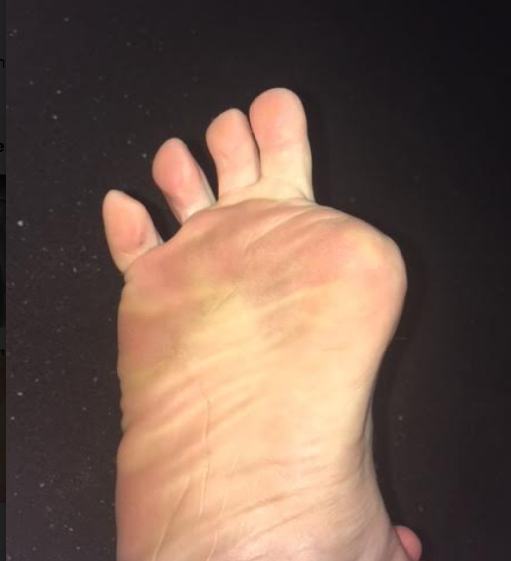
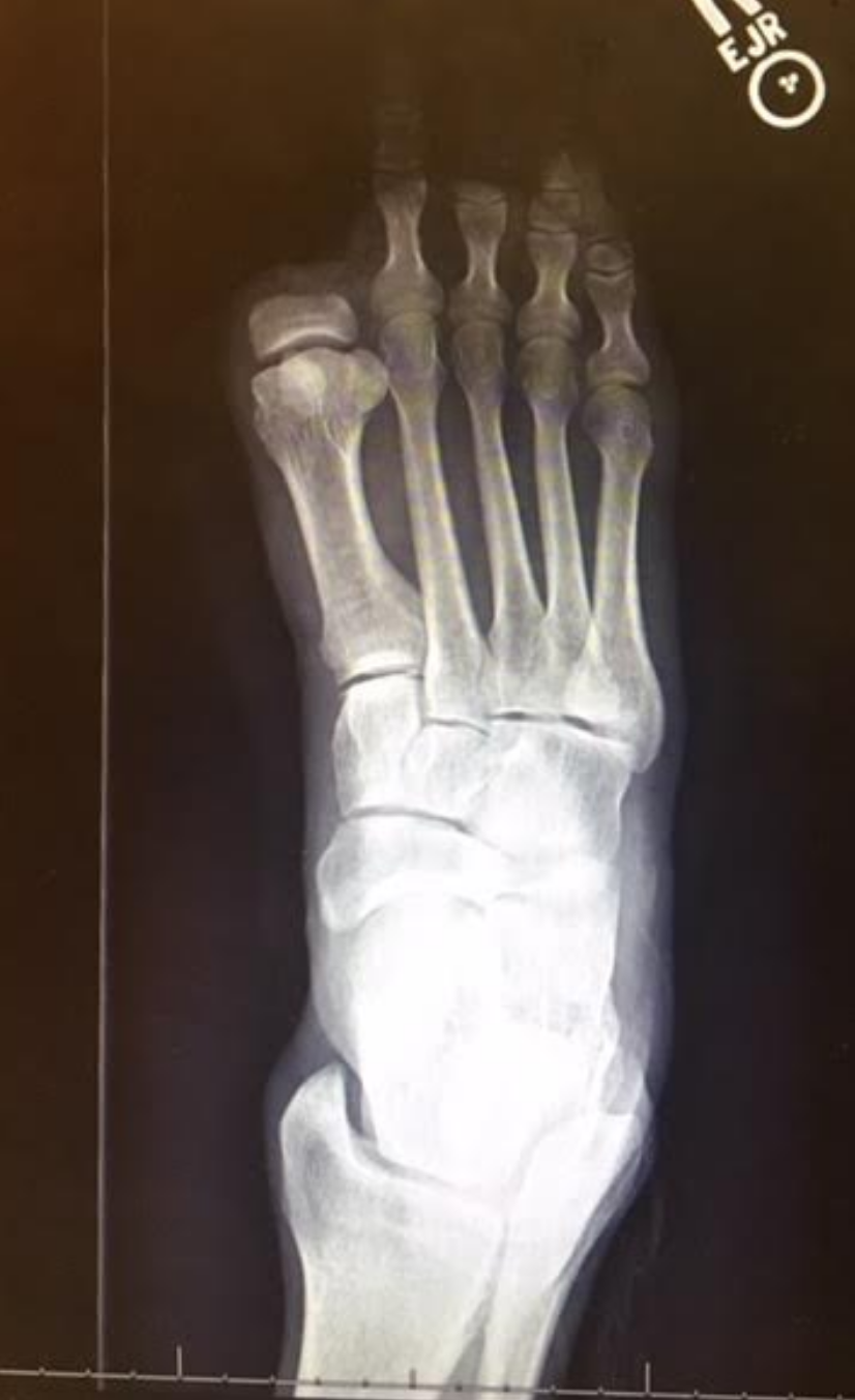
In review, this person (all photos and case premissioned in swap for insight) had the distal hallux removed because of a progressive melanoma on the big toe. Can you believe that ! This is one more reminder that the sun and regular dermatologist screenings are wise.
This person had a complaint of progressing right gluteal and QL pain, spasm, tone and some persistent pain now in the 2nd metatarsal as well as some shoe challenges. We discuss this case briefly in and upcoming podcast, #139 or #140 we believe.

Screen Shot 2018-08-10 at 8.10.19 AM.png

Before we add our final thoughts to this case, lets cap our post from last week.

-Without the hallux, we cannot wind up the windlass and shorten the distance between the first metatarsal and heel, thus the arch will splay (more permanently over time we suspect) and we cannot optimize the arch height.
This will promote more internal spin on that limb because of more midfoot pronation and poor medial foot tripod stabilization.
- More internal limb spin means more internal hip spin, and more demand (which might not be met at the glute level) and thus loads that are supposed to be buffered with hip stabilization, will likely be transferred into the low back, and or into the medial knee. Look for more quad protective tone if they cannot get it from the glutes. Troubles arise when we try to control the hip from quadriceps strategies, it is poorly postured to do so, but people do it everyday, *hint: most cyclists and distance runners to a large degree).
- anterior pelvis posturing on the right, perhaps challenging durability of the lower abdominals, hence suspect QL increased protective tone, possible low back tightness or pain depending on duration of activities
- These factors are likely related to his complaints in the right gluteal and low back/QL area.

Now, onto our next thoughts.

- when the hallux is incompetent, in this case absent, there are few other choices to gain forefoot purchase on the ground other than more flexion gripping of the 2nd toe (then the 3rd, then 4th). This is a progressing "searching" phenomenon for forefoot stability and without the function of the big fella, the 2nd toe will begin a hammering phenomenon, often, but not always. We would not be surprised to see hammer toe development in this case, but this person is now very aware of it, and can at least now fight that battle with increased awareness. There is some mild evidence of this on the side lateral photo.

- We are happy to see that the proximal phalange was spared. The adductor hallucis is inserted medially there, and this will help to reduce bunion generation risk (medial metatarsal drift). Comparing the photo and the radiograph is a great example of how far back/proximal the 1st MTP joint is. One could easily assume that the entire hallux was resected from the photo, but the radiograph shows otherwise.

Screen Shot 2018-08-10 at 8.22.36 AM.png

- Toe off is obviously going to be compromised. The patient cannot adequately stabilize the 1st metatarsal (MET) and this will mean a compromised foot tripod, medial foot/tripod splay, arch pronation control challenges but toe off stabilization is going to have to be met by the 2nd and 3rd digits, as discussed above. They are not suited to be the major players here, they are synergistic to this end. Do not be surprised to see one of 2 strategies at toe off here:

1. heavy medial foot tripod toe off, dropping into the void and this maximize the internal spin challenges and minimizing the requisite foot supination stiffness generation phase that should be normal at toe off

2. avoidance of the above, with a forced conscious forefoot lateral toe off, a supinatory strategy, to avoid internal limb spin, more toe hammering, and the lurch heavily and abruptly off of the right foot and onto the left limb.

Screen Shot 2018-08-10 at 8.10.27 AM.png

3. taking #2 further, any time there is perceived challenges or deficits in strength, endurance, proprioception, balance, power and the like, the brain often will create a premature departure off of said limb, creating a requisite premature loading onto the opposite limb. This can cause a phenomenon well loosely refer to "catching" in the contralateral quadriceps mechanism. These clients, with their abrupt loading pattern onto the opposite limb will most often have troubles getting into initial gluteal hip stabilization strategies, and thus default into a quadriceps strategy, that in time can lead to quad shortness and increased tone, which can cause more compression across the patellofemoral joint and cause knee pain. This is more of a compression/loading response issue rather than tracking phenomenon, which we see at the typical diagnosis. We often look for causes in the opposite limb for contralateral knee pain. IT is quite often there if you are looking hard enough for it. Fix the problem, not the symptom.
There is a long host of other things than can arise from here, including heavy contralateral (in this case left sided) foot loading challenges, often more forefoot initial loading, and all of the problems than can arise when this pattern is cyclical, but that would take this post far too deep and long. So, . . . . another time.

Screen Shot 2018-08-10 at 8.09.47 AM.png

4. Shoe fit, we could make the case that a shoe that nicely hugs the forefoot, as opposed to a wide toe box'ed shoe, could help fight off the risk of 1st metatarsal abduction and thus bunion formation risk. However, one cannot dismiss the wider toe box giving the remaining toes a better environment to engage without hammering with over use of long flexors. We might suggest a trial of an elastic sleeve, one often used for plantar fascitis symptom management, placing a snug one around the forefoot when ambulating. This could help keep that metatarsal snug and stop the bunion-like drift we would be watching for.

have at it gang, cases like this are far and deep and require deep understanding of normal and abnormal biomechanics, and the rabbit hole deep myriad of compensations that can be engaged.

have a great weekend !

Shawn and Ivo

Better gait AFTER rhizotomies?

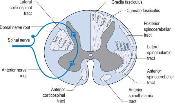
Nothing surprised me more than reading this paper and finding out that folks that have had rhizotomies, which removes the afferent input from the dorsal horn and sensory information from the reflex loops in the cord, actually had better gait. Of course these children had severe spastic diplegia, which means they have lost descending inhibition from higher center's and most likely had increased flexor tone in the lower extremities. 

image credit: http://realtyconnect.me/spinal-cord-cross-section-tracts/background-information-musculoskeletal-key-within-spinal-cord-cross-section-tracts/

image credit: http://realtyconnect.me/spinal-cord-cross-section-tracts/background-information-musculoskeletal-key-within-spinal-cord-cross-section-tracts/

Remember that the fibers entering the dorsal horn not only go to the dorsal columns but also to the spinocerebellar pathways. When someone has spasticity, the feedback loops are skewed and flexor drive coming from the rostral reticular formation generally is increased are often kept in check by the cerebellar and vestibular feedback loops. Perhaps the interruption of this feedback loop and lack of information from type IA and II afferents of the muscle spindles as well as Ib afferents from the globe tendon organs modulated the tone sufficiently to improve gait. This study did a selective dorsal rhizotomy which means only a portion of it was ablated. 

The somatotopic organization  of the dorsal horn of the spinal cord (i.e.: certain areas of the dorsal horn correspond to certain body parts) is well documented in humans; It would make sense that the dorsal root itself (i.e.: the afferent fibers in the nerve going into the dorsal horn) would be as well, as they are that way in murines (2) and felines (3). 

So, how does this apply to gait? People with strokes, cortical lesions, diseases like cerebral palsy and even possibly increased flexor tone, may benefit from altered input into the dorsal horn. It would have been really cool to see if they increased extensor activity in this individuals, if they would be benefited further. 


Abstract

OBJECTIVE: To identify factors associated with long-term improvement in gait in children after selective dorsal rhizotomy (SDR).

DESIGN: Retrospective cohort study SETTING: University medical center PARTICIPANTS: 36 children (age 4-13y) with spastic diplegia (gross motor classification system level I (n=14), II (n=15) and III (n=7) were included retrospectively from the database of our hospital. Children underwent selective dorsal rhizotomy (SDR) between January 1999 and May 2011. Patients were included if they received clinical gait analysis before and five years post-SDR, age >4 years at time of SDR and if brain MRI-scan was available.

INTERVENTION: Selective dorsal rhizotomy MAIN OUTCOME MEASURES: Overall gait quality was assessed with Edinburgh visual gait score (EVGS), before and five years after SDR. In addition, knee and ankle angles at initial contact and midstance were evaluated. To identify predictors for gait improvement, several factors were evaluated including: functional mobility level (GMFCS), presence of white matter abnormalities on brain-MRI, and selective motor control during gait (synergy analysis).

RESULTS: Overall gait quality improved after SDR, with a large variation between patients. Multiple linear regression analysis revealed that worse score on EVGS and better GMFCS were independently related to gait improvement. Gait improved more in children with GMFCS I & II compared to III. No differences were observed between children with or without white matter abnormalities on brain MRI. Selective motor control during gait was predictive for improvement of knee angle at initial contact and midstance, but not for EVGS.

CONCLUSION: Functional mobility level and baseline gait quality are both important factors to predict gait outcomes after SDR. If candidates are well selected, SDR can be a successful intervention to improve gait both in children with brain MRI abnormalities as well as other causes of spastic diplegia.


1. Oudenhoven LM, van der Krogt MM, Romei M, van Schie PEM, van de Pol LA, van Ouwerkerk WJR, Harlaar Prof J, Buizer AI. Factors associated with long-term improvement of gait after selective dorsal rhizotomy. Arch Phys Med Rehabil. 2018 Jul 4. pii: S0003-9993(18)30442-8. doi: 10.1016/j.apmr.2018.06.016. [Epub ahead of print]

2. Wessels WJ1, Marani E. A rostrocaudal somatotopic organization in the brachial dorsal root ganglia of neonatal rats. Clin Neurol Neurosurg. 1993;95 Suppl:S3-11.

3. Koerber HRBrown PB. Somatotopic organization of hindlimb cutaneous nerve projections to cat dorsal horn. J Neurophysiol. 1982 Aug;48(2):481-9.

Surgery vs casting...same results

image source: https://commons.wikimedia.org/wiki/File:Trimalleolar_Ankle_Fracture.jpg

image source: https://commons.wikimedia.org/wiki/File:Trimalleolar_Ankle_Fracture.jpg

We see many people for all types of fractures and rehab. This study looks at folks who had ankle fractures who either got mhm casted or had surgical management. Looks like conservative is just as good in this case.

"In a pre-specified, 3-year extension of a randomized clinical trial of equivalence, close-contact casting maintained equivalence in function compared to surgery in older adults with unstable ankle fracture. Furthermore, no significant differences were reported in quality of life or pain. The authors concluded that the focus of treatment for these patients should be on obtaining and maintaining reduction until union, using the most conservative means possible.

The study enrolled 461 patients; the control group (n=254) had non-diabetes-related foot complications; the study group (n=207) had diabetic foot pathology (including 61 [32%] with diabetic foot ulcer, Charcot neuropathy, foot infection, or acute neuropathic fractures and dislocations).

Researchers found no significant differences between the 2 groups related to fear of blindness, diabetic foot infection, or kidney failure needing dialysis. When compared to those without diabetic foot problems, the authors found that the 32% of the study group with identified diabetic foot disease were 136% more likely to rate LEA as their greatest fear and that 49% were less likely to rate death as their greatest fear. In their conclusion, the authors noted that the presence of a diabetic foot-related complication, having diabetes for more than 10 years, use of insulin, and having peripheral neuropathy were all variables that subjects associated with identifying LEA as the greatest fear.


Wukich DK, Raspovic KM, Suder NC. Patients with diabetic foot disease fear major lower extremity amputation more than death. Foot Ankle Spec. 2018;11(1):17-21."


source: http://lermagazine.com/issues/may/three-year-follow-up-close-contact-casting-vs-surgery

image source: https://commons.wikimedia.org/wiki/File:Trimalleolar_Ankle_Fracture.jpg

Gait: A bottom up process (mostly).

"Bottom up" process of running ? Support that it is not "top down".

"These results support a passive arm swing hypothesis for upper body movement during human walking and running, in which the trunk and shoulders act primarily as elastic linkages between the pelvis, shoulder girdle and arms, the arms act as passive mass dampers which reduce torso and head rotation, and upper body movement is primarily powered by lower body movement."
"Angular acceleration of the shoulders and arm increased with torsion of the trunk and shoulder, respectively, but angular acceleration of the shoulders was not inversely related to angular acceleration of the pelvis or arm."

At The Gait Guys have been saying this for years in our writing based off of the research we have consumed that coaching changes in arm and shoulder swing and posture is not the way the system works, or the way to go with coaching running, not without possible ramifications to the athlete by overriding how the system is wired for locomotion. The arm motor patterns are neurologically driven by the lower limbs. It is a bottom up process as we have always said. The arms are largely subservient to the legs, the work as a team, the but quarterback is the pelvis and legs. Obliquity of the pelvis and the antiphasic principles hold true once again in this study. And monkeying with the shoulders and arm swing has a direct effect on the COM, and can thus impact the brains initial choice of optimal step width in any given contact period. Driving more arm movement can over ride inherently hard wired locomotion processes and that can lead to coaching compensatory patterns that should be first driven by lower limb mechanical changes, or coaching recommendations/exercises.
We have discussed that the arms are ballasts, they are there for balance, but also can assist in producing power, but should not be part of the mainstay and dominantly trained process. The process is only "top down" neurologically, meaning the brain is the CPU.

Control and function of arm swing in human walking and running.
J Exp Biol. 2009 Feb;212(Pt 4):523-34. doi: 10.1242/jeb.024927.
Pontzer H1, Holloway JH 4th, Raichlen DA, Lieberman DE.
J Exp Biol. 2009 Mar;212(Pt 6):894. Holloway, John H 3rd [corrected to Holloway, John H 4th].

In this study the authors
" investigated the control and function of arm swing in human walking and running to test the hypothesis that the arms act as passive mass dampers powered by movement of the lower body, rather than being actively driven by the shoulder muscles. We measured locomotor cost, deltoid muscle activity and kinematics in 10 healthy adult subjects while walking and running on a treadmill in three experimental conditions: control; no arms (arms folded across the chest); and arm weights (weights worn at the elbow). Decreasing and increasing the moment of inertia of the upper body in no arms and arm weights conditions, respectively, had corresponding effects on head yaw and on the phase differences between shoulder and pelvis rotation, consistent with the view of arms as mass dampers. Angular acceleration of the shoulders and arm increased with torsion of the trunk and shoulder, respectively, but angular acceleration of the shoulders was not inversely related to angular acceleration of the pelvis or arm. Restricting arm swing in no arms trials had no effect on locomotor cost. Anterior and posterior portions of the deltoid contracted simultaneously rather than firing alternately to drive the arm. These results support a passive arm swing hypothesis for upper body movement during human walking and running, in which the trunk and shoulders act primarily as elastic linkages between the pelvis, shoulder girdle and arms, the arms act as passive mass dampers which reduce torso and head rotation, and upper body movement is primarily powered by lower body movement."

Hallux amputation: Part 1 . What is next for this person ?

Screen Shot 2018-08-03 at 12.52.22 PM.png

The stuff we get/see.
Hallux amputation.
What would you expect to present in this case ?
We will dive into this one next week, but here are some cursory things to consider:

It is the right foot.
-Without the hallux, we cannot wind up the windlass and shorten the distance between the first metatarsal and heel, thus the arch will splay (more permanently over time we suspect) and we cannot optimize the arch height.
This will promote more internal spin on that limb because of more midfoot pronation and poor medial foot tripod stabilization.
- more internal limb spin means more internal hip spin, and more demand (which might not be met at the glute level) and thus loads that are supposed to be buffered with hip stabilization, will be transferred into the low back, and or into the medial knee. Look for more quad protective tone if they cannot get it from the glutes. Troubles arise when we try to control the hip from quadriceps strategies, it is poorly postured to do so, but people do it everyday, *hint: most cyclists and distance runners to a large degree)
- anterior pelvis posturing on the right, perhaps challenging durability of the lower abdominals, hence suspect QL increased protective tone, possible low back tightness or pain depending on duration of activities

Screen Shot 2018-08-03 at 12.52.29 PM.png


- there is so much more, we are just wetting your appetite here on this one.
see you next week on this one gang !

Screen Shot 2018-08-03 at 12.52.13 PM.png

Ivo and i are in the studio for another podcast this afternoon, hope you got to #137 this week ! lots more goodies to come !

cheers, shawn and ivo

Photo permission by patient

We often don't see people when it counts.

Screen Shot 2018-08-02 at 9.00.28 AM.png

Every morning i wake up, and still imaging and feeling i am Dwayne "the rock" Johnston because i had a good roll in jiujitsu class the night before, battling it out with warriors and surviving the blood and submission attempts on my life, like i was in some blockbuster movie fight. And then i step out of bed and the aches and soreness are there. And then i hobble to the bathroom to empty my bladder, and as i turn on the light i am shockingly, every morning, in the mirror reminded i am not The Rock with glorious tatoos and muscles galore, rather a withering 51 year old greying white fart of a man, just trying to keep it together for another 18 hours. 

Driving to work today and I saw this 40 something lady running. It just might have been the most tortured running i have seen in ages.  It made me wonder, when we see our clients at the office and we asked them to run their fresh state they're going to give us their best running, they are naturally going to put out their best for us, even if we ask them to "just run".  The truth is, they haven't been out there for 10-15 miles slogging it out in the heat and the pain and in the dysfunction. They haven't been out in the elements slowly fatiguing stablizers, slowly layering neuroprotective tightness and strategies to offset the fatiguing structures. Even if that lady came in to see me tomorrow, i am not going to see what i saw in here on the side of the road. The clues i might see will be the muscular inhibition, the neuroprotective tightness, the compensations.  And if i put them through a screening method, i am going to see her strategies in the screen to get around all that i mentioned above.

  I think my question for my clients needs to be, be honest with me how ugly does your run get in the later miles when you're in pain?  But then again, that is an impossible question isn't it !? Self awareness has its limitations.  After all, we are human.

Every morning i wake up, and still imaging i am Dwayne "the rock" Johnston because i had a good roll in jiu jitsu class the night before, battling it out with warriors and surviving like i was in some blockbuster movie fight. And then i step out of bed and the aches and soreness is there. And then i hobble to the bathroom to empty my 51 year old bladder, and as i turn on the light i am sockingly, every morning, reminded i am a withering 51 year old greying white fart of a man, just trying to keep it together for another 18 hours. 

And even so, when asked to run at my doctors office, i am still gonna put on my best run for them. I will hide my flaws, my weaknesses, my tightness, my soreness, and give them the best run i have.  The game is on them. Lets see how good they are.

Good luck today comrades, see what you can find and solve in all those who come begging for help, yet doing their best to hide their worst.

Get your foot in High Gear!

image source: Foot Orthoses and Other Conservative Forms of Foot Care. Michaud 1997; Williams an Wilkins: with permission

image source: Foot Orthoses and Other Conservative Forms of Foot Care. Michaud 1997; Williams an Wilkins: with permission

When it comes to gait, getting the 1st ray to the ground is the name of the game. When weight travels through the medial forefoot and we are able to push off the 1st ray complex, that is called "high gear push off". This was 1st discussed F Bojsen-Møller in this excellent paper (1), that just happens to be a free full text! Craig Payne, The Running Research Junkie has offered and excellent commentary on the topic here as well.

High Gear Push Off can happen when 3 conditions are met: 

  1. we have a intact visual, vestibular and kinesthetic systems that ensure we can remain upright in the gravitational plane.
  2. we have an intact calcanocuboid locking mechanism
  3. we have adequate skill, endurance and strength of our extensor hallucis brevis

The 1st condition is more global and ensures that our cerebellum and vestibular apparatus are playing nice together to create balance, of the literal sort, We seek to keep our visual axes parallel and our center of gravity over our foot. Remember that the BODY will move itself AROUND the visual axes. If the axes are off, the brain will tilt the head and the body will move AROUND the head to accommodate. We have talked about that in these posts here on the blog. 

The 2nd condition, the calcaneo-cuboid locking mechanism, works in the coronal plane and relies on a functioning peroneal group, where the peroneus longus and brevis wrap around the lateral malleolus, cuboid and tail of the 5th metatarsal, crossing the foot diagonally to insert on the base of the 1st metatarsal. When working properly, its actions will be to plantar flex and everting the forefoot, lowering the 1st ray complex down and assisting the shift of the center of gravity more medial for the weight to pass through the medial foot and out through the hallux (ideally). 

The 3rd condition, the ability to descend the 1st ray, relies on the actions of the peroneus, appropriate supination of the forefoot and ability of the extensor hallucis brevis to do its job.  Because the tendon travels behind the axis of rotation of the 1st metatarsal phalangeal joint, in addition to providing extension of the proximal phalynx of the hallux, it can also provide a downward moment on the distal 1st metatarsal (when properly coupled to and temporally sequenced with the flexor hallicus brevis and longus). If the axis of motion for the 1st metatarsal phalangeal joint moves posteriorly, to behind (rather than under) the joint, the plantar pressures increase at MTP’s 4-5 and decrease at the medial mid foot, moving you into low gear push off.  If moved even further posteriorly, the plantar pressures, and contact time in the mid foot and hind foot (2). For more on the extensor hallucis brevis, see our post here.

As you can see, high gear is desirable over low gear push off, but sometimes circumstances or biomechanics do not permit. High gear push off ensures the forefoot is dorsiflexed and everted with respect to the rearfoot and the calcaneocuboid and talonavicular joint axes are perpendicular to one another, giving us a rigid lever to push off of as the center of gravity moves medially across the foot. In low gear push off, the foot is inverted and plantarflexed and the stress falls on the lesser metatarsals and lateral stabilizing complex of the ankle, moving the center of gravity laterally, in addition to the calcaneocuboid and subtalar joint axes being more parallel,  creating a less rigid lever for push off and decreased mechanical efficiency.


1. Calcaneocuboid joint and stability of the longitudinal arch of the foot at high and low gear push off. J Anat. 1979 Aug; 129(Pt 1): 165–176.  link to free full text: https://www.ncbi.nlm.nih.gov/pmc/articles/PMC1233091/

2. van der Zwaard BC1, Vanwanseele B, Holtkamp F, van der Horst HE, Elders PJ, Menz HB Variation in the location of the shoe sole flexion point influences plantar loading patterns during gait. J Foot Ankle Res. 2014 Mar 19;7(1):20.


Posture doesn't matter, until it does.

Screen Shot 2018-07-31 at 10.05.54 AM.png

Awareness is key. Does this person know they are doing this ?  Do they have pain? If they do not have pain or awareness is it a problem and does it need addressed ? These shoes did not make this person load like this, the person broke the shoes into this pattern.  There is something going on, the question is what drove this ?  Most likely, this is not a footwear problem, this is a person problem in the footwear.

What we see is not the problem most of the time. We are seeing their strategies , coping mechanics and their ultimate failure to load more cleanly most of the time.

There are people out in the fields that are saying posture doesn't matter, that your next posture is your best posture. This is a bit curious to us.  Sure, if you are not in pain then is a given posture a problem? One could argue that, but not with a strong argument, we believe.  Take this foot-shoe posturing for example, is this a problem if it is not painful ?  Do we leave it alone if it is not painful? Or do we "head this one off at the pass" before possible pathology or pain presents itself? There are certainly no guarantees, however, one can use some educated and calculated logic and make some reasonable decisions that things are going to go off the rails at some point (or in this case, the heel is going to actually go off the shoe!).  Same for the anti-posture nazi's out there who say posture is not a factor in people's lives.  We are not going to call out people on there personal beliefs, but for us, that is just too extreme thinking. The concept gets some great social media attention, but if you are slouched 90% of your day in a chair or standing at a job, those postures might carry over into other activities and thus matter in time, carrying into aberrant tissue loading.  In our minds, posture does matter, because movement on postures that foster challenging mechanics, lead to challenging loading responses on tissues, and over time, that means change.  Change can be good or bad, sometimes it is how you drive the bus.  Posture can matter in many cases, dismissing if fully is foolish.

So, are you going to change this person's footwear ? Bring it to their awareness ? Look for problems in there body mechanics locally and globally? Or are you just going to say, "foot posturing doesn't matter". Saying it is not a problem, until it is a problem, seems awfully negligent, doesn't it?


Podcast 137: Running: Limitations in thoracic spine function matter

We cover many aspects of human movement on this podcast, the topics are broad ranging on today's show, but they are worthy of your time in our opinion.

direct download: http://traffic.libsyn.com/thegaitguys/pod_138f.mp3

Permalink: http://thegaitguys.libsyn.com/podcast-137-running-limitations-in-thoracic-spine-function-matter

Libsyn: http://directory.libsyn.com/episode/index/id/6866653


Key words:
arm swing, thoracic extension, scapular retraction, arch height, rear foot posting, forefoot loading, ankle dorsiflexion, ankle rocker, shoulder extension, SSEP, F-wave, EMG/NCV testing, gait ataxia
 
Here are some key quotes from today's show:


You may have the range of motion, but are you actually able to use it?
You haven't truly injured yourself, you've just lost your ability to compensate.

And we discuss a case study today, where the following paragraph is germane.

"Abnormal gait changes might be the first signs of an early slow cooking neurologic disorder. Most, not all, pathology is afferent, yet most (not all) EMG/NCV testing is geared towards the efferent pathology (motor end organ disease, not sensory compromise), hence, testing can miss your client's pathology.  We discuss a classic case where the client clearly had the beginnings of a neurologic disorder on our exam (clonus and joint position sense changes and clear ataxic gait) yet the testing "that was done" showed a normal study of this client.  Much pathology is afferent, the input is the problem, so you need to consider requesting Sensory nerve action potentials, SSEP and F-wave testing, because they are difficult to elicit and good technique is paramount. Hence these extra components of the test are not done, and you need to ask for this in your testing.  "Maybe it's not there because you are not looking".  We have much more on this topic, come listen to Podcast 138 and get the full monty."

Our Websites:
www.thegaitguys.com

summitchiroandrehab.com doctorallen.co shawnallen.net

Our website is all you need to remember. Everything you want, need and wish for is right there on the site.
Interested in our stuff ? Want to buy some of our lectures or our National Shoe Fit program? Click here (thegaitguys.com or thegaitguys.tumblr.com) and you will come to our websites. In the tabs, you will find tabs for STORE, SEMINARS, BOOK etc. We also lecture every 3rd Wednesday of the month on onlineCE.com. We have an extensive catalogued library of our courses there, you can take them any time for a nominal fee (~$20).

Our podcast is on iTunes and just about every other podcast harbor site, just google "the gait guys podcast", you will find us.

Gait: When loading COM (center of mass) gets complicated.

When loading COM (center of mass) gets complicated.

Screen Shot 2018-02-04 at 3.10.44 PM.png

Wednesday night on our monthly seminar we discussed some problems with different foot types and how they might load differently compared to others, possible sources of pathology and why someone might have challenges or different strategies in loading the limb over a given foot type. This is just another reason why blanket statements like load the "head over foot" just cannot be made. The COM (center of mass) is subservient to many variables, including how the foot loads, and where its foot loading target is in the step width realm. One just cannot tell or teach everyone to load the head over the foot, it is just foolish to put everyone in that blanket category (besides, it is a category we do not believe is proper or safe). There are just too many variables (genu varum, genu valgum, tibial torsion, femoral torsion, foot types such as rear or forefoot varus or valgus or forefoot supinatus, etc.). The only article we have ever read even suggesting "head over foot", actually said, COM "outside the foot", and that was only during running initiation, the first steps, to get propulsion going, it is not the way we are to run. And over the next few weeks we will continue to write about the problems and repercussions of a COM drift over the foot.

Here was a slide from last night, this is supposedly a drawing of a neutral rearfoot-forefoot relationship, and a relatively neutral pedo mapping of a supposedly neutral foot. But think about this, just because unloaded, during your examination, the foot appears neutral does it mean it will load in that neutral manner. It appears neutral in passive ROM assessment, but under dynamic loading the game can be completely different. For example, what if the tibialis posterior is insufficient? In those cases the rear foot may not support inversion, it might actually valgus load, this can force the loading into premature and sustained pronation issues. Now mind you, if the pedo mapping is dynamic, like we teach it, you will get much of the movement (and compensation) info on the mapping, but many teach static mapping (stand on this, just stand, no walking across it).
But this all comes back to what we say in gait analysis, "what you see is often not the problem, it is the compensation". This is why one must compare one's exam to the dynamic loading challenges your assessment should include, including gait observation. This is also why one is foolish to make gait recommendation off of just what one sees in someone's gait, because you can be recommending changes to an already compensated gait. Just because you think someone will load better in a manner in which you think is better does it mean they can, are able, or capable of doing so without compensation or tissue compromise.
Caveat emptor, and more so, Caveat Venditor (buyer beware, and more so, seller beware).

COM (center of mass) between the feet (inside step width?)

Center of Mass between the feet . . .

The coordinated movement of the spine and pelvis during running. Preece SJ1, Mason D2, Bramah C3.
Hum Mov Sci. 2016 Feb;45:110-8. doi: 10.1016/j.humov.2015.11.014. Epub 2015 Nov 24.

"There appeared to be an anti-phase relationship in the frontal plane between thorax and pelvic motion (Fig. 1b and h)
during stance. Specifically the thorax was laterally flexed towards the stance limb during early stance AND THEN MOVED
TOWARDS A NEUTRAL position during the latter half of stance, as the pelvis became elevated on the contralateral side. The coordination analysis classified the frontal plane pelvis–thorax motion as either anti-phase or pelvis-only during stance (Fig. 4b). This latter classification resulted from the increased motion of the pelvis compared to the thorax which resulted in a more
vertically aligned coupling vector (Seay et al., 2011b) and therefore a coupling angle which was classified as pelvis-only
motion."

This study supports our well founded beliefs that the COM moves within the confines of the step width, it moves TOWARDS, but not over the foot in the early stance phase, and then moves back towards a neutral position during the latter half of stance. One must fully understand the implications of the antiphasic nature in the planes of motion of the body during running (and walking) to understand what the COM is truly doing, what it is not doing, and what it should not be doing.

This study concluded,
"This is the first study to provide an underlying biomechanical rationale for the coordination pattern between the pelvis
and thorax during running in all three body planes. The data showed an anti-phase relationship between these two segments in the sagittal and frontal planes and we suggest that this in a consequence of the requirement to minimise accelerations of the CoM in the AP and ML directions."

The coordinated movement of the spine and pelvis during running. Preece SJ1, Mason D2, Bramah C3.
Hum Mov Sci. 2016 Feb;45:110-8. doi: 10.1016/j.humov.2015.11.014. Epub 2015 Nov 24.

More Flip Flop Madness. Can a flip flop reduce impact forces?

Flip Flop Madness. Can a flip flop reduce loading/impact force? Maybe...

We agree that the increased ankle dorsiflexion moment is to try and keep the flip flop on.  This particular flip flop, the Fit Flop, has different gait parameters (see figure 2 in the article) compared to normal flip flops and barefoot. We hypothesize this is most likely due to the semi rockered design along with the heel cup. Rockered shoes reduce the amount of hallux dorsiflexion needed for forefoot rocker and reduce plantar pressures in the forefoot (1) What surprised us most were the decreased impact forces.

"The current study identified increased ankle dorsiflexor activity in flip-flop style footwear compared to barefoot, coupled with increased dorsiflexion in swing, assumed to be a mechanism to hold the shoe on the foot. The FitFlop limited foot motion in the frontal plane and significantly reduced loading at impact, compared to flip-flop and barefoot. However, it is not clear whether the reductions in these parameters are enough to reduce any potential injury or overuse injuries associated with flip-flop footwear and further, longitudinal, research would be needed to clarify this relationship." (2)

More on the subject with a free, full text below.


1. Sobhani S, van den Heuvel E, Bredeweg S, Kluitenberg B, Postema K, Hijmans JM, Dekker R. Effect of rocker shoes on plantar pressure pattern in healthy female runners. Gait Posture. 2014 Mar;39(3):920-5. doi: 10.1016/j.gaitpost.2013.12.003. Epub 2013 Dec 9.

2.  Price C, Andrejevas V, Findlow AH, Graham-Smith P, Jones R. Does flip-flop style footwear modify ankle biomechanics and foot loading patterns? Journal of Foot and Ankle Research. 2014;7:40. doi:10.1186/s13047-014-0040-y.

link to free full text: https://www.ncbi.nlm.nih.gov/pmc/articles/PMC4182831/

Flip Flops not so bad? We still think they suck and here's why...

journal.pone_.0193653.g001-374x500.jpg

We have talked about the dangers of open back shoes (Including flip-flops)  and loss of ankle rocker as well as changes in forefoot rocker and great toe dorsiflexion on our blog many times.

The findings of this study, with slower cadence and shortened stance are not surprising (especially since you need to fire your long flexors to keep them on!) nor are ankle joint kinematics (flip flops have no heel counter and are not torsionally rigid, so naturally there would be increseased subtalar motion), however we really question the interpretation.

 "Many have long suspected the answer, but a new study would appear to resolve the question: Are flip flops really that bad for your feet? According to Chen and colleagues from the Department of Biomedical Engineering at The Hong Kong Polytechnic University, flip flops are most likely no better than barefoot when it comes to lower-limb co-contraction and joint contact force in the ankle. The authors had hypothesized that the popular rubber footwear would increase co-contraction of the muscles between the knee and ankle joints in what they thought was a compensatory mechanism for the unstable foot–sole interface and would affect gait kinematics and kinetics.

In the study, the researchers had 10 healthy males perform 6 walking trials under 3 conditions: barefoot, sports shoes, and thong-type flip flops. Participants, who reported they were not “regular flip flop wearers,” were fitted with numerous markers that were monitored while they walked on a 10-meter pathway. The study looked at several muscle pairings that stabilize the knee, ankle, and subtalar joints, including vastus lateralis and gastrocnemius medialis; vastus lateralis and biceps femoris; and peroneus longus and tibialis anterior.

In pairwise comparisons, the walking velocity of flip flops was lower than that of sports shoes (p<0.01) but comparable to barefoot (p>0.05), findings that were consistent with the published literature. Although not significant, the minimalist footwear produced a slower cadence and shortened stance phase in walking trials compared to the other 2 types of footwear. Joint kinematics differed significantly in the ankle joint (F[2,18]=6.73, P<.05) and subtalar joint (F[2,18]=4.45; P<.05); Furthermore, ankle and subtalar range of motion was higher for flip flops than for sports shoes. However, co-contraction was not enhanced. The authors propose that walking speed does not need to be consistent for real-world activities and the slower speed could be a natural approach to avoid injury.

The authors conclude that the slowed walking speed of flip flop users could account for the comparable joint biomechanics between flip flop use and barefoot. They note, however, that, for injury prevention, the closed-toe design of the sports shoe would provide better support for joint motion and loading compared to the other 2 options."

Source:

Chen TL, Wong DW, Xu Z, Tan Q, Wang Y, Luximon A, Zhang M. Lower limb muscle co-contraction and joint loading of flip-flops walking in male wearers. PLoS One. 2018;13(3):e0193653."

image and article source: http://lermagazine.com/issues/may/flip-flops-bare-feet-or-sports-shoes-which-are-best-and-which-are-worst

Hamstring injuries and their gluteal relationship, the dialgoue.

Yesterday we talked about hamstring tears. One of the frequent findings we see in our hamstring injured clients is under performance of the gluteal group. We all are well aware that the hamstrings can be an assistive piece of the posterior chain hip extension pattern in gait. When the glutes are underperforming, under protecting hip stability or underperforming in pelvis control, that the hamstrings can be called upon to do more. The loads an move into the low back or into the leg, or both, when the glutes are underperforming. This study is supportive in an indirect manner.

Sagittal trunk flexion and extension in patients with chronic low back pain.
The study found the duration of gluteus maximus activity was shorter in the back pain patients than in controls during the trunk flexion (p<.05), and it ended earlier during extension. Nothing new here for many of our followers, but it is always worth discussing.

We have talked about the fatigue factor and endurance factor of the paraspinals in low back pain in previous podcasts, maybe a year or two ago. But, in looking for something else in particular today, I came across this article from 2000.
It once again suggests the critical function of the glutes, all 3 divisions and that they do play multiple parts other than just hip stability and movement. We see plenty of clients who have poor development of the upper iliac and sacral divisions of the glute max. This could be from anterior pelvis tilt presentations, faulty movement patterning, or even failure to get to end range hip extension to work on developing that portion of the muscle. Regardless, this once again proves that we are an under-developed glute species and all this sitting is a problem, and even the standing desk trend, will not fix this. The body must move, it must be loaded through to the full range of motion and we must incorporate compound movements with load if we are to get even close to the opportunity to see folks with healthy glutes and thus healthy hips and spines.

"RESULTS:
During early flexion, lumbar paraspinal and biceps femoris were activated simultaneously before gluteus maximus. At the end of flexion and during extension all investigated muscles were activated and relaxed in order. Lumbar paraspinal and biceps femoris muscles were activated in a similar order in low back pain patients and healthy controls during flexion and extension. However, the duration of gluteus maximus activity was shorter in the back pain patients than in controls during the trunk flexion (p<.05), and it ended earlier during extension. Active rehabilitation did not change the muscle activities of lumbar paraspinal and biceps femoris in the back pain patients, but in the measurements after rehabilitation the onset of gluteus maximus activity occurred later in flexion and earlier in extension."

"CONCLUSIONS:
The activity of the gluteus maximus muscle during the flexion-extension cycle was reduced in patients with chronic low back pain. The gluteal muscles should be taken into consideration in the rehabilitation of these patients." - Leinonen et al

Arch Phys Med Rehabil. 2000 Jan;81(1):32-7.
Back and hip extensor activities during trunk flexion/extension: effects of low back pain and rehabilitation.

Leinonen V1, Kankaanpää M, Airaksinen O, Hänninen O.
https://www.ncbi.nlm.nih.gov/pubmed/10638873